DOI: 10.1534/genetics.109.113670
Identification of Genes Affecting Wing Patterning Through a
Loss-of-Function Mutagenesis Screen and Characterization
of
med15
Function During Wing Development
Ana Terriente-Fe´lix,
1Ana Lo´pez-Varea and Jose F. de Celis
2Centro de Biologı´a Molecular ‘‘Severo Ochoa,’’ Consejo Superior de Investigaciones Cientı´ficas and Universidad Auto´noma de Madrid, Cantoblanco, Madrid 28049, Spain
Manuscript received December 29, 2009 Accepted for publication March 4, 2010
ABSTRACT
The development of theDrosophila melanogasterwing depends on the correct regulation of cell survival, growth, proliferation, differentiation, and pattern formation. These processes, and the genes controlling then, are common to the development of epithelia in many different organisms. To identify additional genes contributing to wing development we have carried out a genetic screen in mosaic wings carrying clones of homozygous mutant cells. We obtained 12 complementation groups corresponding to genes with a proven role in wing formation such assmoothened,thick veins,mothers against dpp,expanded, andfat and 71 new complementation groups affecting the pattern of veins and the size of wing. We mapped one of these groups to themediator15 gene (med15), a component of the Mediator complex. We show that Med15 and other members of the Mediator complex are required, among other processes, for the transcription ofdecapentaplegictarget genes.
T
HE Drosophila wing imaginal disc is an epithelium of undifferentiated cells that grows and becomes patterned during the larval period and that differ-entiates the fly wing during the pupal stage (Cohen 1993). The patterning of the wing disc involves the activities of several signaling pathways that act in collaboration with sequence-specific transcription fac-tors to define cell fates (reviewed in de Celis 2003). First, the wing blade is specified as a domain ofvestigial (vg) expression in the distal region of the wing disc by the activities of the Wingless (Wg), Epithermal growth factor receptor/Ras (EGFR/RAS), and Decapenteple-gic (Dpp) signaling pathways (Williamset al.1991; Kim et al. 1996). Later in development, the wing blade is subdivided into provein and intervein regions by the activities of the Dpp and Hedgehog (Hh) signaling pathways (reviewed in Bier 2000; de Celis 2003). Adjacent proveins are separated by broader ‘‘intervein’’ regions that correspond to domains of expression of the transcription factor Blistered (Bs) (Fristromet al. 1994; Montagne et al. 1996; Roch et al. 1998). The expressions of vein-specific transcription factors in the proveins and Bs in the interveins regulate theexpression ofrhomboid(rho), leading to the generation of high levels of EGFR/RAS activity in these cells and to their differentiation as veins during pupal development (reviewed in Bier2000; deCelis2003).
Genetic screens have been instrumental in the iden-tification of genes involved in the generation of a wing with a characteristic size and pattern of veins. In general, the imaginal discs are best suited for gain-of-function screens and for screenings carried out in sensitized genetic backgrounds, because these experiments can be done in heterozygous individuals. More conventional screens aiming to identify genes on the basis of their loss-of-function phenotype have not being frequently used, as most of the mutations of interest are likely to be homozygous lethal. These mutations can be identified only in mosaic animals, which with some exceptions (Garcia-Bellido and Dapena 1974) has prevented these experiments until the adoption of the FRT/FLP method to induce mitotic recombination (Xu and Harrison 1994). Since then, several loss-of-function screens have being reported in adult flies, using a heat-shock (hs) promoter to drive the expression of the FLP enzyme (Walshand Brown1998) or directing the ex-pression of FLP to a particular domain of exex-pression in the eye disc (Xuet al.1995). In these cases, the muta-tions are identified in heterozygous animals bearing patches of homozygous cells induced by recombination between homologous FRTelements. These experiments allow the identification of genes required for imaginal development whose mutations are homozygous lethal. Supporting information is available online athttp://www.genetics.org/
cgi/content/full/genetics.109.113670/DC1.
1Present address: Department of Zoology, University of Cambridge, Cambridge CB2 3EJ, United Kingdom.
2Corresponding author:Centro de Biologı´a Molecular ‘‘Severo Ochoa,’’ Consejo Superior de Investigaciones Cientı´ficas and Universidad Auto´noma de Madrid, Cantoblanco, Madrid 28049, Spain.
We have adopted a variation of this method by combining FRT/FLP mitotic recombination with a source of FLP expressed in a broad domain of the wing blade and used aMinutemutation (M) to increase the proportion of homozygous mutants cells that otherwise might be eliminated due to their reduced viability. In our experimental setting, we induced mutations using ethyl nitrous urea (ENU) and selected heterozygous flies with a wing phenotype caused by the presence of M1
clones in the domain of expression ofspalt(sal). In this experiment, carried out in an F1 generation, we isolated 140 mutations affecting the development of the wing. These mutants were classified in phenotypic classes, grouped in complementation groups, and then mapped to chromosomal intervals by complementation with a set of deficiencies covering 83% of the 2L arm. Among the complementation groups identified, 12 correspond to genes already known for their involvement in wing development, such as smoothened (smo), net, thickveins(tkv),mothers against dpp(mad),Star(S),expanded (ex), Suppressor of Hairless [Su(H)], cdc2, echinoid (ed), Protein kinase A(PKA), kuzbanian (kuz), and fat (ft), 16 are new complementation groups composed of at least two mutants, and 55 mutations appear to correspond to single alleles. We present here this screen and the mapping and characterization of one complementation group that corresponds to med15, a gene encoding a component of the Mediator complex (Lewisand R ein-berg 2003; Guglielmi et al. 2004). The Mediator complex (Med) is conserved from yeast to humans and promotes the interaction of the RNApol-II with sequence-specific transcription factors (Kwonet al.1999; Naaret al. 2001). We show that Med15 and other members of the Med complex are required, among other processes, for the transcription of dpp target genes. Interestingly, the med15 homolog in Xenopus laevis, ARC105, regulates specifically the expression of Smad3/4 and Smad2/4 target genes (Katoet al.2002), suggesting a high degree of conservation of Med15-specific functions during evolution.
MATERIALS AND METHODS
Drosophila stocks:Flies were cultured on standard media and crosses were carried out at 25°unless otherwise stated. We used the following stocks:hh-Gal4(Callejaet al.1996),salEPv -Gal4and638-Gal4(Cruzet al.2009),UAS-tauGFP(Itoet al. 1997),UAS-FLP(Duffyet al.1998),nub1,Df(2L)vg,apHGO35,smo3 (Chenand Struhl1996),pkaB3(Liet al.1995),TE35BC-GW24 (su(H)) (Moreland Schweisguth2000), kuz1405(Sotillos et al.1997), Sos34Ea-6 (Rogge et al.1991),spen5 (Kuang et al. 2000), spi1 (Freeman 1994), tkva12 (Nellen et al. 1994), Df(2L)ed-dp(a gift from S. Campuzano),net1(Brentrupet al. 2000),aop1(Roggeet al.1995),fatf18(Mahoneyet al.1991), cass2L-5(Proutet al.1997),Df(2L)LamB1(Urbanoet al.2009), cdc2B47(Clegget al.1993),dppd12(St. Johnstonet al.1990), Df(2L)wgCX3(Baker1987),ex1(Boedigheimeret al.1993),lgl4 (a gift from A. Pe´rez),P{lacZ}bib4163 (Hao et al. 2003), the PyggyBac insertionsd00080andf06555 (Parks et al.2004),
and interference RNA lines against the genesmed15(NIG-Fly 4184R-4), med30(VDRC 32459),med20(NIG-Fly 18780R-3), med27 (NIG-Fly 1245R-1),med19 (NIG-Fly 5546R-1), med10 (NIG-Fly 5057R-1),med12/kto(NIG-Fly 8491R-2),med16 (NIG-Fly 5465R-1), andmed25(NIG-Fly12254R-1).
Generation of mitotic recombination clones: We induced mitotic recombination by Flipase (FLP) at 48–72 hr after egg laying (AEL) in larvae of the following genotypes:
hsFLP1.22 f36a; M(2)z P[f1]30C FRT40A/mut al dp b pr FRT40A (f M1
clones)
hsFLP1.22 f36a; ck P[f1]30C FRT40A/mut al dp b pr FRT40A(ck/f twin clones)
salEPv-Gal4 f36a; M(2)z P[f1] FRT4A/mut FRT40A; UAS-FLP/1 salEPv-Gal4; M(2)z P[f1] FRT40A/mut FRT40A; UAS-FLP/1 w; M(2)z P[f1]30C FRT40A/mut al dp b pr FRT40A; hh-Gal4/
UAS-FLP
638-Gal4; M(2)z P[f1] FRT40A/mut FRT40A; UAS-FLP/1 salEPv-Gal4; M(2)z P{arm-lacZ} FRT40A/mut FRT40A; UAS-FLP/1 638-Gal4; M(2)z P{arm-lacZ} FRT40A/mut FRT40A; UAS-FLP/1. ThesalEPv-Gal4; al dp b FRT40A/FRT40A M(2)zFRT40A; UAS-FLP/1wing disc contains homozygousal dp bM1clones in the wing blade. The number of clones and their sizes increase during the third larval instar. We find clones covering80% of the wing central domain in the corresponding adult wings. The wing blade region of638-Gal4;al dp b FRT40A/FRT40AA M(2)zFRT40A; UAS-FLP/1 discs became entirely mutant in third instar larvae. The dp phenotype is apparent only in mosaic wings generated using the638-Gal4driver, suggesting that it is necessary for a large fraction of dp cells for this phenotype to develop. The presence of thedpallele does not interfere with phenotypes affecting pattern and/or size.
ENU treatment:Groups of 50w; al dp b pr FRT40A; UAS-FLP isogenic males 3 days old were first left starving for 4 hr and then fed during 24 hr with 0.29 mg/ml ENU in a sucrose solution. This concentration is estimated to cause one mutation per chromosomal arm (Ashburner1989). Treated males were crossed with 100 salPE-Gal4; M(2)z FRT40A/CyO females and discarded after 3 days.
Complementation assays: Mutants showing a similar phe-notype in mosaic wings were crossed with each other, and mutations whose combination gave lethality or a visible wing phenotype were considered members of the same comple-mentation group. We also used in these crosses the following alleles of genes localized in the 2L arm: smo1, pkaB3, biglacZ, TE35BC-GW24 (su(H)), kuz14C5, SOS34Ea-b, spen5, SPZ05671, spiEC2, madB1,tkvA12,Df(2L)ed-dp,netJ1,aop1,fatf18,cass2L-5,Df(2L)LamB1, cdc2B47,dppd5,wgCX3,ex1, andlgl4.
single mutants with these deficiencies. Subsequently, all muta-tions belonging to each complementation group were crossed with the corresponding deficiencies. Due to the presence of associated lethals in the treated chromosomes, we can be confident of the mapping data only for complementation groups with more than one allele (seeTable S1andTable S2).
Mapping of the complementation group affecting med15: The77A2and133A1mutants were lethal overDf(2L)aland consequently were mapped to the 21B8–C1;21C8–D1 interval. They were then crossed with a group of smaller and molecular mapped deficiencies, Df(2L)ED5878 (BL9353), Df(2L)ED19 (BL8901), Df(2L)BSC16 (BL6608), Df(2L)BSC106(BL8672), Df(2L)BSC107(BL8673), andDf(2L)ast4(BL6115), resulting in lethality in combination with Df(2L)BSC107. We then generated a new smaller deficiency by FRT recombination between the PBac insertionsXP(1)d00080andWH(1)f06555, allowing the localization of77A2 and 133A1to an interval including eight genes. Finally, both alleles failed to comple-ment with the PBac insertion MED15f04180. To confirm that these mutations aremed15alleles, we amplified and sequenced the genomic region ofmed15from77A2and133A1embryos
using the following primers: med15-1L, TCACACTGTGCT CAGAGAAGAAGA; med15-1R, GTTGCATGGCATTTACGTT; med15-2L, AAATGCCATGCAACAGATGCCT; med15-2R, AAA TGCAATAGCTGCGAAAAA; med15-3L, GATGTGGAGAAGAT GACAAAG; and med15-3R, ACACTTTTTGCCCAGCGTAA.
Immunocytochemistry and in situ hybridization: Wings discs were dissected, fixed, and stained as described in de Celis(1997). To detect apoptotic cells we used anti-activated Caspase3 (1:50; Cell Signaling), rabbit anti-Sal (1:200; Barrio anddeCelis2004), and rabbit anti-PMad (1:1000; a gift from G. Morata). Secondary antibodies were from Jackson Immu-nological Laboratories (used at 1/200 dilution). Pictures were taken with an Axioplan 2 Zeiss microscope and confocal images with a Microradiance–Bio-Rad microscope.
RESULTS
We aimed to identify loss-of-function mutations af-fecting the development of the Drosophila wing. Be-Figure 1.—Crosses and Gal4 lines used to generate homozygous mutant wings in heterozygous flies. (A) Chromosomes and genetic crosses used to generate mosaic flies. Males ofw; al dp b pr FRT40A; UAS-FLP genotype were treated with ENU and crossed in groups of 50 withsalEPv-Gal4; M(2)Z FRT40A/CyO females (first row). The sal-Gal4; M(2)Z FRT40A/al dp b pr FRT40A; UAS-FLP/1 male progeny (13,962 males, second row) was screened for wing phenotypes. Selected males were crossed with w; CyO/ If; UAS-FLP females, and the male progeny of w; al dp b pr FRT40A/CyO; UAS-FLPgenotype were crossed with salEPv-Gal4; M(2)Z FRT40A/CyO females. We established stable w; al dp b pr FRT40A;UAS-FLP/1 stocks when the original phenotype was found in the progeny of this last cross (142 cases). (B) Expression of GFP in the wing blade re-gion of thesalEPv -Gal4/UAS-GFP wing disc. (C and D) Early (C) and late (D) third instar wing discs of salEPv -Gal4; M(2)Z FRT40A tubGFP/al dp b pr FRT40A; UAS-FLP/1genotype, show-ing the clones as black spots. (E) Adult wing off36a salEPv-Gal4; M(2)Z FRT40A Pf1
cause most mutants are expected to be homozygous lethal (Ripolland Garcia-Bellido1979), we developed a method allowing the generation of viable and fertile heterozygous F1animals bearing clones of cells homozy-gous for newly induced mutants in the wing blade (Figure 1A). InsalEPv
-Gal4; al dp b pr mut FRT40A/FRT40A M(2)z; UAS-FLP/1 males, the expression of FLP in the wing blade driven bysalEPv
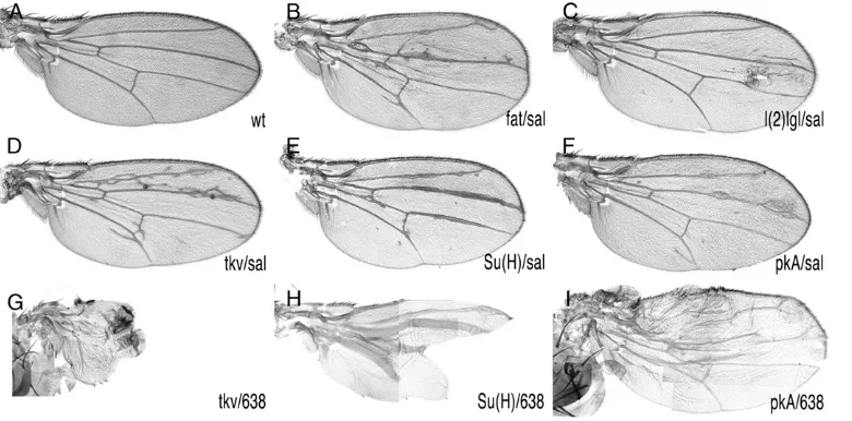
-Gal4 during the third larval instar (Figure 1B) induces FRT-mediated mitotic re-combination in the 2L and results in the generation of cells homozygous for the al dp b pr mut FRT40A chromosomal arm. These cells also lose the Minute mutation [M(2)z] and grow to occupy large extents of the central domain of the wing blade (Figure 1,C–E). We also used638-Gal4 to generate mosaic wings. 638-Gal4 expression starts during the second larval instar and occurs in all wing cells (not shown and Figure 1F). In this case the entire wing blade and hinge became homozy-gous (Figure 1G). To estimate the visibility of phenotypes and the viability and fertility of heterozygous animals with mosaic wings, we induced clones of cells homozygous for mutations in genes whose roles in wing formation are well established. We used lethal alleles offat(ft),lethal (2) giant larvae[l(2)gl],thick veins(tkv),Suppressor of Hairless [Su(H)], andProtein kinase A(PkA). In all cases and for both drivers, salEPv-Gal4 and 638-Gal4, the resulting mosaic animals display wing phenotypes that were easy to identify under the dissecting microscope (Figure 2). The phenotypes of combinations involving638-Gal4were always stronger than those of the corresponding combi-nations usingsalEPv-Gal4(compare Figure 2D with 2G, 2E with 2H, and 2F with 2I). However, the viability of 638-Gal4 combinations was poor, and in many cases adult animals can be recovered only as escapers. For these reasons we used thesalEPv
-Gal4driver for the screen and the638-Gal4driver to analyze the phenotype of homo-zygous mutant wings.
Establishment of complementation groups:We scored 14,000 males of thesalEPv
-Gal4; al dp b pr mut FRT40A/ FRT40A M(2)z; UAS-FLP/1genotype, wheremutmeans a recessive mutation induced in 2L. From the F1males with abnormal wings we established 140 w; al dp b pr mut FRT40A/CyOstocks, and subsequently we used them to generate mosaic animals in which mitotic recombination is driven in the 638-Gal4 domain of expression. We grouped these alleles using the phenotypes of the combinations involving the salEPv
-Gal4 and 638-Gal4 drivers and then crossed all the mutants in each group by each other to establish putative complementation groups. One member of each complementation group was crossed with alleles of known genes showing a similar phenotype in mosaics. The result of this analysis is shown in Table 1 (new complementation groups) and Figure 3A (complementation groups of known genes). As a summary, we identified 83 complementation groups, of which 12 correspond to previously known genes (Figure 3A) and 71 to mutations in other as yet unidentified genes (Figure 3B). The group of ‘‘known’’ genes had the higher number of alleles per complementation group (Figure 3B), whereas of the 71 novel complementation groups only 16 had more than one allele (Table 1 and Figure 3B). Although there is some correlation between the size of the coding region and the number of alleles identified for each known gene (Figure 3A), other aspects of the mutants appear more relevant to determine the probability of their identification.
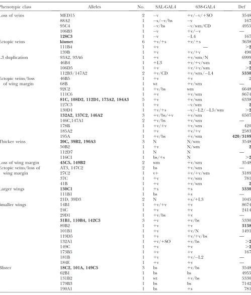
TABLE 1
Mutants identified in the screen grouped in phenotypic classes (first column), indicating the different alleles identified (‘‘Alleles’’ column), the number of alleles included in each complementation group (‘‘No.’’), the phenotype in the
combinationssalEPv-Gal4/1; FRT40A mut al dp b pr/FRT40A M(2)z; UAS-FLP/1(‘‘SAL-GAL4’’) and638-Gal4/1;
FRT40A mut al dp b pr/FRT40A M(2)z; UAS-FLP/1(‘‘638-GAL4’’), and the name of the deficiency they fail to complement (‘‘Def’’)
Phenotypic class Alleles No. SAL-GAL4 638-GAL4 Def
Loss of veins MED15 2 v 1v/v/1SO 3548
88A2 1 s/v/bs v 167
95C4 1 v/bs v/wm/CD 4955
106B3 1 v 1v/v —
128C3 1 v L4 167
Ectopic veins kismet 6 1v/1s 1v/1s 3638
111B4 1 1v — .2
139B 1 1v 1v/1v 490
L3 duplication 93A2, 93A6 1 1v 1v/wm/N 6999
46B4 1 1L3 1v/1v/wm 2
106D5 1 1v 1v/1v/wm .2
112B3/147A2 2 1v/CD 1v/wm/L4 5330
Ectopic veins/loss of wing margin
46B5 1 1v 1v 2
68B 1 wt 1v/wm —
92C2 1 1v/bs wm 6648
111C6 1 1v 1v/wm 8674
81C, 108D2, 112D1, 173A2, 184A3 5 1v 1v/wm 6338
127C3 1 1v v/wm 2
130D1 1 1v/1s v/L2/L5/wm .2
132A2, 137C2, 146A2 3 1v/bs/1v 1v/wm 6507
146C,147A1 2 1v/bs 1v/wm —
178B 1 1v/1v 1v/wm 420
185A2 1 1v 1v/1v 2583
195A 1 1v/bs 1v/wm 420/3189
Thicker veins 20C, 39B2, 190A3 3 N N/wm 3548
50B2 1 1v N/wm 2
112D7 1 N N —
116C1 1 bs/1s N .2
Loss of wing margin 45C5, 149B2 2 wm 1v/wm 3548
Ectopic veins/loss of wing margin
AT3, 147C2 2 bs 1v/wm —
27C2 1 v1 1v/1v/wm 3189
37C 1 1v 1v/wm 781
41B 1 1v 1v/wm 2
Larger wings 130C1 1 1s 1s 5330
111B1 1 bs 1s —
21D, 39D3 2 N 1s/1L3 1045
Smaller wings 14B1 1 1v/1v 1v 8674
24C 1 1v 1v 2414
29D1 1 1v/bs 1v —
31B1, 110B4, 142C3 3 1v 1v/bs 5330
89B2 1 1v 1v 3138
101B1 1 1v 1v/N 1491
119D5 1 1v 1v/1v/bs —
132A1 1 1v/1SO 1v/bs .2
149C 1 1v 1v .2
173B5 1 1v 1v 167
181B 1 1v 1v/L2 —
184E 1 1v 1v —
Blister 18C2, 101A, 149C5 3 bs 1v/bs 3548
62B1 1 bs bs 4955
131B2 1 wt 1v/bs 5330
179B3 1 bs bs 7142
190A1 1 bs 1s 781
the wing margin, causing the loss of wing tissue around the wing margin (Figure 4D) [some of these mutants also affect the distance between the longitudinal veins (Figure 4, E and F)]; (3) mutants affecting mainly the size of the wing with no or only minor effects in the patterning of veins (Figure 5, B and C); (4) mutants affecting the adhesion between the dorsal and ventral wing surfaces (Figure 5, D and E); and (5) mutants affecting the differentiation of trichomes (Figure 5F). Although most mutants can be assigned to one of these classes, in many instances we observed phenotypes with characteristics shared between two or more groups. For example, several mutations affect the size of the wing, the patterning of veins, and the formation of the wing margin (Figures 4, D–F, and 5E). In these cases, we crossed representative alleles with members of several phenotypic classes to establish the complementation groups. The phenotypes of mosaic wings for all new alleles identified and for a representative allele of all known complementation groups isolated in the screen in combination with the Gal4 linessalEPv-Gal4and 638-Gal4are shown insupporting information,Figure S1, Figure S2,Figure S3, andFigure S4.
Cytological mapping of novel mutants:To determine the cytological position of the new complementation groups, we used a collection of 36 chromosomal deficiencies that together cover the majority of the 2L arm (Figure 6A) and crossed them with 1 mutant (16 alleles) representing each complementation group and
with 53 single mutants from the 55 single mutants identified in the screening. To identify gaps in the coverage of the 2L arm by these deficiencies, we also determined the viability oftrans-heterozygous combina-tions between pairs of adjacent delecombina-tions, assuming that combinations between overlapping deficiencies are lethal. Using this criterion, we found that from the possible 35 pairwise combinations between adjacent deficiencies only 13 were lethal (Figure S5), suggesting that at least 22 small chromosomal intervals are not covered by these 36 deficiencies (Figure 6A). Using mainly lethality to define noncomplementation, we mapped to particular cytological intervals 12 of the 16 complementation groups formed by more than one allele (Figure 6B and Table 1). For individual mutations (55) we found 8 that complemented all deficiencies, 30 that failed to complement only one deficiency (Figure 6B and Table 1), and 17 that failed to complement two or more deficiencies (Figure 6C and Table 1). As we know which deficiencies overlap, those mutations that do not complement two adjacent deficiencies (2) were placed in the cytological region of overlap.
The complementation group formed by 77A2 and
133A1 corresponds to med15: We chose the comple-mentation group formed by the alleles77A2and133A1 to carry out an in-depth analysis of the affected gene. These alleles cause in mosaic wings a strong reduction of wing size, accompanied by the loss of the L2 vein and the differentiation of some ectopic bristles along the TABLE 1
(Continued)
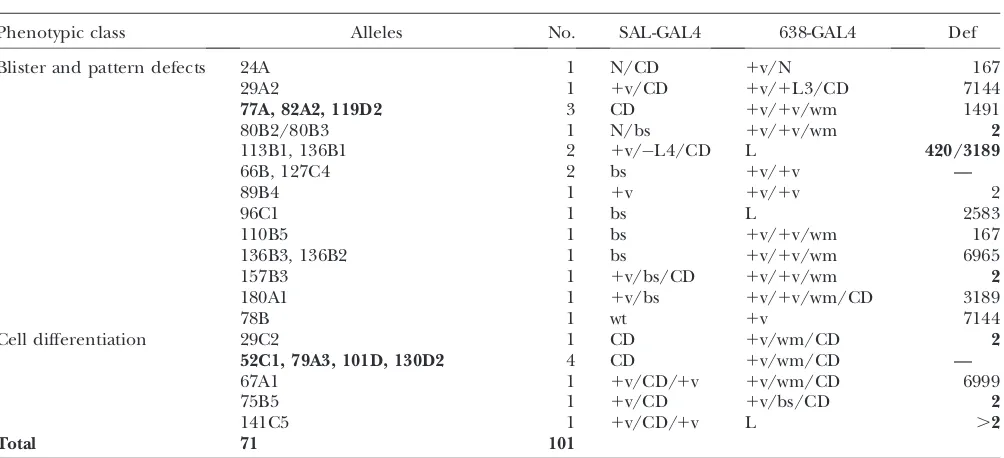
Phenotypic class Alleles No. SAL-GAL4 638-GAL4 Def
Blister and pattern defects 24A 1 N/CD 1v/N 167
29A2 1 1v/CD 1v/1L3/CD 7144
77A, 82A2, 119D2 3 CD 1v/1v/wm 1491
80B2/80B3 1 N/bs 1v/1v/wm 2
113B1, 136B1 2 1v/L4/CD L 420/3189
66B, 127C4 2 bs 1v/1v —
89B4 1 1v 1v/1v 2
96C1 1 bs L 2583
110B5 1 bs 1v/1v/wm 167
136B3, 136B2 1 bs 1v/1v/wm 6965
157B3 1 1v/bs/CD 1v/1v/wm 2
180A1 1 1v/bs 1v/1v/wm/CD 3189
78B 1 wt 1v 7144
Cell differentiation 29C2 1 CD 1v/wm/CD 2
52C1, 79A3, 101D, 130D2 4 CD 1v/wm/CD —
67A1 1 1v/CD/1v 1v/wm/CD 6999
75B5 1 1v/CD 1v/bs/CD 2
141C5 1 1v/CD/1v L .2
Total 71 101
veins L2 and L3 (Figure 7). Both77A2and133Afail to complementDf(2L)al(21B8–C1;21C8–D1), and within the interval covered by this deficiency,77A2and133A1 failed to complementDf(2L)BSC107, which deletes the 21C5;21D1 region (Figure 7A). There are 20 annotated genes in this interval, and we generated a smaller deficiency by FRT-mediated recombination between the Piggybac insertionsXP(1)d00080andWH(-)f06555. This deficiency includes only 8 genes and fails to complement the alleles 77A2 and 133A1 (Figure 7A). Finally, we combined77A2and133A1with mutations in the genescabut(cbt) andmed15and found that both77A2 and133A1are lethal in combination withmed15f04180and viable in combination with acbtallele (Figure 7A).Med15 encodes a 749-amino-acid protein characterized by the presence of a Kix domain, two small poly(Q) stretches, and several LXXLL motifs (Gelbartet al.1997; Figure 7B). We were able to map the133A1allele to themed15 coding region by sequencing genomic regions amplified by PCR from homozygous133A1embryos (see materi-als and methods). This mutation is associated with a C to T transition that introduces a stop codon in the region that corresponds to the N-terminal poly(Q)
localized after the KIX domain (Figure 7C). Using the same approach we could not find any nucleotide change in the coding region ofmed15in the77A2allele. Mitotic recombination clones of themed15f04180allele, generated in 638-Gal4; med15f04180 FRT40A; M(2)z FRT40A; UAS-FLP/1flies, result in smaller than wild-type wings with a normal pattern of veins (Figure 7H), suggesting that this allele is weaker than the novel77A2(Figure 7G) and 133A1(Figure 7F) mutations. Homozygous 133A1die during embryogenesis, and although some homozygous 77A2embryos can hatch, they die during the first larval instar (data not shown). We have not studied the em-bryonic phenotypes ofmed15homozygous alleles.
The most characteristic phenotypes of med15alleles in the wing are loss of the L2 vein and a reduction in the size of the wing (Figure 7, F–H). These phenotypes were observed in mosaic wings generated in638-Gal4; med15 FRT40A/M(2)z FRT40A; UAS-FLP/1flies. We generated mitotic recombination clones ofmed15alleles inhsFLP f36a; med15 al dp b pr FRT40A/M(2)z [f1
] FRT40Amales, because in this case the clones are labeled with the cell marker forked (f), and this allows the study of the autonomy of the clonal phenotypes. We could generate largemed15 M1
mutant clones when they were induced 48–72 h AEL, and in all cases themed15alleles behave in a cell autonomous manner. Thus, when the clones occupy the region between the L3 and L4 veins (n¼ 4), the size of this territory is strongly reduced (compare Figure 7I and 7J), and when the clones include the ventral L2 vein (n¼10) or the dorsal L4 vein (n¼5), these veins are reduced or absent (Figure 7, K and L). Med15clones running along the dorsal L3 vein diminish the pigmentation of the vein, but do not eliminate its differentiation (n¼4, data not shown). Similarly, clones affecting the proximal regions of L3, L4, and L5 do not affect vein differentiation (data not shown). We also studied the phenotypes resulting from the expression in the wing disc of interference RNA directed against med15(med15-i). In these flies, we observe a reduction in the size of the wing, the formation of some ectopic sensory organs, and only in some cases the loss of the L2 vein (Figure 7, N–P). Finally, we also found a require-ment formed15in other tissues such as the thorax and legs. In the first case, the more frequent phenotype was a failure of the left and right hemithoraxes to fuse when one or both hemithoraxes are composed of med15 mutant cells (Figure 7, Q and R). In the legs, we found many cases of legs with severe shortening along their entire length, accompanied by fusions of tarsal seg-ments (not shown). These phenotypes are fully pene-trant when the posterior compartments of the legs are composed ofmed15cells, as happens inw; med1577A2
al dp b pr FRT40A/M(2)z [f1
] FRT40A; UAS-FLP/hh-Gal4 flies (Figure 7, Tand T9, compare with Figure 7S). In general, themed15phenotypes are reminiscent of those caused by reduced dppand TGFb signaling, which consist of loss of veins, failures in dorsal closure, leg morphogenesis Figure3.—General results of the screen. (A) Known genes
defects (Dpp), and reduced wing size (TGFb) (Posakony et al. 1990; de Celis 1997; Lecuit and Cohen 1997; Brummelet al.1999; Harden2002).
We also observed several alterations in gene expres-sion patterns in med15 mutant cells during imaginal development. For example, the expression of Bs is reduced in intervein territories formed bymed15cells (Figure 8, A and B), suggesting that the transcriptional response to Dpp and Hedgehog signaling requires
Med15 activity. Similarly, the expression ofspalt(sal), a direct target of Dpp signaling (de Celis et al. 1996; BarrioanddeCelis2004), is absent inmed15clones localized in the anterior and posterior regions of the Sal domain of expression (Figure 8, C and D) and is reduced in clones localized in the central domain of sal expression (Figure 8, C and D). These effects are always cell autonomous, suggesting that Med15 does not affectdppexpression but compromises the capability of Figure 4.—Representa-tive wings of the pheno-typic classes affecting the formation of veins. (A and A9) Loss of veins and re-duced wing size in salEPv -Gal4; M(2)Z FRT40A/al dp b pr 128C3 FRT40A; UAS-FLP/1 (A) and 638-Gal4; M(2)Z FRT40A/al dp b pr 128C3 FRT40A; UAS-FLP/1 (A9). (B and B9) Loss and gain of veins in salEPv-Gal4; M(2)Z FRT40A/al dp b pr 61C FRT40A; UAS-FLP/1 (B) and 638-Gal4; M(2)Z FRT40A/al dp b pr 61C FRT40A; UAS-FLP/1 (B9). The mutant61Cis an allele of kismet (kis). (C and C9) Thick vein phenotype insalEPv-Gal4; M(2)Z FRT40A/al dp b pr 20C FRT40A; UAS-FLP/1(C) and638-Gal4; M(2)Z FRT40A/al dp b pr 20C FRT40A; UAS-FLP/1(C9). (D and D9) Wing margin phenotype ofsalEPv-Gal4; M(2)Z FRT40A/al dp b pr 45C5 FRT40A; UAS-FLP/1(D) and638-Gal4; M(2)Z FRT40A/al dp b pr 45C5 FRT40A; UAS-FLP/1(D9). (E and E9) Ectopic veins and loss of wing margin insalEPv-Gal4; M(2)Z FRT40A/al dp b pr 81C FRT40A; UAS-FLP/1(E) and638-Gal4; M(2)Z FRT40A/al dp b pr 81C FRT40A; UAS-FLP/1(E9). (F and F9) Ectopic veins and reduction in wing size insalEPv-Gal4; M(2)Z FRT40A/al dp b pr 132A2 FRT40A; UAS-FLP/1(F) and638-Gal4; M(2)Z FRT40A/al dp b pr 132A2 FRT40A; UAS-FLP/1(F9). The phenotypes of other mutations affecting the veins or the wing margin are shown inFigure S1andFigure S3.
Dpp signaling to activate its targets. A reduced response to Dpp signaling might also contribute to the smaller than normal wing size ofmed15mosaic wings and to the partial loss of vein stretches. We could not find changes in the expression of EGFR or Wingless target genes (argos,Delta, anddistalless; data not shown), indicating that Med15 is not required as a general coactivator of transcription, but rather that its function is specific to particular enhancer–promoter interactions.
Other members of the Med complex are required during wing development:The involvement ofmed15in wing disc development suggests that other members belonging to the Med complex would be required in similar processes. Alternatively,med15functions might be independent of its participation in the Med complex. We studied the loss-of-function phenotypes of several Med complex components by driving the expression of specific interference RNAs in the wing disc. In all cases analyzed, we found that the reduction in Med expres-sion gave rise to smaller than normal wings (med10-i, med16-i,med25-i,med27-I, andkto-i; Figure 9), which were extreme in the cases of loss ofmed20andmed30(Figure 9). Only the reduction inmed20andmed30expression resulted in loss of vein phenotypes (Figure 9). In the cases of med20, med30, and med15, the expressions of their interference RNAs induce cell death (see Figure 8, E–H). In summary, and although the phenotypes observed upon a reduction in the expression of various Med components are not identical, they are similar enough to suggest that they could be caused by different degrees of loss of Mediator function.
DISCUSSION
The patterning of the veins and the growth of the wing involve the activities of several conserved signaling pathways and transcription factors, and mutations in these genes result in modifications of vein positioning and wing size (SotillosanddeCelis2005). We have carried out a mosaic screen to search for mutations in the 2L chromosomal arm that modify the pattern of veins and the growth of the wing. In this screen we generated homozygous wing regions in otherwise het-erozygous flies using a combination of the FRT-FLP method and the Gal4-UAS system. We maximize the fraction of the wing occupied by homozygous mutant cells using a Minute mutation. This also allows the survival of mutants cells that otherwise could be out-competed by the surrounding wild-type cells (Morata and Ripoll1975). The inconvenience of using aMinute mutation is that stocks and crosses involving this allele are less healthy. In general, the flies heterozygous for newly induced mutations with mosaic wings were viable and fertile, allowing the screening of a high number of treated chromosomes.
known genes, as only 16 of 71 novel complementation groups were formed by more than one allele. These numbers indicate that the screen is not yet at saturation and that the visibility of the phenotypes is much higher in wings homozygous for mutations in the class of known genes. We were able to map 58% of the complementation groups to individual chromosomal
intervals using a collection of deficiencies that cover an estimated 80% of the 2L arm. However, these data have several caveats due to the high number of complemen-tation groups formed by only one allele and to the presence in the treated chromosomes of associated lethals. Thus, 23% of the novel complementation groups failed to complement with more than one deficiency, and
associated with the loss of the ventral L2 vein. (L) Smallmed15133A1clone in the distal dorsal L4 vein causing the loss of this vein. (M–P) Phenotypes resulting from the expression ofmed15interference RNA (med15i) in the genotypes638-Gal4/1; UAS-med15i (N),salEPv-Gal4/UAS-med15i; UAS-dicer/1(O), andsalEPv-Gal4/UAS-med15i; UAS-dicer/1grown at 29°(P). The wild-type control wing is shown in M. (Q and R) Two examples of thoraxes taken at different magnification showing the failure in the fusion between the left and the right hemithorax when the central region is occupied bymed1577Amutant cells (labeled withforkedinhsFLP1.22 f36a; P[f1]30C M(2)z FRT40A/al med1577A2dp b pr FRT40Aflies). (S) Wild-type male first leg. (T and T9) Two examples of legs taken at different magnifications (T9is34 T) showing the defects in leg morphogenesis and tarsal segmentation inw; M(2)z P[f1]30C FRT40A/al med1577Adp b pr FRT40A; hh-Gal4/UAS-FLPflies.
11% of complementation groups complemented for lethality and phenotype with all deficiencies. In this manner, the cytological localization of all complementa-tion groups composed by only one allele is still tentative. The phenotypes identified in the screen mainly affected the wing veins and wing margin, the size of the wing, the adhesion between the dorsal and ventral wing surfaces, the integrity of the epithelium, and the differentiation of trichomes by wing cells. These phenotypes correspond to alterations in processes that occur during the third larval instar (vein determination, wing disc growth, and wing margin formation) and during pupal development (dorso-ventral adhesion and trichome differentiation). Furthermore, the observed phenotypes are informative about the process, and in some cases the pathways, that might be altered in the mutants. For example, changes in wing size without effects in vein formation are expected by modifications in the insulin and TGFb pathways (Brummel et al. 1999; Johnston and Gallant 2002), alterations in the integrity of the wing margin are a mark of loss of Notch and Wingless signaling at the dorso-ventral boundary (Couso et al. 1994; de Celis and Garcia-Bellido 1994), changes in the formation of veins are expected by modifications in the Dpp, EGFR, and Notch pathways (SotillosanddeCelis2005), and the formation of blistered wings is typical of defects in Integrin and Laminin functions (Walshand Brown1998; Urbanoet al.2009). Future work will aim to unambigu-ously map the different complementation groups to individual genes and to identify the developmental func-tions they affect.
We chose to analyze in some detail the complemen-tation group formed by the77A and 133A1 mutants. These mutations are alleles ofmed15, a gene encoding one component of the Mediator complex (Guglielmi
et al.2004). Thus, they fail to complement othermed15 alleles, and med15133A1
is associated with a stop codon that could truncate the protein in the N-terminal region after the KIX domain. The Mediator multiprotein complex promotes the transcription of inducible genes, acting as a link between the RNApolII holoenzyme and several sequence-specific transcription factors (Naar et al. 2001; Lewis and Reinberg 2003; Taatjes et al. 2004). The human homolog of Med15, MED105, is included in all Mediator complexes identified so far and forms part of a module named the tail that is the main target for the transcriptional activators (Guglielmiet al. 2004; Taatjeset al.2004). Thus, Med15 homologs can bid to different transcription factors such as Gcn4 and Gal4 in Saccharomyces cerevisiae (Fishburn et al. 2005; Reeves and Hahn 2005), SREBP in Caenorhabditis elegans (Taubert et al. 2006; Yang et al. 2006), and, more interesting from the perspective of our data, to Smad2/3 and Smad4 in Xenopus (Katoet al. 2002). Other members of the Mediator complex that were previously analyzed are kohtalo and skuld(Med12 and Med13, respectively), which form part of the conserved Cdk8 module (Bourbon et al. 2004; Conaway et al. 2005; Kim and Lis 2005). Interestingly, mouse Cdk8 and Cdk9 phosphorylate Smad proteins, regulating their transcriptional activity and turnover (Alarco´ n et al. 2009). However, kohtalo and skuld are required for sensory organ development, for some aspects of Notch and Hedgehog signaling, and for the transcrip-tion of Wingless downstream genes ( Janodyet al.2003; Carreraet al.2008).
expression of other components of the Mediator com-plex, most notablymed20,med27, andmed30, also results in smaller than normal wings and failures in vein differentiation, in addition to causing some levels of cell death. Although these phenotypes were similar, they are not identical, which might indicate specific require-ments of these subunits or, alternatively, a different degree in the effectiveness of each interference RNA used. Mutantmed15cells display specific defects in gene expression, suggesting a requirement limited to partic-ular enhancer–promotor interactions. In particpartic-ular, the expression ofspalt, a direct target of Dpp signaling, is compromised in med15 mutant cells. There are no known transcriptional targets of TGFbsignaling in the wing, and consequently we could not determine directly whether the activity of this pathway is diminished in med15mutants. A direct requirement of Med15 for the transcription of TGFbtarget genes is nonetheless sug-gested by the similar phenotypes of wing size reduction observed in med15 mutants and in baboon mutations (Brummelet al.1999).
We are very grateful to Rosario Herna´ndez and Cristina Prieto for their technical assistance. This work was supported by grants BFU2006-06501 and Consolider CSD-2007-00008 from the Spanish Ministry of Research and Innovation. An institutional grant from the Ramo´n Areces Foundation to the Centro de Biologı´a Molecular ‘‘Severo Ochoa’’ is also acknowledged.
LITERATURE CITED
Alarco´ n, C., A.-I. Zaromytidou, Q. Xi, S. Gao, J. Yu et al., 2009 Nuclear CDKs drive Smad transcriptional activation and turnover in BMP and TGF-b pathways. Cell139:757–769. Ashburner, M., 1989 Drosophila. A Laboratory Manual.Cold Spring
Harbor Laboratory Press, Cold Spring Harbor, NY.
Baker, N. E., 1987 Molecular cloning of sequences from wingless a segment polarity gene in Drosophila the spatial distribution of a transcript in embryos. EMBO J.6:1765–1774.
Barrio, R., and J. F.deCelis, 2004 Regulation ofspaltexpression in theDrosophilawing blade in response to the Decapentaplegic signaling pathway. Proc. Natl. Acad. Sci. USA101:6021–6026. Bier, E., 2000 Drawing lines in theDrosophilawing: initiation of wing
vein development. Curr. Opin. Genet. Dev.10:393–398. Boedigheimer, M., P. Bryantand A. Laughon, 1993 Expanded,
a negative regulator of cell proliferation in Drosophila, shows homology to the NF2 tumor suppressor. Mech. Dev.44:83–84. Bourbon, H. M., A. Aguilera, A. Z. Ansari, F. J. Asturias, A. J.
Berket al., 2004 A unified nomenclature for protein subunits of mediator complexes linking transcriptional regulators to RNA polymerase II. Mol. Cell14:553–557.
Brentrup, D., H. P. Lerch, H. Jackle and M. Noll, 2000 Regulation ofDrosophilawing vein patterning:netencodes a bHLH protein repressingrhomboidand is repressed by rhom-boid dependent EGFR signalling. Development127:4729–4741. Brummel, T., S. Abdollah, T. E. Haerry, M. J. Shimell, J. Merriam
et al., 1999 The Drosophila Activin receptor Baboon signals through dSmad2 and controls cell proliferation but not pattern-ing durpattern-ing larval development. Genes Dev.13:98–111. Calleja, M., E. Moreno, S. Pelazand G. Morata, 1996 Visualization
of gene expression in living adultDrosophila.Science274:252–255. Carrera, I., F. Janody, N. Leeds, F. Duveauand J. E. Treisman, 2008 Pygopus activates Wingless target gene transcription through the mediator complex subunits Med12 and Med13. Proc. Natl. Acad. Sci. USA105:6644–6649.
Chen, Y., and G. Struhl, 1996 Dual roles for Patched in sequester-ing and transducsequester-ing Hedgehog. Cell87:553–563.
Clegg, N. J., I. P. Whitehead, J. A. Williams, G. B. Spiegelmanand T. A. Grigliatti, 1993 A developmental and molecular analysis ofCdc2mutations in Drosophila melanogaster. Genome36:676– 685.
Cohen, S. M., 1993 Imaginal Disc Development.Cold Spring Harbor Laboratory Press, Cold Spring Harbor, NY.
Conaway, R. C., S. Sato, C. Tomomorisato, T. Yao and J. W. Conaway, 2005 The mammalian Mediator complex and its role in transcriptional regulation. Trends Biochem. Sci. 30: 250–255.
Couso, J. P., S. A. Bishopand A. MartinezArias, 1994 The wing-less signalling pathway and the patterning of the wing margin in
Drosophila.Development120:621–636.
Cruz, C., A. Glavic, M. Casadoand J. F.deCelis, 2009 A gain of function screen identifying genes required for growth and pat-tern formation of the Drosophila melanogaster wing. Genetics 183:122.
deCelis, J. F., 1997 Expression and function ofdecapentaplegicand
thick veinsin the differentiation of the veins in theDrosophilawing. Development124:1007–1018.
deCelis, J. F., 2003 Pattern formation in theDrosophilawing: the development of the veins. BioEssays25:443–451.
deCelis, J. F., and A. Garcia-Bellido, 1994 Roles of theNotchgene inDrosophilawing morphogenesis. Mech. Dev.46:109–122. deCelis, J. F., R. Barrioand F. C. Kafatos, 1996 A gene complex
acting downstream of Dpp in Drosophilawing morphogenesis. Nature381:421–424.
Duffy, J. B., D. A. Harrisonand N. Perrimon, 1998 Identifying loci required for follicular patterning using directed mosaics. Development125:2263–2271.
Fishburn, J., N. Mohibullah, and S. Hahn, 2005 Function of a eu-karyotic transcription activator during the transcription cycle. Mol. Cell18:369–378.
Freeman, M., 1994 Thespitzgene is required for photoreceptor de-termination in theDrosophilaeye where it interacts with the EGF receptor. Mech. Dev.48:25–33.
Fristrom, D., P. Gotwals, S. Eaton, T. Kornberg, M. A. Sturtevant
et al., 1994 blistered; a gene required for vein/intervein formation in wings ofDrosophila.Development120:2661–2686.
Garcia-Bellido, A., and J. Dapena, 1974 Induction, detection and characterization of cell differentiation mutants in Drosophila.
Mol. Gen. Genet.128:117–130.
Gelbart, W. M., M. Crosby, B. Matthews, W. P. Rindone, J. Chillemi
et al., 1997 FlyBase: aDrosophiladatabase. The FlyBase consor-tium. Nucleic Acids Res.25:63–66.
Guglielmi, B., N. L. V. Berkum, B. Klapholz, T. Bijma, M. Boube
et al., 2004 A high resolution protein interaction map of the yeast Mediator complex. Nucleic Acids Res.32:5379–5391. Hao, I., R. B. Green, O. Dunaevsky, J. A. Lengyeland C. Rauskolb,
2003 The oddskipped family of zinc finger genes promotes
Drosophilaleg segmentation. Dev. Biol.263:282–295.
Harden, N., 2002 Signaling pathways directing the movement and fusion of epithelial sheets: lessons from dorsal closure in Dro-sophila. Differentiation70:181–203.
Ito, K., W. Awano, K. Suzuki, Y. Hiromi and D. Yamamoto, 1997 TheDrosophilamushroom body is a quadruple structure of clonal units each of which contains a virtually identical set of neurones and glial cells. Development124:761–771. Janody, F., Z. Martirosyan, A. Benlali and J. E. Treisman,
2003 Two subunits of theDrosophilamediator complex act to-gether to control cell affinity. Development130:3691–3701. Johnston, L. A., and P. Gallant, 2002 Control of growth and
organ size in Drosophila. BioEssays24:54–64.
Kato, Y., R. Habas, Y. Katsuyama, A. M. Naarand X. He, 2002 A component of the ARC/Mediator complex required for TGF beta/Nodal signalling. Nature418:641–646.
Kim, J., A. Sebring, J. J. Esch, M. E. Kraus, K. Vorwerket al., 1996 Integration of positional signals and regulation of wing formation by Drosophila vestigial gene. Nature 382: 133–138.
Kim, Y. J., and J. T. Lis, 2005 Interactions between subunits of
Dro-sophilaMediator and activator proteins. Trends Biochem. Sci.30: 245–249.
Kuang, B., S. C. Wu, Y. Shin, L. Luoand P. Kolodziej, 2000 split
fate and axon extension in theDrosophilaembryo. Development 127:1517–1529.
Kwon, J. Y., J. M. Park, B. S. Gim, S. J. Han, J. Lee et al., 1999 Caenorhabditis elegans mediator complexes are required for developmental specific transcriptional activation. Proc. Natl. Acad. Sci. USA96:14990–14995.
Lecuit, T., and S. M. Cohen, 1997 Proximal distal axis formation in theDrosophilaleg. Nature388:139–145.
Lewis, B. A., and D. Reinberg, 2003 The mediator coactivator com-plex: functional and physical roles in transcriptional regulation. J. Cell Sci.116:3667–3675.
Li, W., J. Talavera, M. Laneand D. Kalderon, 1995 Function of protein kinase A in hedgehog signal transduction andDrosophila
imaginal disc development. Cell80:553–562.
Mahoney, P. A., U. Weber, P. Onofrechuk, H. Biessmann, P. J. Bryant
et al., 1991 Thefattumor suppressor gene inDrosophilaencodes a novel member of the cadherin gene superfamily. Cell67:853–868. Montagne, J., J. Groppe, K. Guillemin, M. A. Krasnow, W. J. Gehring
et al., 1996 The Drosophila serum response factor gene is re-quired for the formation of intervein tissue of the wing and is allelic toblistered.Development122:2589–2597.
Morata, G., and P. Ripoll, 1975 Minutes: mutants ofDrosophila au-tonomously affecting cell division rate. Dev. Biol.42:211–221. Morel, V., and F. Schweisguth, 2000 Repression by Suppressor of
Hairless and activation by Notch are required to define a single row of singleminded expressing cells in theDrosophilaembryo. Genes Dev.14:377–388.
Naar, A. M., B. D. Lemonand R. Tjian, 2001 Transcriptional coac-tivator complexes. Annu. Rev. Biochem.70:475–501.
Nellen, D., M. Affolterand K. Basler, 1994 Receptor serine/ threonine kinases implicated in the control of Drosophila body pattern by decapentaplegic. Cell78:225–237.
Parks, A. L., K. R. Cook, M. Belvin, N. A. Dompe, R. Fawcettet al., 2004 Systematic generation of high resolution deletion cover-age of theDrosophila melanogastergenome. Nat. Genet.36:288– 292.
Posakony, L. G., L. A. Rafteryand W. M. Gelbart, 1990 Wing for-mation in Drosophila melanogaster requires decapentaplegic gene function along the anterior posterior compartment boundary. Mech. Dev.33:69–82.
Prout, M., Z. Damania, J. Soong, D. Fristromand J. W. Fristrom, 1997 Autosomal mutations affecting adhesion between wing surfaces inDrosophilamelanogaster. Genetics146:275–285. Reeves, W. M., and S. Hahn, 2005 Targets of the Gal4 transcription
activator in functional transcription complexes. Mol. Cell Biol.25: 9092–9102.
Ripoll, P., and A. Garcia-Bellido, 1979 Viability of homozygous deficiencies in somatic cells ofDrosophila melanogaster.Genetics 91:443–453.
Roch, F., A. Baonza, E. Martin-Blanco and A. Garcia-Bellido, 1998 Genetic interactions and cell behaviour in blistered
mu-tants during proliferation and differentiation of theDrosophila
wing. Development125:1823–1832.
Rogge, R., P. J. Green, J. Urano, S. Hornsaban, M. Mlodziket al., 1995 The role of yan in mediating the choice between cell division and differentiation. Development121:3947–3958. Rogge, R. D., C. A. Karlovichand U. Banerjee, 1991 Genetic
dis-section of a neurodevelopmental pathway: Son of sevenless func-tions downstream of the sevenless and EGF receptor tyrosine kinases. Cell64:39–48.
Sotillos, S., and J. F. de Celis, 2005 Interactions between the Notch, EGFR, and decapentaplegic signaling pathways regulate vein differentiation duringDrosophilapupal wing development. Dev. Dyn.232:738–752.
Sotillos, S., F. Rochand S. Campuzano, 1997 The metallopro-tease disintegrin Kuzbanian participates in Notch activation during growth and patterning ofDrosophilaimaginal discs. Devel-opment124:4769–4779.
St. Johnston, R. D., F. M. Hoffmann, R. K. Blackman, D. Segal, R. Grimailaet al., 1990 Molecular organization of the
decapenta-plegicgene inDrosophila melanogaster.Genes Dev.4:1114–1127. Taatjes, D. J., M. T. Marrand R. Tjian, 2004 Regulatory diversity
among metazoan co-activator complexes. Nat. Rev. Mol. Cell Biol. 5:403–410.
Taubert, S., M. R. V. Gilst, M. Hansen and K. R. Yamamoto, 2006 A Mediator subunit, MDT15, integrates regulation of fatty acid metabolism by NHR49 dependent and independent path-ways inC. elegans.Genes Dev.20:1137–1149.
Urbano, J. M., C. Torgler, C. Molnar, A. Lo´ pez-Varea, N. Brown
et al., 2009 Drosophilalaminins act as key regulators of basement membrane assembly and morphogenesis. Development 136: 4165–4176.
Walsh, E. P., and N. H. Brown, 1998 A screen to identifyDrosophila genes required for integrin mediated adhesion. Genetics150: 791–805.
Williams, J. A., J. B. Belland S. B. Carroll, 1991 Control of
Dro-sophilawing and haltere development by the nuclear vestigial gene product. Genes Dev.5:2481–2495.
Xu, T., and S. D. Harrison, 1994 Mosaic analysis using FLP recom-binase. Methods Cell Biol.44:655–681.
Xu, T., W. Wang, S. Zhang, R. A. Stewart and W. Yu, 1995 Identifying tumor suppressors in genetic mosaics: the Dro-sophila latsgene encodes a putative protein kinase. Development 121:1053–1063.
Yang, F., B. W. Vought, J. S. Satterlee, A. K. Walker, Z. Y. JimSun
et al., 2006 An ARC/Mediator subunit required for SREBP control of cholesterol and lipid homeostasis. Nature442:700– 704.
Supporting Information
http://www.genetics.org/cgi/content/full/genetics.109.113670/DC1
Identification of Genes Affecting Wing Patterning Through
a Loss-of-Function Mutagenesis Screen and Characterization
of
med15
Function During Wing Development
Ana Terriente-Félix, Ana López-Varea and Jose F. de Celis
Copyright © 2010 by the Genetics Society of America
A. Terriente-Félix et al.
2 SI
FIGURE S1.—Mosaicwings for mutations isolated in the screen in flies of salEPv-Gal4/+; FRT40A mut al dp b pr/FRT40A
A. Terriente-Félix et al. 3 SI
FIGURE S2.—Mosaic wings for mutations isolated in the screen in flies of salEPv-Gal4/+; FRT40A mut al dp b pr/FRT40A
M(2)z; UAS-FLP/+ (B-R) and 638-Gal4/+; FRT40A mut al dp b pr/FRT40A M(2)z; UAS-FLP/+ (B’-R’). A and A’ correspond to control wings of salEPv-Gal4/+; FRT40A al dp b pr/FRT40A M(2)z; UAS-FLP/+ (A) and 638-Gal4/+; FRT40A al dp b
A. Terriente-Félix et al. 4 SI
FIGURE S3.—Mosaic wings for mutations isolated in the screen in flies of salEPv-Gal4/+; FRT40A mut al dp b pr/FRT40A
A. Terriente-Félix et al. 5 SI
FIGURE S4.—Mosaic wings for mutations isolated in the screen in flies of salEPv-Gal4/+; FRT40A mut al dp b pr/FRT40A
A. Terriente-Félix et al.
6 SI
FIGURE S5.—Results of the combinations between the deficiencies used for the cytological mapping of the
A. Terriente-Félix et al. 7 SI
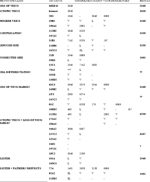
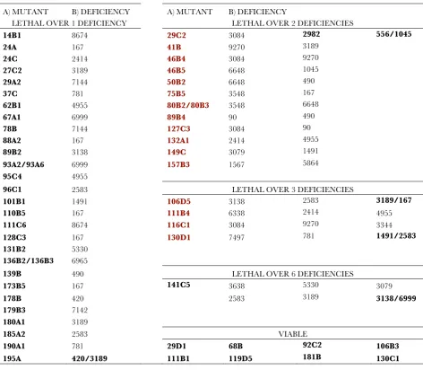
TABLE S1
Results of the mapping of complementation groups composed by two or more alleles
PHENOTYPE CLASS MUTANTS COMPLEMENTATION WITH DEFICIENCIES RESULT
LOSS OF VEINS MED15 3548 3548
ECTOPIC VEINS kismet 3638 3638
20C 1045 - 3548 5869
39B2 V V L V
THICKER VEINS
190A3 V 2892 - V
3548
112B3 3548 5330
L3 DUPLICATION
147A2 V L 5330
31B1 7142 5330 V 167
110B4 - L V -
REDUCED SIZE
142C3 V SL V V
5330
21D 1045 5869
AUGMENTED SIZE
39D3 L V 1045
52C1 1045 7142 2583
79A3 +V L
101D V V -
CELL DIFFERENTATION
130D2 V V -
??
45C5 3548 3079 3344 6999
LOSS OF WING MARGIN
149B2 L V V V 3548
AT3 2583 6374
147C2 V V ??
81C V 6338 179 V 6965
108D2 490 L V - 167
112D1 490 L 2892 V
173A2 V - V V
184A3 - - V -
6338
146A2 3084 6507
137C2 V L
132A2 V -
6507
146C -
ECTOPIC VEINS + LOSS OF WING MARGIN
147A1 - ?
18C2 3548 3189
101A L V
BLISTER
149C5 L V
3548
77A 1491 3638 3138 6965
82A2 SL V V V
BLISTER + PATHERN DEFFECTS
119D2 SL - - -
A. Terriente-Félix et al.
8 SI
113B1 420 3189
136B1 L L 420/3189
66B 5869
127C4 - ?
A. Terriente-Félix et al. 9 SI
TABLE S2
Results of the mapping of complementation groups formed by one single allele
A)MUTANT B) DEFICIENCY A) MUTANT B) DEFICIENCY
LETHAL OVER 1 DEFICIENCY LETHAL OVER 2 DEFICIENCIES
14B1 8674 29C2 3084 2982 556/1045
24A 167 41B 9270 3189
24C 2414 46B4 3084 9270
27C2 3189 46B5 6648 1045
29A2 7144 50B2 6648 490
37C 781 75B5 3548 167
62B1 4955 80B2/80B3 3548 6648
67A1 6999 89B4 90 490
78B 7144 127C3 3084 90
88A2 167 132A1 2414 4955
89B2 3138 149C 3079 1491
93A2/93A6 6999 157B3 1567 5864
95C4 4955
96C1 2583 LETHAL OVER 3 DEFICIENCIES
101B1 1491 106D5 3138 2583 3189/167
110B5 167 111B4 6338 2414 4955
111C6 8674 116C1 3084 9270 3344
128C3 167 130D1 7497 781 1491/2583
131B2 5330
136B2/136B3 6965
139B 490 LETHAL OVER 6 DEFICIENCIES
173B5 167 141C5 3638 5330 3079
178B 420 2583 3189 3138/6999
179B3 7142
180A1 3189
185A2 2583 VIABLE
190A1 781 29D1 68B 92C2 106B3
195A 420/3189 111B1 119D5 181B 130C1