_____________________________________________________________________________________________________ *Corresponding author: E-mail: [email protected];
www.sciencedomain.org
SEM (Scanning Electron Microscope) Evaluation of
Surface Roughness after Proximal Stripping of
Teeth Followed by Application of Fluoride Varnish
and Bonding Agent
Nishil Agrawal
1*, Hina Desai
1, Kalpesh Patel
1, Nikunj Patel
1,
Rahul Aghera
1and Nirav Dholakiya
11
Department of Orthodontics and Dentofacial Orthopaedics, Manubhai Patel Dental College, Vadodara, India.
Authors’ contributions
This work was carried out in collaboration between all authors. Author NA designed the study, performed the statistical analysis, wrote the protocol and wrote the first draft of the manuscript. Authors HD, KP and NP managed the analyses of the study. Authors RA and ND managed the literature searches. All authors read and approved the final manuscript.
Article Information
DOI: 10.9734/BJMMR/2017/30295 Editor(s): (1) Emad Tawfik Mahmoud Daif, Professor of Oral & Maxillofacial Surgery, Cairo University, Egypt. Reviewers: (1) Ibraheem A. Samotu, Ahmadu Bello University, Nigeria. (2)Bouchaib Zazoum, Royal Military College of Canada, Kingston, Ontario, Canada. (3)Vesna M. Maksimovic, University of Belgrade, Serbia. (4)Mahmood Ahmad, The Islamia University of Bahawalpur, Pakistan. Complete Peer review History:http://www.sciencedomain.org/review-history/17970
Received 31st October 2016 Accepted 20th February 2017 Published 27th February 2017
ABSTRACT
Aims: The objective of this SEM study was to compare the surface roughness after proximal
stripping with tungsten carbide bur and hand pulled strip then again followed by application of fluoride varnish and bonding agent.
Place and Duration of Study: Department of Orthodontics and Dentofacial Orthopaedics,
Manubhai Patel Dental College, Vadodara, between September 2015 and March 2016.
Methodology: 30 healthy human teeth were used which required removal for orthodontic purpose.
Teeth were randomly selected and divided into 6 group of 5 each. Group I – stripping with Hand pulled strips(Ortho Organizers), Group II – stripping with tungsten carbide bur ( SS White), Group
III - stripping with single sided hand pulled strips
application ,Group IV - stripping with tungsten carbide bur ( application ,Group V- stripping with Sin
varnish application, Group VI - stripp varnish application.
Results: The SEM investigations demonstrated that small number of furrow
group ii when compared to group i, the bonding agent formed a smooth layer over rough area produced after stripping in group iii and group iv, while a continuous uniform layer of fluoride varnish was formed in group v and group vi
Conclusion: It can be said that the bonding agent and fluoride varnish form a protective layer,
which will make the area less favourable for plaque accumulation or prevent its demineralization from oral acids and thereby reduce the incidence of caries, which is
stripping of teeth. Tungsten carbide bur produces a surface which is smoother when compared to the deep uniform furrows formed with hand pulled proximal strip. So It can be said that proximal stripping with tungsten carbide bur f
preferable.
Keywords: Scanning electron microscope; proximal stripping; fluoride varnish; bonding agent.
1. INTRODUCTION
Interproximal enamel reduction is also known as interdental stripping, enamel approximation, or slenderizing. Clinicians have practiced stripping to be an attractive alternative to transverse or antero-posterior expansion and to extractions. Several procedures are used in daily orthodontic practice to perform precise interdental stripping as part of the treatment plan[1].
Stripping of anterior teeth in cases which exhibit mild crowding is increasing in popularity [2]. Some orthodontists also advocate stripping to improve stability or to reshape the morphology of the teeth for aesthetic reasons.
Proximal stripping can be achieved with hand held or motor-driven abrasive strips and also with disks or burs mounted on a hand piece. Nowadays AIROTOR STRIPPING
most commonly used method for reduction of enamel [3-5].
According to Hudson [6], mesiodistal diameter reduction increased the risk of caries by roughening the enamel thus producing a tendency to retain debris. Several reported data suggest that the burs used to reduce interproximal enamel create furrows and scratches that can lead to carious lesions Danesh et al. [7] concluded that the use of coarse strips or burs for interproximal
left irregular surfaces that cannot be smoothed effectively by subsequent polishing.
study done by Srivastava et al
striping increases the chance of demineralization
stripping with single sided hand pulled strips (Ortho Organizers) followed by bonding agent ng with tungsten carbide bur (SS White) followed by bonding agent stripping with Single sided hand pulled strips(Ortho Organizers) with fluoride stripping with tungsten carbide bur (SS white) followed by fluoride
The SEM investigations demonstrated that small number of furrows were formed in group ii when compared to group i, the bonding agent formed a smooth layer over rough area produced after stripping in group iii and group iv, while a continuous uniform layer of fluoride varnish was formed in group v and group vi.
It can be said that the bonding agent and fluoride varnish form a protective layer, which will make the area less favourable for plaque accumulation or prevent its demineralization from oral acids and thereby reduce the incidence of caries, which is always seen after proximal Tungsten carbide bur produces a surface which is smoother when compared to the deep uniform furrows formed with hand pulled proximal strip. So It can be said that proximal stripping with tungsten carbide bur followed by application of fluoride varnish or bonding agent is
Scanning electron microscope; proximal stripping; fluoride varnish; bonding agent.
Interproximal enamel reduction is also known as interdental stripping, enamel approximation, or Clinicians have practiced stripping to be an attractive alternative to transverse or posterior expansion and to extractions. Several procedures are used in daily orthodontic practice to perform precise interdental stripping
Stripping of anterior teeth in cases which exhibit in popularity [2]. Some orthodontists also advocate stripping to improve stability or to reshape the morphology of
Proximal stripping can be achieved with hand-driven abrasive strips and also with rs mounted on a hand piece. Nowadays AIROTOR STRIPPING (ARS) are most commonly used method for reduction of
, mesiodistal diameter reduction increased the risk of caries by roughening the enamel thus producing a Several reported data suggest that the burs used to reduce interproximal enamel create furrows and scratches that can lead to carious lesions [1]. concluded that the use of coarse strips or burs for interproximal reduction left irregular surfaces that cannot be smoothed effectively by subsequent polishing. According to study done by Srivastava et al. [8] airrotar increases the chance of demineralization
and the chance of developing caries increases by many fold. In a study done by Alessandra and Federico [9] they concluded that when human enamel surfaces were stripped and finished under in vitro condition, it was not possible to produce an enamel surface free of furrows when evaluated under scanning electron microscope (SEM). According to Piacentini and Sfondrini tungsten carbide bur produces smoother surface when compared to diamond bur, so we have used tungsten carbide bur in this study
Rogers and Wagner [10] studied that the enamel treated with a single application of fluoride had a significantly lower rate of decalcification. Sheridan and LeDoux [11] concluded that when Sealant resins were applied to mechanically roughened surfaces, it produces a no
smoother surface than the unaltered enamel surface. In a study done by Shah et al enamel surfaces were rougher after stripping with diamond coated bur compared to surface after stripping with tungsten carbide bur.
Various method such as digital subtraction radiography, profilometry and scanning electron microscopy are available to visualize and compare the surface structures after the treatment. In our study, Scanning electron microscopy was used to visualize grooves and trenches on the surface as it a reliable method used to see surface morphological changes in many published orthodontic literature.
So the Aim of the present study was to determine the change in surface roughness after stripping with single sided hand pulled strip and tungst
(Ortho Organizers) followed by bonding agent SS White) followed by bonding agent gle sided hand pulled strips(Ortho Organizers) with fluoride SS white) followed by fluoride
s were formed in group ii when compared to group i, the bonding agent formed a smooth layer over rough area produced after stripping in group iii and group iv, while a continuous uniform layer of fluoride
It can be said that the bonding agent and fluoride varnish form a protective layer, which will make the area less favourable for plaque accumulation or prevent its demineralization always seen after proximal Tungsten carbide bur produces a surface which is smoother when compared to the deep uniform furrows formed with hand pulled proximal strip. So It can be said that proximal ollowed by application of fluoride varnish or bonding agent is
Scanning electron microscope; proximal stripping; fluoride varnish; bonding agent.
and the chance of developing caries increases In a study done by Alessandra and they concluded that when human enamel surfaces were stripped and finished under in vitro condition, it was not possible to produce an enamel surface free of furrows when on microscope According to Piacentini and Sfondrini [3] tungsten carbide bur produces smoother surface when compared to diamond bur, so we have used tungsten carbide bur in this study.
studied that the enamel treated with a single application of fluoride had a significantly lower rate of decalcification.
concluded that when Sealant resins were applied to mechanically roughened surfaces, it produces a noticeably smoother surface than the unaltered enamel In a study done by Shah et al. [12] enamel surfaces were rougher after stripping with diamond coated bur compared to surface after stripping with tungsten carbide bur.
ital subtraction radiography, profilometry and scanning electron microscopy are available to visualize and compare the surface structures after the In our study, Scanning electron to visualize grooves and ace as it a reliable method used to see surface morphological changes in many published orthodontic literature.
carbide bur and to study the change in surface roughness followed by application of bonding agent or fluoride varnish on stripped surface.
2. MATERIALS AND METHODS
For this In-vitro study 30 permanent extracted human teeth (15 incisors and 15 premolars) requiring orthodontic extraction were used for the study (5 per each group).
2.1 Inclusion Criteria
a) Non-carious permanent teeth b) Sound undamaged proximal surfaces
2.2 Exclusion Criteria
a) Grossly carious teeth b) Teeth with enamel hypoplasia c) Abraded teeth.
d) Teeth with cervical caries.
e) Enamel cracks on the proximal surface f) Restoration of any surfaces
2.3 Methodology
Caries-free intact human teeth that had been extracted because of periodontal involvement or for orthodontic extraction were included in this study. The soft tissues and the calculus were removed. The teeth were rinsed with water and stored in distilled water [7]. To simulate in situ conditions, the teeth were mounted on a acrylic base creating a contact at interproximal surfaces of 2 teeth placed adjacently (Fig. 1).
Fig. 1. Two premolar mounted in acrylic base
Teeth were randomly selected and divided into 6 group of 5 each based on the method of proximal stripping and the material of application after proximal stripping.
carbide bur and to study the change in surface roughness followed by application of bonding agent or fluoride varnish on stripped surface.
HODS
30 permanent extracted human teeth (15 incisors and 15 premolars) requiring orthodontic extraction were used for the
Sound undamaged proximal surfaces
Teeth with enamel hypoplasia
Enamel cracks on the proximal surface
free intact human teeth that had been extracted because of periodontal involvement or for orthodontic extraction were included in this study. The soft tissues and the calculus were removed. The teeth were rinsed with water and . To simulate in situ teeth were mounted on a acrylic base creating a contact at interproximal surfaces
1).
Two premolar mounted in acrylic base
Teeth were randomly selected and divided into 6 of 5 each based on the method of proximal stripping and the material of application after
Group I: stripping with single sided Hand pulled strips.
Group II : stripping with tungsten carbide bur . Group III: stripping with single sided
strips followed by bonding agent application without etching.
Group IV: stripping with tungsten carbide bur followed by bonding agent application without etching.
Group V: stripping with Single sided hand strips followed by fluoride varnish application.
Group VI: stripping tungsten carbide bur followed by fluoride varnish application.
The interproximal reduction procedures were carried out to achieve comparable amounts of enamel reduction using the different methods with an interproximal enamel reduction of at least 0.25 mm [7].
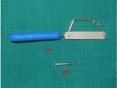
Stripping was performed with a single side cutting hand pulled strip(medium single sided blade ,manufactured by Ortho organizers) and a 169 L tungsten carbide bur (Shape: T
diameter: 0.5 mm, manufactured by SS white) at 4,000-6,000 rpm (Fig. 2). Before and after stripping, the mesio-distal diameter of each tooth was measured with a sliding digital calliper.
Fig. 2. (a) Single sided hand pulled strip (above); (b) Tungsten carbide bur
After completion of the stripping,
bonding agent (TransbondTM XT, light cure adhesive, manufactured by 3M Unitek) and fluoride varnish(Slow release topical dental fluoride, 50 mg sodium fluoride, manufactured Fluoritop SR) was done on the teeth of respective group by the same operator
The teeth were removed from their supports for further SEM evaluation.
stripping with single sided Hand pulled
stripping with tungsten carbide bur . stripping with single sided hand pulled strips followed by bonding agent application without etching.
stripping with tungsten carbide bur followed by bonding agent application
stripping with Single sided hand pulled strips followed by fluoride varnish
stripping tungsten carbide bur followed by fluoride varnish application.
The interproximal reduction procedures were carried out to achieve comparable amounts of different methods with an interproximal enamel reduction of at least
Stripping was performed with a single side cutting hand pulled strip(medium single sided blade ,manufactured by Ortho organizers) and a 169 L tungsten carbide bur (Shape: Taper, Tip diameter: 0.5 mm, manufactured by SS white) at 2). Before and after distal diameter of each tooth was measured with a sliding digital calliper.
(a) Single sided hand pulled strip Tungsten carbide bur (below)
Fig. 3. (a) bonding agent (b) fluoride varnish
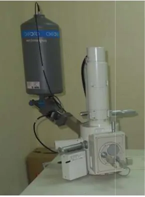
Each sample was then mounted on metallic supports. A 20kV scanning electron microscope (JEOL JSM-5610LV) was used at various magnifications (250 x, 550 x and 1000 x) to see the degree of roughness and the characteristics of furrows (Fig. 4).
Fig. 1. Scanning electron microscope JSM-5610LV)
3. RESULTS AND DISCUSSION
The images of the proximally stripped surface captured by scanning electron microscope (at 3 magnification showed that:
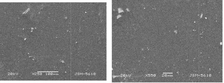
• The surface after stripping with hand pulled strip (Group I) shows furrows uniformly distributed over the entire surface (Fig. 5).
(a) bonding agent (b) fluoride varnish
Each sample was then mounted on metallic supports. A 20kV scanning electron microscope 5610LV) was used at various 550 x and 1000 x) to see the degree of roughness and the characteristics
microscope (JEOL
SSION
The images of the proximally stripped surface captured by scanning electron microscope (at 3
The surface after stripping with hand strip (Group I) shows furrows uniformly distributed over the entire
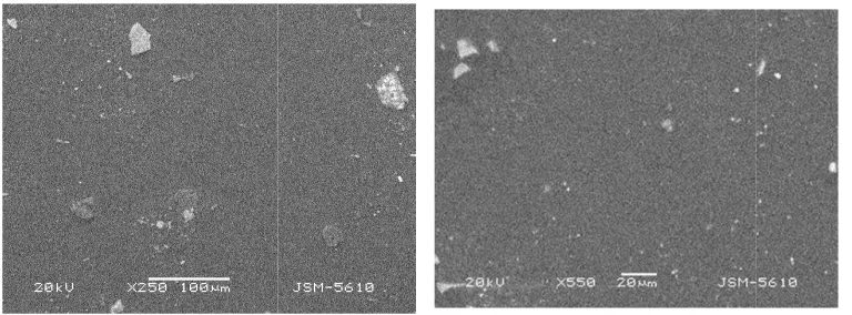
• Group II which contains surface stripped with tungsten carbide bur shows a small number of furrows interspersed with rough area. Surface is relatively smothe
compared to those with hand pulled strip (Fig. 6).
• Surface roughness after application of bonding agent in Group III and Group IV were markedly reduced when compared to Group I and Group II respectively. A smooth layer of sealant is formed over the stripped surface
• Proximal surface of Group V and Group VI shows a continuous outer layer of fluoride formed on the stripped surface. The surface roughness is covered with a uniform layer of fluoride
4. DISCUSSION
Enamel reduction with bur or interproximal s leads to furrows of a size which creates the site for plaque accumulation. So it is necessary to treat the stripped surface. Finishing the stripped surface with polishing strip or bur may smoother, the surface but even the maximum effort to polish interdentally stripped enamel fails to eliminate all furrows [1,13,14].
In the present study SEM images showed deep furrows were formed on the proximal surface stripped with abrasive proximal strip. Similar results were seen in a study done by Joseph et al. [15] in which they concluded that mechanical stripping with coarse abrasive may have advantage of removing the crowding quickly but this produces deep furrows which remains permanently. In our study the SEM images of the surface after stripping tungsten carbide bur appeared to be smother when compared to that of hand pulled strip. Our findings are also supported by findings of Piacentini et al. [3] who found that tungsten carbide bur allows a very precise first strip and leave a very fine furrow when compared to deep and irregular furrow formed by coarse diamond abrasive. Flouride varnish has been considered as an important material in preventing demineralization. In our study, a layer of fluoride can be seen on the surfaces stripped with bu and hand pulled strip after fluoride varnish application, which forms an outer protective layer. Rogers et al. [10] found that the roughened surface is less resistant to lactate buffer and so a fluoride treatment is inevitable. Topical fluoride application produces a initial reduction in the penetration rate of oral acids by forming a Group II which contains surface stripped with tungsten carbide bur shows a small
ws interspersed with Surface is relatively smother compared to those with hand pulled strip
Surface roughness after application of bonding agent in Group III and Group IV were markedly reduced when compared to Group I and Group II respectively. A smooth layer of sealant is formed over the
Proximal surface of Group V and Group VI shows a continuous outer layer of fluoride formed on the stripped surface. The surface roughness is covered with a
Enamel reduction with bur or interproximal strip leads to furrows of a size which creates the site for plaque accumulation. So it is necessary to treat the stripped surface. Finishing the stripped surface with polishing strip or bur may smoother, the surface but even the maximum effort to erdentally stripped enamel fails to
In the present study SEM images showed deep furrows were formed on the proximal surface stripped with abrasive proximal strip. Similar results were seen in a study done by Joseph
resistant outer layer [6]. In the present study, when the sealant was applied on proximally stripped surface by bur or hand pulled strip showed marked reduction in the roughness and the surface was remarkably smother when compared to surface without application of sealant, this indicates that sealant application reduces surface roughness and thereby can
Fig. 5. Group I: SEM micrograph showing deep furrows uniformly distributed.
Fig. 6. Group II: SEM micrograph showing small no of furrows interspersed with rough area
Fig. 7. Group III: SEM micrograph showing a smooth layer formed over the enamel surface
In the present study, when the sealant was applied on proximally stripped surface by bur or hand pulled strip showed marked reduction in the roughness and the surface was remarkably smother when compared to surface without application of cates that sealant application reduces surface roughness and thereby can
reduce plaque accumulation and can decrease the risk of dental caries. This findings are similar to those of Sheridan et al. [11] who documented that when bonding agent were applied proximally grinded surface, the surface roughness was markedly reduced and the sealed surface area were as smooth as the unaltered enamel.
Group I: SEM micrograph showing deep furrows uniformly distributed.
Group II: SEM micrograph showing small no of furrows interspersed with rough area
Group III: SEM micrograph showing a smooth layer formed over the enamel surface
reduce plaque accumulation and can decrease the risk of dental caries. This findings are similar who documented that when bonding agent were applied to
the surface roughness was markedly reduced and the sealed surface area were as smooth as the unaltered
Group I: SEM micrograph showing deep furrows uniformly distributed.
Group II: SEM micrograph showing small no of furrows interspersed with rough area
Fig. 8. Group IV: SEM micrograph showing
Fig. 9. Group V: SEM micrograph showing a uniform layer formed over the stripped surface
Fig. 10. Group VI: SEM micrograph showing that the stripped surface is covered by a uniform
5. CONCLUSION
The morphological analysis with scanning electron microscope (SEM) of the surface shows that the results obtained with use of different stripping method are as follow:
• Tungsten carbide bur produces a finely rough surface which is smoother
Group IV: SEM micrograph showing smooth layer formed over the roughened stripped area
Group V: SEM micrograph showing a uniform layer formed over the stripped surface
Group VI: SEM micrograph showing that the stripped surface is covered by a uniform layer of fluoride
The morphological analysis with scanning electron microscope (SEM) of the surface shows that the results obtained with use of different
Tungsten carbide bur produces a h is smoother
when compared to the deep uniform furrows formed with hand pulled proximal strip.
• A smooth layer is formed after bonding agent application on the rough stripped surface, which acts as a protective coating and makes the area less favourable fo plaque accumulation and reduces the incidence of caries.
roughened stripped
Group V: SEM micrograph showing a uniform layer formed over the stripped surface
Group VI: SEM micrograph showing that the stripped surface is covered by a uniform
when compared to the deep uniform furrows formed with hand pulled proximal
• A outer uniform layer in formed after fluoride varnish application on the stripped surface which prevents its demineralization from oral acid. Interproximal stripping is a highly useful tool as long as it is done judiciously. so it can be said that proximal stripping with tungsten carbide bur followed by application of fluoride varnish or bonding agent is preferable.
CONSENT
It is not applicable.
ETHICAL APPROVAL
Institutional Ethics Committee (IEC) For Research Manubhai Patel Dental College, Hospital and Oral Research Institute, Vadodara REF. NO.: IEC/ MPDC_068/ORTHO-15/15
COMPETING INTERESTS
Authors have declared that no competing interests exist.
REFERENCES
1. Radlanski RJ, Jäger A, Schwestka R, Bertzbach F. Plaque accumulations caused by interdental stripping. Am. J. Orthod. 1988;94:416-420.
2. Di Paolo RJ, Boruchov MJ, Thoughts on stripping of anterior teeth. J. Clin. Orthod. 1971;5:510-511.
3. Piacentini C, Sfondrini G. A scanning electron microscopy comparison of enamel polishing methods after air-rotor stripping. Am. J. Orthod. 1996;109:57-63.
4. Zhong M, Jost-Brinkmann PG, Radlanski RJ, Miethke RR. SEM evaluation of a new
technique for interdental striping. J. Clin. Orthod. 1999;33:286-292.
5. Sheridan JJ. Air-rotor stripping. J. Clin. Orthod. 1985;19:43-59.
6. Hudson AL. Study of the effects of mesiodistal reduction of mandibular anterior teeth. Am. J.Orthod. 1956;42:615-624.
7. Danesh G, Hellak A, Ziebura T, Schafer E. Enamel surfaces following interproximal reduction with different methods. Angle Orthodontist. 2007;77(6).
8. Srivastava S, Verma V, Panda S. Current status of interproximal enamel reduction in orthodontic treatment. Pakistan Oral & Dental Journal. 2012;32(2).
9. Alessandra L, Federico P. Effects of various stripping techniques on surface enamel. J. Clin. Orthod. 2001;11:691-695. 10. Rodgers GA, Wagner MJ. Protection of
stripped enamel surfaces with topical fluoride applications. Am. J. Orthod. 1969; 56:551-559.
11. Sheridan JJ, LeDoux PM. Air-rotor stripping and proximal sealants: An SEM evaluation. J. Clin. Orthod. 1989;23:790-794.
12. Shah L, Desai H, Arora P, Patel N, Harsora V. Manual vs air rotor stripping“ Just Do It with Care”- SEM evaluation. International Journal of Engineering Research and Reviews. 2014;2:119-126. 13. Tavaries C, Juchem CO. Anchorage
control after air-rotor stripping. J. Clin. Orthod. 2004;XXXVII(7).
14. Sheridan JJ. Air-rotor stripping update. J. Clin. Orthod. 1987;21:781-788.
15. Joseph VP, Rossouw PE, Basson NJ. Orthodontic microabrasive reproximation. Am. J. Orthod. 1992;102:351-359.
_________________________________________________________________________________ © 2017 Agrawal et al.; This is an Open Access article distributed under the terms of the Creative Commons Attribution License (http://creativecommons.org/licenses/by/4.0), which permits unrestricted use, distribution, and reproduction in any medium, provided the original work is properly cited.
Peer-review history: