_____________________________________________________________________________________________________
www.sciencedomain.org
In vitro
Bacteriostatic Effect of a Scaffold with a
Mixture of
Hypericum perforatum
and
Azadirachta indica
Oil Extracts
Maria Letizia Iabichella
1*1
HELIOS MED ONLUS, International Health Cooperation, Italy.
Author’s contribution
Author MLI has designed all studies, collected and revised the data, written the manuscript and approved it.
Article Information
DOI:10.9734/BJMMR/2015/14258 Editor(s): (1) Arun Kumar Nalla, College of Medicine, University of Illinois, Peoria, IL, USA. Reviewers: (1) Anonymous, Turkey. (2)Anonymous, Brasil. Complete Peer review History:http://www.sciencedomain.org/review-history.php?iid=723&id=12&aid=7489
Received 23rd September 2014 Accepted 30th October 2014 Published 26th December 2014
ABSTRACT
Background: Clinical evidences suggest antimicrobial activities of a new oil extract mixture of
Hypericum perforatum and Azadirachta indica, included in a polymeric scaffold (or Hyperoil™ Polimeric Substrate - HPS).
Methods: Bacteriostatic activity was investigated on selected strains of Staphylococcus aureus (ATCC® 12598™), Pseudomonas aeruginosa (ATCC® 10145™) and Klebsiella ozaenae AM through standardized in vitro tests.
Results and Conclusion: All bacterial strains were stabilized or reduced in size after 3 and 24 hours contact with the HPS if compared to the empty polymeric scaffolds.
Results showed the bacteriostatic effect of HPS that, added with its anti-inflammatory properties, could explain its tissue repair effects observed in vivo.
Keywords: Hypericum perforatum; Azadirachta indica; Staphylococcus aureus (ATCC® 12598™); Klebsiella ozaenae AM and Pseudomonas aeruginosa (ATCC® 10145™); bacteriostatic effect; scaffold; Hyperoil™ polymeric substrate.
1. BACKGROUND
Nimh (Azadirachta indica) extracts have been used for centuries in the traditional Indian medicine; its oil, obtained with cold extraction from its berries, is also included in the Ayurvedic Pharmacopoeia of India [1]. Nimh oil contains diterpenoids and triterpenoids having cicatrizing [2], bacteriostatic [3], antifungal [4], immune-modulatory [5] and anti-inflammatory properties [6]. Nimh oil resulted relatively safe for external application in wounds [7].
The oil extract of Hypericum perforatum flowers was esteemed as one of the most popular remedy for excoriations, wounds and bruises [8]. This product showed anti-infective [9], antiphlogistic and cicatrizing activity [10] while anti-inflammatory effects of H. perforatum
extracts were recently demonstrated [11] thus providing the rationale for using these preparations in legs wounds, together with nimh oil extract [12].
Hyperoil™ is a mixture of hypericum flowers extract (H. perforatum) and nimh oil (A. indica) produced by RIMOS S.r.L. Mirandola (MO) - Italy (Medical Device Class IIB CE0476), available as oil, gel, cream and gauze gel, that was recently used in complicated diabetic foot ulcers [13]. RIMOS developed a Hyperoil™ Polymeric Substrate (HPS) where Hyperoil™ oil was included in a polymeric scaffold, constituted by a fibrous membrane of poli (L-lactic) acid (PLLA), reproducing the biologic nano-structured fibrous matrix (extracellular matrix), whose fibers have a diameter from 10 to 300 nm. PLLA was chosen as it is a biocompatible, biodegradable and bio-absorbable product and, thus, used for the production of several biomaterials used for tissues regeneration [14].
Hyperoil™, together with improved metabolic control, was observed having recovered diabetic foot ulcers [13]. In patients with diabetes mellitus, foot infections in foot ulcers pose a significant risk, especially when patients are hospitalized for surgical treatments. These are complex infections commonly caused by Staphylococcus aureus, Pseudomonas aeruginosa and A. baumannii, being the Gram-positive
Staphylococcus aureus and the Gram-negative
Pseudomonas aeruginosa the most common and difficult to be treated [15] especially when nosocomial infections occur [16]. Klebsiella ozaenae is an enterobacterial, frequent in nosocomial infections [17], continuously evolving
to increase its resistance [18], thus representing a potentially difficult to be treated pathogen.
The aim of this paper is to present the results of
in vitro evaluations of the inhibitory bacteriostatic effect of HPS on S. aureus (ATCC® 12598™), P. aeruginosa (ATCC® 10145™) and K. ozaenae
AM strains, to confirm bacteriostatic effects of its components, being maintained when included in PLLA, on bacteria possibly related with diabetic foot infections.
2. MATERIALS AND METHODS
2.1 Chemicals
All chemicals used, including solvents, were of analytical grade. In details, PLLA (Lacea H.100-E) (average molecular weight by GPC = 8.4 x 10⁴ g/mol, polydispersity index, PDI = 1.7) was supplied by Mitsui Fine Chemicals (Dusseldorf,
Germany); dichloromethane (DCM) and
dimethylformamide (DMF) were purchased from Sigma-Aldrich and used without further purification [19].
2.2 HPS
The Hyperoil™ oil mixture was incorporated in PLLA during the electrospinning polymeric scaffold production process to have HPS.
HPS was produced through an original electrospinning process, licensed by RIMOS S.r.L. Mirandola (MO) – Italy, to create a tissue-non-tissue, with fibers having a diameter of 600±200 nm (Fig. 1), simulating the extracellular matrix to build the environment where cells live. This biological fibrous matrix actively modulates the main processes that regulate cellular survival, proliferation and differentiation. The HPS had an average thinning of 50 micron to ensure its simplest usability.
The produced HPS was, then, meshed (by Mash-Graft II Tissue Expansion System, Zimmer Surgical, Dover OH, USA) to ensure surface expandability, tissue repair and the passage of the exudate through the HPS when used as matrix in patients with cutaneous ulcers.
The weight percentage of Hyperoil™ oil included in the polymeric scaffolds was confirmed through a Differential Scanning Calorimetry (DSC) exam and resulted 0.0864 mg/cm2 for HPS-1.5, 0.218 mg/cm2 for HPS-2.5, 0.432 mg/cm2 for HPS-5, 0.864 mg/cm2 for HPS-10, and 2.160 mg/cm2 for HPS-25.
HPS were sterilized with ethylene oxide and kept at + 4ºC before performing the experiments.
2.3 Bacterial Strains
2.3.1 In vitro bacterial assays on HPS
2.3.1.1 Bacterial strains and culture conditions
The microorganisms used in this study were
Staphylococcus aureus (ATCC® 12598™) (S. aureus), Pseudomonas aeruginosa (ATCC® 10145™) (P. aeruginosa) and Klebsiella ozaenae
AM (K. ozaenae AM). K. ozaenae AM was a laboratory isolate not-labelled strain provided by the “Zooprofilattico Institute of Pavia”, Pavia, Italy.
P. aeruginosa (ATCC® 10145™) and K. ozaenae
AM were routinely grown in Luria Bertani Broth (LB) (Difco, Detroit, MI, USA) whereas S. aureus (ATCC® 12598™) grew in Brain Heart Infusion (BHI) (Difco) overnight under aerobic conditions at 37ºC using a shaker incubator (New Brunswick Scientific Co., Edison, NJ, USA).
These cultures, used as source for the experiments, were reduced at a final density of 1x10¹⁰ cells/mL as determined by comparing the OD600 of the sample with a standard curve
relating OD600 to cell number [20,21]. We used
the cultures at the bacterial cell cycle indicated as the period required for replication (known as the C period) [22]. An overnight culture of each bacterial strain was diluted in fresh medium and cultured for 2-3 hrs in order to reach the OD600 around 0.6-0.7, indicative of a growth exponential phase.
2.3.1.2 Antibacterial activity
To examine the antimicrobial activity of each HPS sterilized, 200 µl (1x10³ cells) of a growth exponential phase of an overnight diluted suspension of S. aureus (ATCC® 12598™), P. aeruginosa (ATCC® 10145™) and K. ozaenae
AM cells was seeded on the surface of no Hyperoil™ oil (Control) or HPS-1.5, HPS-2.5, HPS-5, HPS-10 or HPS-25 and incubated for 1, 3 and 24 hrs, respectively [21].
Fig. 1. Scanning electron microscopy picture of fibrous membrane of poli (L-lactic) acid (PLLA) constituting the polymeric scaffold
structure
Tissue culture wells (TCPS) used as internal control of bacterial growth were incubated with the same cell suspension for 1, 3 and 24 hrs, respectively.
At the end of each incubation time, the bacterial suspension was serially diluted in its specific culture medium and plated on the Muller Hinton (Difco) agar plates, respectively. The plates were incubated for 24-48 hrs at 37ºC. No significant difference was observed in bacterial growth between cells grown on TCPS and the empty polymeric scaffolds. The data are expressed as number of CFU/mL.
2.4 Statistical Analysis
Results were expressed as mean number of bacterial colonies and standard error using Microsoft Excel worksheet. No formal statistical analyses were performed.
3. RESULTS AND DISCUSSION
The tested bacterial strains grew on the empty polymeric scaffolds without any significant difference if compared to the bacterial growth on the TCPS (data not shown). On the contrary, a variable reduction was observed after various times (1, 3 and 24 hours) of incubation on strains cultured with HPS containing different concentrations of Hyperoil™ (Figs. 2, 3 and 4) and compared to the polymeric substrate not containing the Hyperoil (Figs. 2, 3 and 4).
results are related to the exponential phase of each bacterial culture. As previously reported, the bacterial cell cycle is traditionally divided into three stages: the period between division (birth) and the initiation of chromosome replication (the B period); the period required for chromosome replication (the C period); and the time between the completion of chromosome replication and the completion of cell division (the D period) [22]. To evaluate the efficacy of antibacterial activity of the HPS-Hyperoil it is important to perform the incubation with bacterial cells which are all in the growth exponential phase.
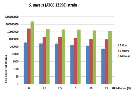
Antibacterial activity resulted dose-dependent through time for S. aureus (ATCC® 12598™) (Fig. 2) and K. ozaenae AM (Fig. 4) and, at a lesser extent, for P. aeruginosa (ATCC® 10145™) (Fig. 3). The maximal inhibition in the growth of bacterial strains was consistently observed after 3 hours, being for K. ozaenae AM the highest values at all tested doses if compared to S. aureus (ATCC® 12598™) and P.
aeruginosa (ATCC® 10145™). The percent of reduction of cell viability on P. aeruginosa
(ATCC® 10145™) (Table 2) and S. aureus 8325-4 (Table 1), resulted slightly lower in the 1st hour if compared to K. ozaenae AM (Table 3).
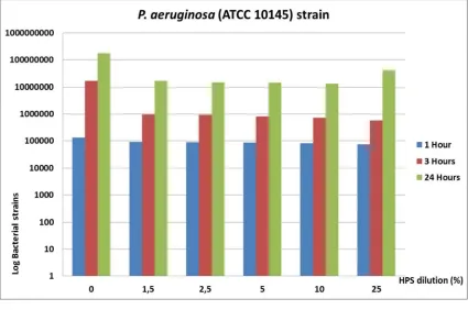
On the contrary, at higher incubation time (24h), the percent of reduction was kept higher on P. aeruginosa (ATCC® 10145™) (around 90-92%) and S. aureus (ATCC® 12598™) (around 90-94%) and resulted slightly decreased on K. ozaenae AM (between 80-88%). In particular, the effect on P. aeruginosa (ATCC® 10145™) was not dose-dependent after 24 hours being the HPS-25 the less effective on bacterial growth control (Fig. 3).
Because of these different effects, the results of this study cannot sustain the bactericide effect, defined as the capacity of killing bacteria [23], of Hyperoil™ on S. aureus (ATCC® 12598™), P. aeruginosa (ATCC® 10145™) and K. ozaenae
AM, but strongly sustain a bacteriostatic effect.
Fig. 3. P. aeruginosa strain (Log) by time and HPS dilution
436
Table 1. Average (±SE) S. aureus strain by time and HPS dilution
HPS dilution (%)
S. aureus (ATCC® 12598™) strain 1 hour
SE % reduction vs control
S. aureus (ATCC® 12598™) strain 3 hours
SE % reduction vs control
S. aureus (ATCC® 12598™) strain 24 hours
SE % reduction vs control
0 345333 37351 N/A 26800000 208167 N/A 226000000 3785939 N/A
1,5 264067 93544 23,5% 1903333 17638 92,9% 21233333 384419 90,6%
2,5 247033 87469 28,5% 1820000 10000 93,2% 20066667 88192 91,1%
5 151550 56706 56,1% 1463333 33333 94,5% 19400000 208167 91,4%
10 133000 34078 61,5% 944000 406041 96,5% 14600000 360555 93,5%
25 53250 6083 84,6% 928000 15308 96,5% 12666667 384419 94,4%
HPS: Hyperoil Polimeric Substrate; Control = PS polymeric substrate without Hyperoil; N/A Not Applicable; % percentage; SE Standard Error
Table 2. Average (±SE) P. aeruginosa strain by time and HPS dilution
HPS dilution (%)
P. aeruginosa (ATCC® 10145™) strain
1 hour
SE % reduction vs control
P. aeruginosa (ATCC® 10145™) strain
3 hours
SE % reduction vs control
P. aeruginosa (ATCC® 10145™) strain
24 hours
SE % reduction vs control
0 130333 10333 N/A 16466667 3286504 N/A 175333333 2905933 N/A
1,5 91400 305 29,9% 970666 6333 94,1% 16633333 409606 90,5%
2,5 88766 2533 31,9% 923666,7 31991 94,4% 15000000 152752 91,4%
5 84700 3572 35,0% 808000 44635 95,1% 14400000 709459 91,8%
10 81200 2886 37,7% 728333 14813 95,6% 13333333 328295 92,4%
25 72933 5944 44,0% 574666 63878 96,5% 40236667 29387860 77,1%
HPS: Hyperoil Polimeric Substrate; Control = PS polymeric substrate without Hyperoil; N/A Not Applicable; % percentage; SE Standard Error
Table 3. Average (±SE) K. ozaenae strain by time and HPS dilution
HPS dilution (%)
K. ozaenae AM strain 1 hour
SE % reduction vs control
K. ozaenae AM strain
3 hours
SE % reduction vs control
K. ozaenae AM strain 24 hours
SE % reduction vs control
0 228667 4702 N/A 43033333 1049338 N/A 110333333 3179797 N/A
1,5 17067 698 92,5% 375667 14193 99,1% 21233333 384419 80,8%
2,5 16633 669 92,7% 314667 40043 99,3% 20066667 88192 81,8%
5 16100 1000 93,0% 302667 41914 99,3% 19400000 208167 82,4%
10 14067 1510 93,8% 246667 32028 99,4% 17733333 371184 83,9%
25 11800 551 94,8% 186000 9165 99,6% 12666667 384419 88,5%
The activity against the Gram positive S. aureus
(ATCC® 12598™), the Gram negative
P. aeruginosa (ATCC® 10145™) being common and often resistant strains in skin and hospital infections [24], where Hyperoil™ scaffolds are expected to be used, could explain the successful results observed while suing Hyperoil™ on diabetic foot ulcers [13]. On the other hand, the activity on K. ozaenae AM, has a particular importance considering the wide number of identified antibiotic-resistant strains of this enterobacterium, frequently linked with opportunistic infections of opened wounds [25]. The particular structure of the polymeric scaffold might not ensure a-priori the efficacy of Hyperoil™ when meshed with the PLLA structure. The results of this study support in-vitro the observed clinical results.
4. CONCLUSION
HPS, being Hyperoil™ included in a highly biocompatible scaffold of poly-lactic acid, showed bacteriostatic effects on the tested S. aureus
(ATCC® 12598™), P. aeruginosa (ATCC® 10145™) and K. ozaenae AMstrains.
ACKNOWLEDGEMENTS
The author thanks Andrea Rossi for his friendly support for medical writing; Livia Visai PhD, of the “A. Castellani" Biochemistry Unit of the Department of Molecular Medicine of Pavia University-Italy, for supervising the microbial tests and Maria Letizia Focarete PhD, of the Chemistry Department of Bologna University-Italy, for supervising the electrospinning process and scaffold fabrication and for providing the SEM image of the scaffold.
COMPETING INTERESTS
Maria Letizia Iabichella acts as research consultant for RIMOS S.r.L. Mirandola (MO) - Italy.
REFERENCES
1. Government of India Ministry of Health and Family Welfare Department of Ayush. The Ayurvedic Pharmacopoeia of India. The Ayurvedic Pharmacopoeia of India. 2012;5:119. 1. part- I, Access date
October 28, 2012.
Available:http://www.ayurveda.hu/api/API-Vol-5.pdf
2. Dos Santos ACG, Rodrigues OG, De Araujo LVC, Dos Santos SB, De C. Guerra RDMSN, Feitosa MLT, Teixeira WC, Santos-Ribeiro A. Use of neem extract in the control of acariasis by Myobia musculi
Schranck (Acari: Miobidae) and Myocoptes musculinus Koch (Acari: Listrophoridae) in mice (Mus musculus var. albina L.).
Neotropical Entomology. 2006;35:269-272. 3. Narayanan AS, Raja SSS, Ponmurugan K, Kandekar SC, Natarajaseenivasan K, Maripandi A, Mandeel QA. Antibacterial activity of selected medicinal plants against multiple antibiotic resistant uropathogens: a study from Kolli Hills, Tamil Nadu, India. Beneficial Microbes. 2011;2:235-243. 4. Murthy SP, Sirsi M. Pharmacological
studies on Melia Azadirachta indica. Indian Journal of Physiology and Pharmacology. 1958;2:387-396.
5. Upadhyay SN, Dhawan S, Garg S, Talwar GP. Immunomodulatory effects of neem (Azadirachta indica) oil. Int J Immunopharmacol. 1992;14:1187-93. 6. Athar A, Saikat H, Hirekodathakallu T, et
al. Novel Anti-inflammatory Activity of Epoxyazadiradione against macrophage migration inhibitory factor. Journal of Biological Chemistry. 2012;287:24844-24861.
7. Tandan SK, Gupta S, Chandra S, Lal J, Singh R. Safety evaluation of Azadirachta indica seed oil, a herbal wound dressing agent.Fitoter. 1995;66:69-72.
8. Fernie WT. Herbal simples. Bristol: John Wright & Co.; 1897.
9. Aizenman BE. Antibiotic preparations from
Hypericum perforatum.Kiev: Mikrobiol. Zh. 1969;31:128-33.
10. Bombardelli E, Morazzoni P. Hypericum perforatum. Fitoterapia. 1995;62:43-68. 11. Koeberle A, Rossi A, Bauer J, Dehm F,
Verotta L, Northoff H, Sautebin L, Werz O.
Hyperforin, an anti-inflammatory
constituent from St. John's Wort, inhibits microsomal prostaglandin E(2) synthase-1 and suppresses prostaglandin E(2) formation in vivo. Front Pharmacol. 2011;2. 12. Läuchli S, Hafner J, Wehrmann C, Frenc LE. Post-surgical scalp wounds with exposedbone treated with a plant-derived wound therapeutic. J Wound Care. 2012;21:228-33.
13. Iabichella ML. The use of an extract of
unexpected outcome. BMJ Case Reports; 2013. DOI: 10.1136/bcr-2012-007299. 14. Jagur-Grodzinski J. Polymers for tissue
engineering, medical devices, and regenerative medicine. Concise general review of recent studies. Polymers for Advanced Technologies. 2006;17(6):395-418.
15. Mendes JJ, Leandro C, Mottola C, Barbosa R, Silva FA, Oliveira M, Vilela CL, Melo-Cristino J, Górski A, Pimentel M, São-José C, Cavaco-Silva P, Garcia M. In vitro design of a novel lytic bacteriophage cocktail with therapeutic potential against organisms causing diabetic foot infections. J Med Microbiol; 2014. PII: jmm.0.071753-0. DOI: 1jmm.0.071753-0.1099/jmm.jmm.0.071753-0.071753-jmm.0.071753-0.
16. Nguyen TK, Milch H. Klebsiella and
Enterobacter strains derived from hospital infections. I. Correlation between species, phage type and antibiotic sensitivity. Acta Microbiol Acad Sci Hung. 1981;28(1):67-81.
17. Ji X1, Jin P, Chu Y, Feng S, Wang P. Clinical characteristics and risk factors of diabetic foot ulcer with multidrug-resistant organism infection. Int J Low Extrem Wounds. 2014;13(1):64-71.
DOI: 10.1177/1534734614521236. Epub 2014 Feb 10.
18. Brisse S1, Fevre C, Passet V, Issenhuth-Jeanjean S, Tournebize R, Diancourt L, Grimont P. Virulent clones of Klebsiella pneumoniae: identification and evolutionary scenario based on genomic and phenotypic characterization. PLoS One. 2009;4(3):e4982.
DOI: 10.1371/journal.pone.0004982. Epub 2009 Mar 25.
19. Saino E, Focarete ML, Gualandi C, Emanuele E, Cornaglia AI, Imbriani M, Visai L. Effect of electrospun fiber diameter and alignment on macrophage activation and secretion of proinflammatory cytokines and chemokines. Biomacromolecules. 2011;12(5):1900-11.
20. Del Curto B, Brunella MF, Giordano C, Pedeferri MP, Valtulina V, Visai L, Cigada A. Decreased bacterial adhesion to surface-treated titanium.Int J Artif Organs. 2005;28:718-30.
21. Petrini P, Arciola CR, Pezzali I, Bozzini S, Montanaro L, Tanzi MC, Speziale P, Visai L. Antibacterial activity of zinc modified titanium oxide surface. Int J Artif Organs. 2006;29:434-42.
22. Wang JD, Levin PA. Metabolism, cell growth and the bacterial cell cycle. Nat Rev Microbiol. 2009;7(11):822-7.
23. Pankey GA, Sabath LD. Clinical relevance of bacteriostatic versus bactericidal mechanisms of action in the treatment of Gram-positive bacterial infections. Clin. Infect. Dis. 2004;38(6):864–70.
24. Mohammadzadeh R1, Ahmadiyan N. Skin infection management using novel antibacterial agents. Adv Pharm Bull. 2013;3(1):247-8.
25. Livermore DM, Yuan M. Antibiotic resistance and production of
extended-spectrum beta-lactamases amongst
Klebsiella spp. from intensive care units in
Europe. J Antimicrob Chemother.
1999;38(3):409-24.
© 2015 Iabichella; This is an Open Access article distributed under the terms of the Creative Commons Attribution License (http://creativecommons.org/licenses/by/4.0), which permits unrestricted use, distribution, and reproduction in any medium, provided the original work is properly cited.
Peer-review history: