Breast Cancer - Targets and Therapy 2016:8 183–197
Breast Cancer - Targets and Therapy
Dove
press
183 R E V I E W open access to scientific and medical research
Open Access Full Text Article
submit your manuscript | www.dovepress.com
Current advances in biomarkers for targeted
therapy in triple-negative breast cancer
Brett Fleisher1
Charlotte Clarke2
Sihem Ait-Oudhia1
1Department of Pharmaceutics,
Center for Pharmacometrics and Systems Pharmacology, College of Pharmacy, University of Florida, Orlando, FL, 2Department of
Translational Research, UT MD Anderson Cancer Center, Houston, TX, USA
Abstract: Triple-negative breast cancer (TNBC) is a complex heterogeneous disease
characterized by the absence of three hallmark receptors: human epidermal growth factor receptor 2, estrogen receptor, and progesterone receptor. Compared to other breast cancer subtypes, TNBC is more aggressive, has a higher prevalence in African-Americans, and more frequently affects younger patients. Currently, TNBC lacks clinically accepted tar-gets for tailored therapy, warranting the need for candidate biomarkers. BiomarkerBase, an online platform used to find biomarkers reported in clinical trials, was utilized to screen all potential biomarkers for TNBC and select only the ones registered in completed TNBC tri-als through clinicaltritri-als.gov. The selected candidate biomarkers were classified as surrogate, prognostic, predictive, or pharmacodynamic (PD) and organized by location in the blood, on the cell surface, in the cytoplasm, or in the nucleus. Blood biomarkers include vascular endothelial growth factor/vascular endothelial growth factor receptor and interleukin-8 (IL- 8); cell surface biomarkers include EGFR, insulin-like growth factor binding protein, c-Kit, c-Met, and PD-L1; cytoplasm biomarkers include PIK3CA, pAKT/S6/p4E-BP1, PTEN, ALDH1, and the PIK3CA/AKT/mTOR-related metabolites; and nucleus biomarkers include BRCA1, the gluco-corticoid receptor, TP53, and Ki67. Candidate biomarkers were further organized into a “cellular protein network” that demonstrates potential connectivity. This review provides an inventory and reference point for promising biomarkers for breakthrough targeted therapies in TNBC.
Keywords: anti-cancer directed pharmacotherapy, difficult-to-treat breast cancer, biological
markers
Introduction
Breast cancer is the most common cancer in women, and the second most frequent cause of cancer-related deaths in women worldwide.1 Approximately 20% of all breast
cancers are referred to as triple-negative breast cancer (TNBC) due to a lack of three proteins: estrogen receptor (ER), progesterone receptor (PR), and human epidermal growth factor receptor 2 (HER2).2 TNBC tends to be more aggressive than other breast
cancer subtypes and has a higher prevalence in African-Americans, more frequently affects younger patients (average age <50 years), and is associated with a greater risk of mortality.1,3 Currently, breast cancer treatment options and outcome are highly
dependent on targeting ER, PR, or HER2. As a result, the American Society of Clini-cal Oncology (ASCO) does not currently recommend tailoring therapy for TNBC but instead recommends a general chemotherapy treatment based on the combination of an anthracycline with a taxane.4
Correspondence: Sihem Ait-Oudhia Department of Pharmaceutics, Center for Pharmacometrics and Systems Pharmacology, College of Pharmacy, University of Florida, 6550 Sanger Road, Room 469, Orlando, FL 32827, USA Tel +1 407 313 7037
Fax +1 407 313 7030
Email sihem.bihorel@cop.ufl.edu
Journal name: Breast Cancer - Targets and Therapy Article Designation: REVIEW
Year: 2016 Volume: 8
Running head verso: Fleisher et al
Running head recto: Current advances in biomarkers for targeted therapy in TNBC DOI: http://dx.doi.org/10.2147/BCTT.S114659
Breast Cancer: Targets and Therapy downloaded from https://www.dovepress.com/ by 118.70.13.36 on 20-Aug-2020
For personal use only.
This article was published in the following Dove Press journal: Breast Cancer - Targets and Therapy
6 October 2016
Dovepress Fleisher et al
Although physicians have found that TNBC will initially respond to the combination of anthracycline and taxanes, treatment failure and disease recurrence continue to be clini-cally challenging.5 Clinicians have attempted to tailor therapy
for TNBC by monitoring the Food and Drug Administration (FDA)-approved biomarkers which can be classified as sur-rogate, prognostic, predictive, or pharmacodynamic (PD).6
Surrogate biomarkers may predict an outcome by acting as a substitute for a clinical endpoint.7 For example, evidence
suggests that interleukin-8 (IL-8) blood levels are linked to breast cancer resistance protein (BCRP), a transient efflux transporter8 that confers resistance to cytotoxic drugs;9
therefore, IL-8 could be measured as a surrogate biomarker for BCRP levels in TNBC. Prognostic biomarkers suggest survival probability for cancer patients with and without drug therapy and would be used to ascertain whether patients require additional therapy.6 For example, one study found
that the presence of c-Kit, a protein expressed during cell replication, may determine the progression of TNBC.10
Pre-dictive biomarkers (also known as companion biomarkers) identify subpopulations of patients who are most likely to benefit from a specific drug therapy and form the basis for tailored-TNBC therapies. The HER2 protein is an example of a predictive biomarker that indicates a breast cancer that is more likely to have a favorable response to the drug trastuzumab (Herceptin; Genetech, Inc., South San Francisco, CA, USA).4 Finally, PD biomarkers assess which molecular
indicators are linked to a drug regimen, target effect, or tumor response, thereby providing rationale for new drug targets, drug combinations, or assay development. S6 and P4E-BP1 are potential PD biomarkers as their expression in in vitro TNBC experiments reflects activity in the TNBC chemotherapy-resistance pathway, PIK3CA/AKT/mTOR.11
Although these four types of biomarkers provide a cornerstone of modern cancer therapeutics, they have not demonstrated significant clinical utility in the treatment of TNBC,4 likely due to its highly complex biology and
interpatient heterogeneity.4 TNBC often exhibits resistance
to standard combination chemotherapy through multiple interacting pathways with feedback and cross-talk loops.6
These loops, in turn, may be altered by mutations in coding regions, regulatory elements, and noncoding sequences in the tumor DNA and gene expression,12 which leads to
down-stream protein–protein, protein–gene, and other interactions which cannot be represented by single protein biomarker.
In the last decade, -omics technologies have been used to analyze the complex biology of TNBC by examining several forms of macromolecules (eg, DNA, RNA, proteins, and
car-bohydrates) that may shed light on TNBC signaling pathways.13
These -omics technologies include proteomics, metabolomics, genomics, and epigenomics. Proteomic studies are used to measure the changes in tumor-associated proteins, which can differentiate between samples of normal tissue, benign, and malignant tumor or as any of the biomarkers described above. For example, the modification of specific proteins in patients with TNBC differentiates TNBC pathology and physiology from that of other forms of breast cancer.14 In metabolomics,
one analyzes how cells utilize small molecules (lipids, small peptides, and vitamins) to meet the energy requirements for sustaining life, and as building blocks for cellular division. For example, unlike healthy tissue, tumors perform aerobic glycolysis, thereby increasing lactate concentrations, which is known as the Warburg effect. In TNBC, this process appears to be interconnected with tumorigenic pathways PI3K/mTOR15
and EGFR,16,17 and metabolomic assays may be used to
mea-sure the response of TNBC to specific therapies that target these pathways.13 Genomic studies of cancer cells examine genes
specific to the tumor pathology which can include gene copy number, single nucleotide polymorphisms (SNPs), mutations, and loss of heterogeneity.12 For example, deleterious BRCA1
mutations are found in high-risk TNBC population18–20 and
may increase tumor susceptibility to DNA-damaging and PARP inhibitor therapies.21 Epigenomics is the examination
of changes in cell phenotype that are the result of gene modi-fication, such as DNA methylation, rather than changes in the DNA sequence itself.22 For example, a significant proportion of
TNBC may have BRCA1 promotor site hypermethylation;18,23,24
although epigenetic silencing creates a similar protein profile to the loss-of-function BRCA1 mutation,25 therapeutic efficacy
may differ.26
Aside from the complexity of TNBC, finding new and improved TNBC biomarkers is logistically challenging for several reasons. Centralized tumor specimen banks require proper sample collection, processing, and storage, which add financial burden27 and may deter candidate institutions from
investing the necessary start-up capital. Following sample collection, data mining for novel biomarkers is time con-suming and requires substantial input from data managers, bioinformaticians, and biostatisticians to correctly interpret the results.6 Additionally, the biomarker discovery process
is not always straightforward.28 For example, because most
cancer treatments use combination therapy rather than mono-therapy, it can be difficult to connect the identified biomarker to a single drug or target.6 Before a new biomarker can be
implemented in the clinic, newly discovered TNBC biomark-ers must be thoroughly examined and validated in order to
Breast Cancer: Targets and Therapy downloaded from https://www.dovepress.com/ by 118.70.13.36 on 20-Aug-2020
Dovepress Current advances in biomarkers for targeted therapy in TNBC
potentially fill the gaps in our understanding of TNBC treat-ment and patient survival. In this work, biomarkers that have been studied in late-stage clinical trials were reviewed and were classified according to its biological location as blood (plasma or serum), cell surface, cytoplasm, or nucleus bio-markers. How recently published -omics studies may provide useful information on TNBC biomarkers is also discussed, and these markers are connected through an evidence-based molecular pathway landscape.
Methodology of data mining for
biomarkers in TNBC
There are many preclinical study publications on TNBC bio-markers; a recent search in PubMed Central using the words “triple negative breast cancer and biomarker” returned over 2300 search results. In order to select only biomarkers with the most clinician-backed support, biomarkers associated with completed TNBC trials were chosen to be focused on by using BiomarkerBase, a biomarker knowledgebaseTM
devel-oped by Amplion. BiomarkerBase uses a comprehensive list of synonyms to identify biomarkers registered in the records of clinical trials via the government website clinicaltrials.gov. With BiomarkerBase, breast cancer biomarkers were first found through the search engine. Then, for each breast cancer biomarker, subsearches were conducted for clinical trials that explicitly used TNBC (or the full name, triple-negative breast cancer) in the title of the study. If the breast cancer biomarker was registered in at least one completed TNBC study, the biomarker was analyzed (with the exceptions of HER2, ER, and PR). Of note, most clinical trials surveyed for the work presented in this review completed Phase II or III. Current literature about the biomarkers was further examined using PubMed. Papers that studied one of the biomarkers as a general-disease biomarker, explored how -omics studies further characterized these biomarkers, and examined how the biomarker pathways may interact were sought.
Current advances in clinical
biomarkers for TNBC patients
The following sections examine biomarkers found in the blood, on the cell surface, in the cytoplasm or nucleus in TNBC samples. Circulating blood biomarkers include vascular endothelial growth factor (VEGF), its receptor, VEGFR, and interleukin-8 (IL-8). The cell surface recep-tors include endothelial growth factor receptor (EGFR), insulin-like growth factor binding proteins (IGFBP), c-Kit, and PD-L1. All the plasma and cell surface biomarkers used in this review are associated with completed-TNBC clinical
trials. Cytoplasm biomarkers include PIK3CA, pAKT/S6/ p4E-BP1, PTEN, and PIK3CA/AKT/mTOR metabolites, in addition to ALDH1. PIK3CA, PTEN, ALDH1, and p4E-BP1 were registered in completed TNBC clinical trials, whereas pAKT/S6 biomarkers and the PIK3CA/AKT/mTOR metabolites were not. Nuclear biomarkers include BRCA1, the glucocorticoid receptor (GR), TP53, and Ki67 and were all studied in completed TNBC trials.
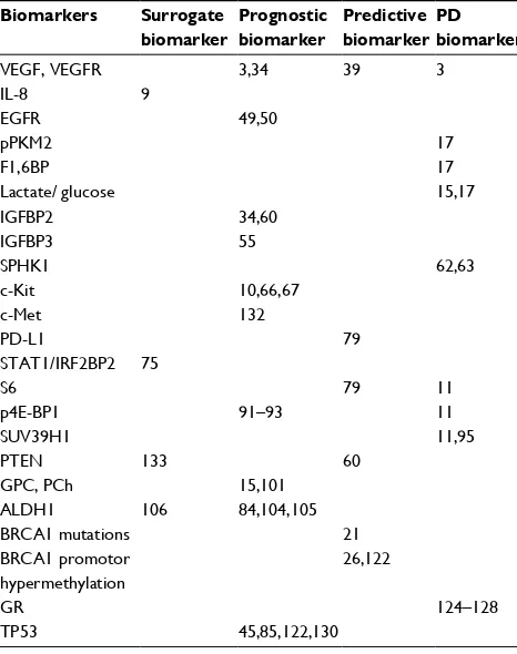
Figure 1 shows the system biology of TNBC. It summa-rizes all identified biomarkers through a “cellular protein net-work” that demonstrates the potential connectivity between the different subtypes of biomarkers. Table 1 provides an overview of the examined surrogate, prognostic, predictive, and PD biomarkers in TNBC in addition to current literature found in PubMed that provides support for their potential use.
TNBC biomarkers in blood
Tumors or neighboring cells have been shown to release proteins that influence the survival of tumor cells,3,29 thereby
leading to a worse outcome in TNBC patients. Only few biomarkers were reported in the serum or plasma of TNBC patients in late-stage clinical trial, and so far, none has been approved by the FDA, yet. They include VEGF, VEGFR, and IL-8.
VEGF/VEGFR
VEGF is typically found in blood as a disulfide-linked homodimer. Created from a family of six proteins, these VEGF isoforms include VEGF-A, VEGF-B, VEGF-C, VEGF-D, VEGF-E, and placental growth factor (PlGF). Glycosylated VEGF binds the VEGFR, a family of recep-tor tyrosine kinases (RTKs) including VEGFR1, VEGFR2, and VEGFR3, which upon stimulation lead to an increase of angiogenesis and the permeability of nearby vessels and lymphatics;3,30,31 the end result of which is improved
oxygen/nutrient transport, as well as increased tumor metastasis.32,33
Roberti et al34 demonstrated the potential use for VEGF
as a prognostic biomarker in TNBC. Briefly, animal models were implanted with either metastatic or nonmetastatic TNBC cells. The metastatic TNBC cells tended to express higher levels of PlGF and VEGF-A than the nonmetastatic counterparts, suggesting that overexpression of these mark-ers correlated with metastatic potential for TNBC. Later, in a clinical study conducted by Bahhnassy et al,35 VEGF-A
expression was examined before and after standard chemo-therapy. In agreement with Roberti’s study, the expression of VEGF-A correlated with a poorer overall survival and a less-favorable response to chemotherapy.
Breast Cancer: Targets and Therapy downloaded from https://www.dovepress.com/ by 118.70.13.36 on 20-Aug-2020
Dovepress Fleisher et al
Overwhelming preclinical evidence predicted a positive response to bevacizumab (anti-VEGFA therapy) in TNBC patients, and thus, the potential for VEGF as a predictive biomarker for VEGF-tailored treatment;36,37 unfortunately,
the clinical results were not positive. Bear et al38 examined
breast cancer response to the combination of bevacizumab and standard chemotherapy. The addition of bevacizumab significantly increased the overall rate of pathological com-plete response (pCR), but after subset analysis, bevacizumab was found to be slightly more effective in the HER2-positive group than the TNBC group.
Shortly after Bear et al released their results, Chen et al21 released a meta-analysis examining eleven studies
focused on standard neoadjuvant chemotherapy with- and without bevacizumab in TNBC patients. The meta-analysis revealed that, overall, TNBC treated with bevacizumab resulted in an improvement in pCR.21 Interestingly, Chen
et al found no clear homogeneity between the studies, suggesting that the current studies do not provide enough patient details including VEGF-A expression to deter-mine which TNBC subgroups most benefit from VEGF therapy.
VEGFR levels may be indicative of successful anti-VEGF-A therapy in TNBC. Recently, Tolaney et al39
con-ducted a Phase II study to examine the effect of bevacizumab on TNBC patients and found that TNBC with high VEGFR1
GC
HGF
IGFBP3 IGFBP2
IGF1 IGF2
c-Met
IGF-R1
Plasma
Cell surface
Cell survival
SPHK1
Glucose F1,6BP Cell
proliferation
G6B HK2 miR-143
Sphingosine
PEP
Cytoplasm
Nucleus
GR-GC GR
P38
c-Kit
BRCA1
S1P
DNA replication
DNA repair
SUV39H1 PKM2
Pyruvate
PIK3CA
T-Cell mediated
cell death
Lactate
EGF VEGF-A PIGF T-Cell
mediated cell death
IL-8 VEGF-C
EGFR VEGFR1 PD-L1 BCRP VEGFR3
pPKM2
Anglogenesis
PIK3CA
PTEN
PIP2
PIP3
PDK1
S6
mTOR
p53 eIF4E p4E-BP1 4E-BP1 mTORC2
mTORC1 mTOR inhibition
increases EGFR expression
Drug resistance
Tumor metastasis
AKT
pAKT
PTEN synthesisProtein TP53
Figure 1 Biomarker pathways summarized through a “cellular protein network” that demonstrates potential connectivity.
Notes: BiomarkerBase, a biomarker knowledgebaseTM developed by Amplion, was used to find registered biomarkers in completed TNBC trials through clinicaltrials.
gov with the exception of HER2 and ER/PR. Current literature about the biomarkers was located using PubMed. The protein map is sectioned into plasma (blood), cell surface, cytoplasm, and nucleus. Biomarkers shown in cylinders represent markers found on the cell surface, rounded-edged boxes and circles represent other proteins
or metabolites, and curved boxes represent DNA or RNA. Biomarkers examined more thoroughly throughout the review are filled in with gray. A black line with an
arrow represents increase, expression, or activation; a dotted lined connected to an X represents inhibition. Star figures denote action such as tumor suppression and cell proliferation.
Abbreviations: EGF, endothelial growth factor; ER, estrogen receptor; GC, glucocorticoid; GR, glucocorticoid receptor; HER2, human epidermal growth factor receptor 2; HGF, hepatocyte growth factor; HK2, hexokinase 2; IGFBP, insulin-like growth factor binding protein; IGF-R1, IGF receptor 1; IL-8, interleukin-8; mTOR, mammalian target of rapamycin; PDK1, phosphoinositide-dependent kinase 1; PIP2, phosphatidylinositol 4,5-biphosphate; PIP3, phosphatidylinositol 3,4,5-triphosphate; PKM2, pyruvate
kinase M2; pPKM2, phosphorylated version of pyruvate kinase M2; PR, progesterone receptor; SPHK1, sphingosine kinase 1; VEGF, vascular endothelial growth factor; BCRP, breast cancer resistance protein.
Breast Cancer: Targets and Therapy downloaded from https://www.dovepress.com/ by 118.70.13.36 on 20-Aug-2020
Dovepress Current advances in biomarkers for targeted therapy in TNBC
had the greatest response to anti-VEGF-A therapy. The therapy appeared to prune vessels high in VEGFR1 while normalizing the other vessels, suggesting that high baseline VEGFR1 microvascular density may be required for success-ful neoadjuvant anti-VEGF-A therapy in TNBC. TNBC may be dependent on both VEGF and VEGFR analysis for effec-tive treatment, and VEGF and VEGFR together as prediceffec-tive biomarkers may demonstrate improved clinical utility for tailored TNBC therapy.
IL-8
IL-8 (or CXCL8) is a major CXC motif cytokine encoded by
IL-8 on chromosome 4q13-q21. IL-8 is produced by TNBC
tumors in response to hypoxic conditions and is thought to recruit mesenchymal stem cells (MSCs) to the TNBC loca-tion.9 MSCs normally reside in the bone marrow and adipose
tissue, but when recruited by TNBC, they home to the breast cancer tumor location and create a microenvironment around the tumor hypothesized to increase the stem-cell-like charac-teristics of the TNBC,29 thereby increasing TNBC multidrug
resistance (MDR) and metastatic risk.34
TNBC often becomes resistant to doxorubicin, a standard anthracycline used in TNBC treatment.40 TNBC resistance to
doxorubicin may be due to the ability of IL-8 to upregulate the BCRP found on the surface of TNBC cells. BCRP is a 72 kDa transmembrane protein responsible for removing doxorubicin from a tumor cell.8,41–43 In vitro data suggest
that baseline levels of BCRP expression on the cell surface is very high in TNBC, and the expression can be further upregulated in response to drug therapy.8 Importantly, BCRP
upregulation is transient, lasting only for a few hours after exposing the tumor to doxorubicin8 while IL-8 expression
can last for several days.44 The potential usefulness of IL-8 as
a biomarker was demonstrated by an in vitro study by Chen et al,9 which demonstrated that IL-8 increased BCRP
expres-sion without affecting the expresexpres-sion of other hallmark efflux transporters correlating with increased TNBC resistance to doxorubicin. IL-8 may serve to protect the TNBC from doxorubicin-induced killing by increasing BCRP levels in TNBC and may function as a surrogate biomarker for BCRP expression in TNBC.
TNBC biomarkers on the cell
surface membrane
Various membrane-bound receptors have the potential function as biomarkers for TNBC as they were shown to increase anti-apoptotic signals in TNBC cell lines; thus, their blockade would be expected to increase tumor death either alone or as part of combination therapy. These biomarkers include EGFR, IGFBP, C-kit, and PD-L1.46–48 In this section,
each of these membrane-bound candidate biomarkers are described along with the clinical trials in which they were investigated.
EGFR
The EGFR family consists of four similarly structured RTKs that have important roles in tumor proliferation: EGFR (HER1), HER2, HER3, and HER4. After EGF or a related ligand such as tumor growth factor α binds to the EGFR, the EGFR dimerizes resulting in activation of the EGFR intracellular tyrosine kinase (TK) domain, which in turn recruits additional linker molecules and intracellular TKs. This induces an intracellular signaling cascade that promotes cell proliferation, angiogenesis, metastatic spread, and apop-totic inhibition. Of the four EGFR receptor family members, TNBC most frequently expressed EGFR/HER1.46
Significant evidence shows that EGFR overexpression in TNBC makes TNBC more difficult to treat49 and significantly
lowers the 10-year survival rate in breast cancer patients.50
Although based on preclinical data, anti-EGFR therapy would
Table 1 Potential triple-negative breast cancer biomarkers
Biomarkers Surrogate biomarker
Prognostic biomarker
Predictive biomarker
PD biomarker
VEGF, VEGFR 3,34 39 3
IL-8 9
EGFR 49,50
pPKM2 17
F1,6BP 17
Lactate/ glucose 15,17
IGFBP2 34,60
IGFBP3 55
SPHK1 62,63
c-Kit 10,66,67
c-Met 132
PD-L1 79
STAT1/IRF2BP2 75
S6 79 11
p4E-BP1 91–93 11
SUV39H1 11,95
PTEN 133 60
GPC, PCh 15,101
ALDH1 106 84,104,105
BRCA1 mutations 21
BRCA1 promotor hypermethylation
26,122
GR 124–128
TP53 45,85,122,130
Notes: An overview of the examined surrogate, prognostic, predictive, or PD TNBC biomarkers in addition to current literature found in PubMed that provides support for their potential use.
Abbreviations: EGFR, endothelial growth factor receptor; GPC,
glycerophosphocholine; GR, glucocorticoid receptor; IGFBP, insulin-like growth factor binding protein; IL-8, interleukin-8; pPKM2, phosphorylated version of pyruvate kinase M2; SPHK1, sphingosine kinase 1; VEGF, vascular endothelial growth factor; VEGFR, vascular endothelial growth factor receptor.
Breast Cancer: Targets and Therapy downloaded from https://www.dovepress.com/ by 118.70.13.36 on 20-Aug-2020
Dovepress Fleisher et al
seem to have an application in TNBC, clinical results have so far been disappointing in trials,51 and as such, the role of
EGFR in TNBC therapy has been a hot topic for debate for the past two decades.49 The Asian population might be a
special case; however, the studies making these claims do not have an agreed upon mechanism of action. In a genomic analysis of 110 Japanese TNBC patients, 30% of the TNBC had EGFR overexpression, with 21% having an EGFR copy number increase. Interestingly, there was no correlation between EGFR expression and EGFR copy number.52 A
later study by Teng et al53 examined 70 Asian TNBC tumors
(predominantly Chinese) and found 12% of the tumors to carry an EGFR mutation. At first glance, the EGFR mutations found in Teng et al’s53 study appeared to be unique to Asian
TNBC as they were not found in any European TNBC study conducted during the same year.54 However, like the EGFR
copy number changes in the previous study, EGFR mutations did not appear to account for the increased EGFR expression in Asian patients. The biology behind EGFR expression may not be completely explained by genomics. Nevertheless, the EGFR may represent a useful target in Asian patients as EGFR mutations, but not EGFR levels, signal responsive-ness to EGFR-targeted therapy in non-small cell lung cancer. A recent landmark preclinical study by Lim et al17 utilized
metabolomics and proteomics to shed light on an EGFR unique mechanism of action in TNBC. Unlike the metabolism in healthy tissue, many tumors metabolize glucose via the aerobic glycolysis pathway. The study by Lim et al17 examined
two vital isozymes that regulate aerobic glycolysis in tumor proliferation: hexokinase 2 (HK2) and pyruvate kinase M2 (PKM2). HK2 catalyzes glucose phosphorylation, the first step in the aerobic glycolysis pathway. EGFR “removes the breaks” from HK2 expression by downregulating miR-148, a microRNA that normally silences HR2 expression. Increased HK2 expression increases glucose utilization. PKM2, an embryonic isozyme, phosphorylates phosphoenolpyruvate into pyruvate, thereby accelerating the last step in aerobic glycolysis. EGFR activity leads to PKM2 phosphorylation at the Tyr148 residue, forming a slightly less active phosphory-lated version (pPKM2)17 and thereby slowing down the last
step in the aerobic glycolytic pathway.
Increased HK2 activity combined with decreased pyru-vate production via pPKM2 creates a “glycolytic jam” where intermediate aerobic glycolytic metabolites accumulate and have the potential to drive the production of nonessential amino acids needed for cellular proliferation. Interestingly, the metabolite fructose 1,6-bisphosphate (F1,6BP) was found to independently alter the phosphorylation of EGFR,
thereby creating a positive feedback loop. Because the aerobic glycolytic pathway is active, there is an increase in extracellular lactate production. Lim et al17 found that pPKM2
was positively correlated with increased EGFR expression and increased Ki67 levels, a generalized biomarker for cell proliferation. Because these characteristics were found only in the TNBC lines examined,17 extracellular lactate, F1,6BP,
and pPKM2 may have roles as predictive biomarkers in EGFR expressing TNBC.
IGFBP
IGFBPs are a family of six receptors (IGFBP1–IGFBP6) that regulate tumorigenesis through binding to insulin-like growth factors (IGF) both increasing IGF half-life and sequestering IGF.55 IGF, in turn, binds and stimulates IGF receptor 1
(IGF-R1), which leads to proliferative and antiapoptotic effects through activating the PIK3/AKT pathway.47,56 IGFs are
secreted by adipocytes as well as cancer cells and have been proposed to increase the risk of breast cancer metastasis.57
African-Americans have a higher prevalence of obesity and TNBC risk compared to Caucasians,1,58 which may be due to
a contribution of the IGFBP/IGF pathway in TNBC. IGFBP2 is a fetal growth factor overexpressed in neo-plastic cells,56 especially in HER2-negative breast cancer.59
Mechanistic studies examining the role of IGFBP2 in TNBC are limited, but current evidence suggests that IGFBP2 binds both IGF1 and IGF2 and increases the opportunity for IGFs to bind to IGF-1R.47
There is conflicting evidence on IGFBP2 as a TNBC marker. Preclinical evidence suggests that IGFBP2 may function as a potential prognostic biomarker.34 In addition,
IGFBP2 was found to be a predictor of recurrence-free sur-vival (RFS) when measured along with four other proteins in TNBC patients receiving post-neoadjuvant chemotherapy.60
However, a report by Hernandez et al61 in 2015 disagreed
with the findings. Hernandez et al examined the relationship between IGF and IGFBP expression with survival rates in Asian, Pacific Islander, and Caucasian patients and found that increased IGFBP2 expression correlated to a decreased breast cancer survival rate, and the increased IGFBP2 expression varied between racial/ethnic groups. Additionally, this study showed that TNBC was associated with decreased IGF1 and IGFBP2 expression.61 The African-Americans, the
demo-graphic with the highest TNBC risk, were not tested. IGFBP2 may have utility in combination with other biomarkers as a prognostic biomarker in ethnic populations; however, more evidence for an IGFBP2 correlation with outcomes in the TNBC in the African-American population is needed.
Breast Cancer: Targets and Therapy downloaded from https://www.dovepress.com/ by 118.70.13.36 on 20-Aug-2020
Dovepress Current advances in biomarkers for targeted therapy in TNBC
IGFBP3 has a similar mechanism of action to IGFBP2 by binding to IGF-1 and IGF-2 and increasing its half-life. IGF, in turn, bind to IGF-1R, leading to increased tumor survival. Studies disagree whether IGF-1 and IGF-1R are correlated with the presence of TNBC.35,61 Because of the
uncertainty of this mechanism, IGFBP3/IGF-1 pathway may be a part of a larger cellular process as IGFBP3 appears to be independently associated with aggressive TNBC.55 Increased
IGFBP3 expression appears to increase EGFR expression through sphingosine kinase 1 (SPHK1) activity in TNBC,62,63
and as mentioned in the EGFR section, the EGFR can lead to increased cell proliferation, angiogenesis, and tumor metasta-sis. As such, IGFBP expression may function as a prognostic biomarker, and SPHK1 may function as a PD biomarker.
c-Kit
c-Kit (also called CD117 and stem cell factor receptor) is an RTK encoded by the 21 exon proto-oncogene, c-Kit, located on chromosome 4q12. c-Kit is found on the surface of hematopoietic stem cells, and after binding to its substrate cytokine, it increases cell survival proliferation and chemo-taxis.48 The use of c-Kit as a TNBC biomarker has been
sug-gested, but is unclear. c-Kit may be expressed in ~25%–45% of TNBC,64,65 but studies disagree as to whether c-Kit levels
predict the overall survival of TNBC patients.10,66,67 A Phase II
trial examined sunitinib (a targeted agent that inhibits c-Kit as well as multiple other kinases) as monotherapy in patients with advanced TNBC as compared to standard chemothera-peutic regimen and found that sunitinib was not an effective treatment.68 Because of the controversial evidence of c-Kit
in TNBC, more studies are needed in order to determine its potential usefulness as a TNBC biomarker.
c-Met
c-Met (also called hepatocyte growth factor receptor [HGFR]) is an RTK that, after binding to its substrate hepa-tocyte growth factor (HGF), increases cell survival. c-Met is encoded by the proto-oncogene c-Met on chromosome 7q21-31, and, while c-Met mutation, amplification, or c-Met overexpression leads to increased proliferation, motility, and invasion of cancerous tissue,69 increased c-Met copy number
appears to be the most common origin of c-Met aberrations in TNBC.70,71
Contrary to preclinical evidence, recent clinical data suggest that c-Met inhibition may not demonstrate utility as a TNBC treatment option.39,72,73 Dieras et al73 examined
metastatic TNBC treated with the addition a c-Met inhibi-tor, onartuzumab, alongside bevacizumab or paclitaxel. The
addition of the c-Met inhibitor did not improve patients’ progression-free survival nor their overall survival. Similarly, Tolaney et al39 examined metastatic TNBC, except the
sub-jects that were treated solely with the oral c-Met inhibitor, tivantinib. Toxicity from the c-Met inhibitor was minimal, but the c-Met inhibitor monotherapy treatment was not effica-cious. Although treating TNBC inhibitor does not appear to be as effective as once thought, a meta-analysis conducted by Yan et al74 demonstrated that overexpression of c-Met
increases the risk of RFS in TNBC. c-Met may, therefore, function as a prognostic biomarker in TNBC patients.
PD-L1
Programmed cell death 1 ligand 1 (PD-L1, B7-H1, CD274) is a transmembrane protein encoded by CD274 that functions as a key checkpoint regulator in the immune response.75,76
PD-L1 is typically found in B cells, natural killer cells, and vascular endothelial cells and binds the programmed cell death protein 1 (PD-1) found on activated cytotoxic T-cells. PD-L1/PD-1 binding prevents the release of IL-2, T-cell activation, and proliferation, thereby serving as an important regulatory checkpoint preventing excessive adaptive immune responses.75
PD-L1 expression on tumor cells appears to be higher in TNBC than non-TNBC75,77 and is estimated to occur in ~20%
of TNBC.78 A recent clinical trial79 used pembrolizumab,
a high-affinity anti-PD-L1 antibody in metastatic, PD-L1 expressing TNBC patients. The overall survival in patients on pembrolizumab monotherapy was 18.5%, demonstrating the potential role of PD-L1 as a predictive TNBC biomarker in tailoring immune checkpoint therapy.
The exact pathway by which PD-L1 is upregulated in TNBC is not fully understood. A preclinical study by Mit-tendorf et al78 suggests that PD-L1 expression may be
asso-ciated with PTEN loss. Mittendorf et al78 compared PD-L1
expression in PTEN-knockdown TNBC lines to non-PTEN knockdown lines. As mentioned in the “PIK3CA” section, two downstream targets of PTEN are AKT and mTOR.80
When non-PTEN knockdown lines were treated with either AKT inhibitor or mTOR inhibitor, they exhibited inhibited PD-L1 expression while the PTEN knockdown demonstrated increased PD-L1.78 As a result, PD-L1 expression may be
linked to the activation state of the PIK3CA pathway. Another potential mechanism by which PD-L1 is upregu-lated in TNBC is through interferon γ (IFNγ), an inflamma-tory mediator.75,81 Soliman et al75 noted two proteins that
positively and negatively regulate IFNγ expression, STAT1 and interferon regulatory factor 2 binding 2 protein 2
Breast Cancer: Targets and Therapy downloaded from https://www.dovepress.com/ by 118.70.13.36 on 20-Aug-2020
Dovepress Fleisher et al
(IRF2BP2), respectively. The study found that breast cancer cell lines with the highest PD-L1 expression tended to have higher levels of STAT1 and lower levels of IRF2BP2 expres-sion. Although not yet tested clinically, a high STAT1 to low IRFBP2 ratio may identify TNBC that have higher PD-L1 upregulation potential and may function as a surrogate bio-marker for response to checkpoint inhibitors.
TNBC biomarkers in the cell
cytoplasm
Majority of the biomarkers in the cytoplasm of TNBC cells have been shown to confer TNBC resistance to drug therapy in interventional studies.82–84 These biomarkers include
pro-teins in the PIKCA/AKT/mTOR pathway such as PIK3CA, PTEN, pAKT/pS6/p4E-BP1, and associated metabolites in addition to ALDH1. In this section, these proteins are all detailed along with the clinical trials in which they were investigated.
PIK3CA/AKT/mTOR
The PIK3CA/AKT/mTOR pathway has gained popularity as a potential route for TNBC resistance to chemotherapy.82,83
PIK3CA, at the top of the pathway, is located in a span of
DNA on chromosome 3q that encodes for the p110α cata-lytic subunit of the phosphoinositide 3-kinase 3A (PIK3CA) among other mediators and is amplified in a significant frac-tion of TNBC. PIK3A is an upstream catalytic enzyme that, when active, leads to cell growth and proliferation and in particular inhibition of cell death. PIK3CA activating muta-tions, as well as general dysregulation of the PIK3CA/AKT/ mTOR pathway, are associated with TNBC.85,86 PIK3CA
phosphorylates phosphatidylinositol 4,5-biphosphate (PIP2) to form 3,4,5-triphosphate (PIP3), which activated multiple proteins containing PH and other domains that are recruiting to PIP3, thereby leading to the activation of a downstream signaling cascade that mediates the effects of PIK3CA including cell proliferation. PIP3 recruits phosphoinositide-dependent kinase 1 (PDK1) and AKT to the cell membrane among other PH domain proteins, and when PDK1 and AKT are in close proximity, PDK1 phosphorylates AKT (pAKT) thereby increasing AKT activity.87
pAKT/pS6/p4E-BP1
pAKT activates mammalian target of rapamycin (mTOR), a key serine/threonine kinase that is vital for TNBC cell sur-vival46 and proliferation.88 mTOR regulates three important
proteins in the PIK3CA/AKT/mTOR pathway: S6, eukaryotic translation initiation factor 4e binding protein (4E-BP1), and SUV39H1. S6 is a 40S ribosomal protein that regulates
translation and is frequently used in determining the down-stream activity of an important therapeutic target, the mTOR complex (mTORC1).11
Activated mTOR leads to downstream 4E-BP1 phos-phorylation (p4E-BP1), which stimulates cap-independent translation.89 Cap-independent translation is a potential
mechanism by which large breast tumors stimulate angio-genesis under hypoxic conditions.90 Studies on large breast
tumors found that 4E-BP1 expression was positively asso-ciated with cell survival, demonstrating p4E-BP1’s role as a potential prognostic biomarker.91–93 p4E-BP1 can also be
used as an indication of the activity of the therapeutic target mTOR complex 1 (mTORC1). When mTORC1 is inhibited, mTORC2 activity increases and vice versa. pS6 and p4E-BP1 have demonstrated usefulness as dual PD biomarkers for determining mTOR pathway activity.11
Finding TNBC-specific biomarkers through the mTOR pathway has been challenging, as mTOR expression and activity seem to be similar in TNBC and non-TNBC.94
Nascent chromatin capture is a relatively new biochemical process that recently uncovered SUV39H1, a methyltransfer-ase, as a potential protein biomarker.95 SUV39H1 modulates
DNA expression through histone modification, an epigen-etic route of action, which seems to play a major role in homologous recombination (HR), a key DNA damage repair pathway and a determinant of sensitivity to PARP inhibitors and platinum-based chemotherapy.96 SUV39H1 may bridge
mTOR activity with BRCA1 activity. As mentioned in the “BRCA1” section, many TNBCs have aberrant BRCA1 and BRCA2 function with these proteins playing a critical role in HR in double-strand break repair.97 Inhibiting mTOR leads
to SUV39H1 suppression, and the decreased SUV39H1 has been correlated to decreased DNA repair in BRCA1-mutated TNBC samples, independent of pS6 and p4E-BPT.11
SUV39H1 may prove to be useful proteomic/epigenomic PD biomarker in tailored TNBC therapy targeting DNA repair.
PTEN
PTEN also represents the key negative regulator of PIK3CA/ AKT/mTOR signaling. PTEN is a nine-exon tumor suppres-sor gene found on chromosome 10q23 that encodes PTEN, a dual acting phosphatase.80 PTEN dephosphorylates the
3-phosphoinositide products of PI3Ks, acting as a break for the PI3K/AKT/mTOR pathway, and thus inhibits TNBC metastasis and replication.80 PTEN is considered a
high-penetrance breast cancer predisposing gene because PTEN mutations found in Cowden’s syndrome are associated with and increased lifetime breast cancer incidence.98 PTEN SNPs
have also been associated with TNBC incidence.99 PTEN
Breast Cancer: Targets and Therapy downloaded from https://www.dovepress.com/ by 118.70.13.36 on 20-Aug-2020
Dovepress Current advances in biomarkers for targeted therapy in TNBC
loss, which can occur through multiple mechanisms, has been associated with breast cancer tumor size, grade, reoc-currence, drug resistance, and worse prognosis.100
A proteomic study on 76 breast cancer biomarkers examined proteins that could predict RFS in TNBC patients. The study created a risk score (RS) module composed of six proteins. Three were proteins from the PIK3/AKT/mTOR pathway including AKT, S6, and PTEN. The other three proteins were IGFBP, stathmin, a regulator of cell division, and LKB1, a kinase-activating kinase.60 As mentioned
previ-ously in the “PD-L1” section , PTEN loss may be associated with greater PD-L1 expression,78 which may help to explain
the role of PTEN in TNBC outcomes.99,100 Metabolites
Metabolomics may play an important role in examining the functional consequences of PIK3CA/AKT/mTOR pathway activity. The PIK3CA/AKT/mTOR pathway is a key regula-tor of glucose uptake. As discussed in the “EGFR” section, tumors frequently have high levels of aerobic glycolysis unlike normal tissues, thereby increasing extracellular lactate concentrations. Moestue et al15 found that inhibition of PI3K/
mTOR in TNBC decreased lactate production and increased glucose levels. In addition, Chen et al16 found that inhibiting
mTOR also leads to EGFR upregulation in TNBC, which has shown to increase lactate production in a recent study by Lin et al17 albeit in the absence of PI3K pathway inhibition.
This evidence suggests that lactate/glucose levels may serve as an important metabolomic PD biomarker in monitoring PI3KCA/AKT/mTOR activity, as well as its relationship to EGFR signaling in TNBC-directed therapy.
Choline metabolism may also be selectively important for TNBC. Moestue et al15,101 found that PI3K/mTOR
inhibi-tion leads to altered choline metabolism in basal-like breast cancer. Glycerophosphocholine (GPC) to phosphocholine (PChO) conversion was significantly higher in aggressive basal-like breast cancer.101 Because of the high overlap
between TNBC and basal-like breast cancer,50 GPC and PCh
may function as prognostic biomarkers in TNBC.
ALDH1
ALDH1 catalyzes the oxidation of endogenous and exogenous aldehydes into inactive carboxylic acid species.102 ALDH1 is
a cytoplasmic stem cell-related marker found in a number of breast cancers and is associated with tumor initiating cells.103
ALDH1 expression is significantly correlated with tumor grade metastasis104 and may be associated with increased
resistance to taxane- and epirubicin-based chemotherapy.84
ALDH1 may have promise as a TNBC-specific marker. Ohi et al105 examined 107 TNBC tumors that express EGFR
and cytokeratin 5/6 (this phenotype was described as basal-cell TNBC by Kashiwagi et al10). The study found that
relapse-free survival was lower in ALDH1-positive tumors, suggesting its potential role as a prognostic biomarker in TNBC. Another study examined 147 invasive breast tumors from African patients from Ghana and found an association between the prevalence of ALDH1 expression in TNBC and androgen receptor (AR) expression.106 AR expressing
TNBC cell lines are more sensitive to AR antagonists lead-ing to trials explorlead-ing the role of AR antagonists in TNBC. ALDH1 is thus a potential predictive biomarker for AR-targeted therapy in TNBC107 as well as a likely prognostic
marker.
TNBC biomarkers in the cell nucleus
A number of nuclear biomarkers including ER and PR have a well-established role as prognostic and predicted biomarkers in breast cancer. The role of nuclear proteins as biomarkers in TNBC is less well developed. A number of nuclear biomark-ers including BRCA1/2, GR, and TP53 have been clinically validated as risk factors for cancer development,108 tumor
survivability,85 and tumor proliferation.109 In this section, each
of these genes and the clinical trials in which they served as biomarkers of disease were described.
BRCA1 and BRCA2
BRCA1 and BRCA2 are protein-expressing spans of DNA
found on chromosome 17q21 and 13:12.3, respectively. As noted earlier, these proteins are critical components of the HR DNA repair pathway. Mutations in BRCA1 and BRCA2 have been linked to increased lifetime breast cancer inci-dence, independent of other breast cancer-related genes.110
BRCA1 and BRCA2 are considered a high-penetrance breast
cancer-predisposing gene because of the strong correlation between BRCA1 aberrant genetic changes (either genomic or epigenomic) and TNBC risk.108
Evidence suggests that BRCA1-mutated breast cancer has significant overlap with TNBC. BRCA1-related breast cancers share pathological features with TNBC (a phenotype called “BRCAness”) including low or changed expression of ER/PR/HER2, EGFR expression, TP53 mutation, extreme genomic instability from HR deficiency, and a high mitotic index.111,112 BRCA1-associated breast cancer also shares two
unique metastasis characteristics with TNBC. Although most breast cancer metastasis risks typically correlate with increasing tumor size, there is no apparent association
Breast Cancer: Targets and Therapy downloaded from https://www.dovepress.com/ by 118.70.13.36 on 20-Aug-2020
Dovepress Fleisher et al
between BRCA1-related breast cancer metastasis risk and tumor size, which is also seen in TNBC. In addition, most breast cancer metastasis risk remains constant over time, but in BRCA1-negative breast cancer and TNBC, metastasis risk seems to increase significantly after 3 years, then decline rapidly thereafter.113,114
The exact mechanism by which BRCA1 and BRCA2 loss contributes to breast cancer predisposition is still unknown and in particular why individuals with germline mutations are prone to specific subset of tumors such as breast and ovarian cancer. However, the major role is probably due to the defects in HR and gene transcription, and thereby a decreased efficiency in repairing double-strand DNA breaks.108 BRCA1 and BRCA2 loss leads to non-HR repair,111
thereby increasing genome instability and tumor mutation.97
Because BRCA1 and BRCA2 seem to be heavily involved in DNA repair, aberrations in BRCA1 and BRCA2 appear to sen-sitize TNBC to DNA-damaging platinum agents and PARP inhibitors. A meta-analysis from Chen et al21 examined the
risk of remission rate in TNBC using standard neoadjuvant therapy with- or -without carboplatin. The results suggest that carboplatin improves pCR over than other agents used in TNBC treatment.
BRCA1- and BRCA2-related TNBC is categorized into
two groups, familial or sporadic, both of which exhibit the “BRCAness” phenotype. The term “familial” makes the assumption that the cancer occurs due to a germline pre-disposition aberration.115 Clinically, TNBC is classified as
familial if the TNBC patient meets the following criteria: the patient has at least three breast and/or ovarian cancer cases in the family, two TNBC cases in close relatives with at least one diagnosed before 50 years old, or two breast cancer cases in the family before 40 years old, Ashekenazi Jewish ancestry, or has ovarian cancer in addition to their TNBC.108 Germline BRCA1 and BRCA2 mutations represent
the majority of BRCA1- and BRCA2-related TNBC.116–118
Majority of familial TNBC may be caused by deleterious germline BRCA1 and BRCA2 mutations18 as seen in young
TNBC patients (<40 years old) with aggressive TNBC19 and
Ashkenazi Jewish TNBC patients.20 However, current data
suggest that the potential likelihood of an individual with a relatively early onset TNBC having a germline aberration in
BRCA1 and BRCA2 is sufficiently high that genetic
counsel-ing and genomic testcounsel-ingare warranted.
Only a small proportion of TNBC can be explained by germline aberrations or family history.115 Approximately
7% of breast cancers have somatic mutations in BRCA1 and
BRCA2.99,119 Epigenetic silencing of BRCA1 but not BRCA2
may contribute to sporadic TNBC. Epigenetic BRCA1 silencing occurs through hypermethylation of CpG islands in the BRCA1 promotor sequence.23 Hsu et al24 examined
139 early stage Taiwanese breast cancer patient tissues and demonstrated an association between hypermethylation of the BRCA1 promoter and TNBC. This idea is reinforced with evidence from a recent study by Zhu et al18 that found
50% of sporadic TNBC cases also demonstrated promotor site hypermethylation.120 However, there is a conflicting
evidence regarding the significance of CpG island methyla-tion in TNBC.121
Epigenetics may be an important factor in tailoring treat-ment options. One study suggests that a standard anthracy-cline plus taxane regimen may be more effective in sporadic TNBC with BRCA1 promotor site hypermethylation than those without hypermethylation.26 In contrast, another study
suggests that the taxane regimens may not be effective in BRCA1 promotor site hypermethylated TNBC but that plati-num agents are more effective in treating this subgroup.122
Because epigenetic silencing BCRA1 silencing creates a similar BRCA1 expression profile as BRCA1-mutant breast cancer,25 epigenetic tests are needed to verify BRCA1 activity
and tailor therapy for TNBC. Although mutations in BRCA1 and BRCA2 are clearly associated with prognosis and likely prediction of responsiveness to platinum and PARP inhibitor therapy, it remains unclear whether BRCA1 silencing plays a similar role potentially due to silencing being reversible under therapeutic stress.
GR
GR is encoded by a nine-exon span of DNA located on 5p31q.123 The GR ligand, glucocorticoid (GC), is a
protein-bound plasma hormone released from the adrenal cortex dur-ing times of stress. Ligand-activated GR translocates to the nucleus where it dimerizes and increases the transcription of GC-inducible genes,124 which leads to antiapoptotic activity
and resistance to chemotherapy in TNBC.125,126
The mechanism by which GR acts has been debated. Pre-clinical evidence suggests that GR antiproliferative effects are mediated by BRCA1, where BRCA1 activity leads to downstream phosphorylation of the MAPK p38, which, in turn, phosphorylates GR to GR-active and GC independent form (P-Ser211).124 However, some studies suggest that
GR long-term activity decreases BRCA1 expression while free GR increase BRCA1 expression.127,128 More evidence
is required for the specifics of this mechanism, but GR and P-Ser211 may be useful proteomic PD biomarkers in TNBC therapy.
Breast Cancer: Targets and Therapy downloaded from https://www.dovepress.com/ by 118.70.13.36 on 20-Aug-2020
Dovepress Current advances in biomarkers for targeted therapy in TNBC
TP53
TP53 is coded by a gene located on the chromosome 17p13,
encoding the tumor suppression protein p53. Cellular stress induces p53 expression, which induces cell cycle arrest, apoptosis, DNA repair, and cell-metabolism changes.85
TP53 that is mutated in the germline of Li Fraumeni
fami-lies is a high-penetrance breast cancer disposing gene with mutations in this segment being associated with a high risk of breast cancer and in particular very early onset breast cancer.129 Genomic TP53 alterations are prevalent in TNBC
tumors99,122 with up to 85% of basal breast cancers having
TP53 mutations.45,130
There has been a debate over the characteristics of TP53 mutations in TNBC. A study by Kim et al45 found that neither
frameshift, nonsense, nor splicing TP53 mutations were associated with breast-specific survival in TNBC. Rather, the study reported that missense DNA binding motif mutations and non-DNA-binding motif mutations were associated with a higher rate for disease relapse. Other studies how-ever propose that loss of TP53 function regardless of the underlying genomic event is associated with worse clinical outcome. Indeed, these studies found no significance in the type of TP53 mutation.122,130 Foedermayr et al122 found that
most of the TP53 mutations in TNBC were localized in the DNA-binding domain. A number of recent studies suggest that decreased TP53 function is associated with worse overall survival in TNBC patients130 and increased metastatic risk.85
A recent study by Powell et al85 suggests that TP53 interacts
with the BTG2 promoter, which functions to enhance tumor proliferation. A BTG2 role as a PD biomarker is unclear; however, TP53 mutation may be important as a prognostic biomarker in aggressive TNBC.45 The discrepancies between
studies on the role of p53 as a prognostic marker in TNBC may arise from suggestions that the function of the p53 pathway is abnormal in the majority of basal breast cancer due to aberrations in TP53 as well as other critical members of the pathway. If tumors with TP53 mutations represent only a subset of tumors where the p53 pathway is aberrant, TP53 mutation alone would not be expected to be a power-ful biomarker
Conclusion
Breast cancer is a complex, heterogeneous disease currently categorized by the expression of three predominate receptors, namely ER, PR, and/or HER2, as prognostic and predictive biomarkers. The current recommendations by the ASCO suggest to treat postmenopausal women with ER-expressing metastatic tumors with an aromatase inhibitor such as
anastrozole, letrozole, or exemestane, and premenopausal patients with ER-expressing metastatic tumors with a selec-tive ER modulator such as tamoxifen, raloxifene, or toremi-fene. Likewise, the ASCO suggests to treat HER2-expressing metastatic tumors with trastuzumab, an anti-HER2 mono-clonal antibody.4 To date, there are no clinically validated
biomarkers for TNBC, which has hindered the development4
of tailored therapy for both chemosensitive and refractory TNBC.5
TNBC biology is a complex interplay of protein–protein, protein–DNA, or other component–component interactions. Indeed, TNBC is the most heterogenous of all types of breast cancer being composed of multiple different subtypes.107
Omics technologies provide cutting-edge tools for an in-depth understanding of the molecular landscape of TNBC, where data are extracted from a clinical source, a network between these pathways is inferred, and -omic biomarkers developed through the process of top-down modeling.2,131 However,
validation of these biomarkers must occur, and stringent-clinical criteria met before we can rely on their routine use in the clinical setting. The list provided is an inventory and reference point for promising biomarkers for breakthrough, targeted therapies in TNBC.
Acknowledgments
We want to thank Dr Gordon Mills at MD Anderson Cancer Center for reviewing this manuscript and sharing his knowl-edge of biomarkers and TNBC.
Disclosure
The authors report no conflicts of interest in this work.
References
1. Siegel R, Ma J, Zou Z, Jemal A. Cancer statistics, 2014. CA Cancer
J Clin. 2014;64(1):9–29.
2. Podo F, Buydens LM, Degani H, et al. FEMME Consortium. Triple-negative breast cancer: present challenges and new perspectives. Mol
Oncol. 2010;4(3):209–229.
3. Bender RJ, Mac Gabhann F. Expression of VEGF and semaphorin genes define subgroups of triple negative breast cancer. PLoS One. 2013;8(5):e61788.
4. Van Poznak C, Somerfield MR, Bast RC, et al. Use of biomarkers to guide decisions on systemic therapy for women with metastatic breast cancer: American society of clinical oncology clinical practice guideline. J Oncol Pract. 2015;33(24):2695–2704.
5. Perou CM. Molecular stratification of triple-negative breast cancers.
Oncologist. 2011;16(Suppl 1):61–70.
6. de Gramont A, Watson S, Ellis LM, Rodón J, Tabernero J, de Gramont A, Hamilton SR. Pragmatic issues in biomarker evaluation for targeted therapies in cancer. Nat Rev Clin Oncol. 2015;12(4):197–212. 7. Lips EH, Michaut M3, Hoogstraat M, et al. Center for Personalized
Cancer Treatment. Next generation sequencing of triple negative breast cancer to find predictors for chemotherapy response. Breast Cancer
Res. 2015;17(1):134.
Breast Cancer: Targets and Therapy downloaded from https://www.dovepress.com/ by 118.70.13.36 on 20-Aug-2020
Dovepress Fleisher et al
8. Dufour R, Daumar P, Mounetou E, et al. BCRP and P-gp relay overexpression in triple negative basal-like breast cancer cell line: a prospective role in resistance to Olaparib. Sci Rep. 2015;5:12670. 9. Chen DR, Lu DY, Lin HY, Yeh WL. Mesenchymal stem cell-induced
doxorubicin resistance in triple negative breast cancer. Biomed Res
Int. 2014;2014:532161.
10. Kashiwagi S, Yashiro M, Takashima T, et al. c-Kit expression as a prognostic molecular marker in patients with basal-like breast cancer.
Br J Surg. 2013;100(4):490–496.
11. Mo W, Liu Q1, Lin CC, et al. mTOR inhibitors suppress homologous recombination repair and synergize with PARP inhibitors via regulat-ing SUV39H1 in BRCA-proficient triple-negative breast cancer. Clin
Cancer Res. 2016;22(7):1699–1712.
12. Judes G, Rifaï K, Daures M, Dubois L, Bignon YJ, Penault-Llorca F, Bernard-Gallon D. High-throughput «Omics» technologies: New tools for the study of triple-negative breast cancer. Cancer Lett. 2016.pii: S0304–S3835(16)30137–30139.
13. Claudino WM, Quattrone A, Biganzoli L, Pestrin M, Bertini I, Di Leo A. Metabolomics: available results, current research projects in breast cancer, and future applications. J Clin Oncol. 2007;25(19):2840–2846. 14. Gast MC, Schellens JH, Beijnen JH. Clinical proteomics in breast
cancer: a review. Breast Cancer Res Treat. 2009;116(1):17–29. 15. Moestue SA, Dam CG, Gorad SS, et al. Metabolic biomarkers for
response to PI3K inhibition in basal-like breast cancer. Breast Cancer
Res. 2013;15(1):R16.
16. Chen SM, Guo CL, Shi JJ, et al. HSP90 inhibitor AUY922 abrogates up-regulation of RTKs by mTOR inhibitor AZD8055 and potentiates its antiproliferative activity in human breast cancer. Int J Cancer. 2014;135(10):2462–2474.
17. Lim SO, Li CW, Xia W, et al. EGFR Signaling enhances aerobic gly-colysis in triple-negative breast cancer cells to promote tumor growth and immune escape. Cancer Res. 2016;76(5):1284–1296.
18. Zhu X, Shan L, Wang F, et al. Hypermethylation of BRCA1 gene: implication for prognostic biomarker and therapeutic target in spo-radic primary triple-negative breast cancer. Breast Cancer Res Treat. 2015;150(3):479–486.
19. Young S, Pilarski RT, Donenberg T, et al. The prevalence of BRCA1 mutations among young women with triple-negative breast cancer.
BMC Cancer. 2009;9:86.
20. Nanda R, Schumm LP, Cummings S, et al. Genetic testing in an eth-nically diverse cohort of high-risk women: A comparative analysis of brca1 and brca2 mutations in american families of european and african ancestry. JAMA. 2005;294(15):1925–1933.
21. Chen XS, Yuan Y, Garfield DH, Wu JY, Huang O, Shen KW. Both carboplatin and bevacizumab improve pathological complete remis-sion rate in neoadjuvant treatment of triple negative breast cancer: a meta-analysis. PLoS One. 2014;9(9):e108405.
22. Egger G, Liang G, Aparicio A, Jones PA. Epigenetics in human disease and prospects for epigenetic therapy. Nature. 2004;429(6990):457–463. 23. Jaenisch R, Bird A. Epigenetic regulation of gene expression: how
the genome integrates intrinsic and environmental signals. Nat Genet. 2003;33 Suppl:245–254.
24. Hsu NC, Huang YF, Yokoyama KK, Chu PY, Chen FM, Hou MF. Methylation of BRCA1 promoter region is associated with unfavor-able prognosis in women with early-stage breast cancer. PLoS One. 2013;8(2):e56256.
25. Toffoli S, Bar I, Abdel-Sater F, et al. Identification by array compara-tive genomic hybridization of a new amplicon on chromosome 17q highly recurrent in BRCA1 mutated triple negative breast cancer.
Breast Cancer Res. 2014;16(6):466.
26. Xu Y, Diao L, Chen Y, et al. Promoter methylation of BRCA1 in triple-negative breast cancer predicts sensitivity to adjuvant chemotherapy.
Ann Oncol. 2013;24(6):1498–1505.
27. Saini KS, Saini ML, Marbaix E. Biobanking in the era of precision oncology. Indian J Med Paediatr Oncol. 2015;36(1):1–2.
28. Wagner PD, Srivastava S. New paradigms in translational science research in cancer biomarkers. Transl Res. 2012;159(4):343–353.
29. Houthuijzen JM, Daenen LGM, Roodhart JML, Voest EE. The role of mesenchymal stem cells in anti-cancer drug resistance and tumour progression. Br J Cancer. 2012;106(12):1901–1906.
30. Xin Y, Li J, Wu J, et al. Pharmacokinetic and pharmacodynamic analy-sis of circulating biomarkers of anti-NRP1, a novel antiangiogeneanaly-sis agent, in two phase I trials in patients with advanced solid tumors.
Clin Cancer Res. 2012;18(21):6040–6048.
31. Achen MG, Stacker SA. The vascular endothelial growth factor family; proteins which guide the development of the vasculature. Int J Exp
Pathol. 1998;79(5):255–265.
32. Nishida N, Yano H, Nishida T, Kamura T, Kojiro M. Angiogenesis in cancer. Vasc Health Risk Manag. 2006;2(3):213–219.
33. Ning Q, Liu C, Hou L, et al. Vascular endothelial growth factor receptor-1 activation promotes migration and invasion of breast cancer cells through epithelial-mesenchymal transition. PLoS One. 2013;8(6):e65217.
34. Roberti MP, Arriaga JM, Bianchini M, et al. Protein expression changes during human triple negative breast cancer cell line progression to lymph node metastasis in a xenografted model in nude mice. Cancer
Biol Ther. 2012;13(11):1123–1140.
35. Bahhnassy A, Mohanad M, Shaarawy S, et al. Transforming growth factor-β, insulin-like growth factor I/insulin-like growth factor I recep-tor and vascular endothelial growth facrecep-tor-A: prognostic and predictive markers in triple-negative and non-triple-negative breast cancer. Mol
Med Rep. 2015;12(1):851–864.
36. Ghosh S, Sullivan CA, Zerkowski MP, et al. High levels of vascular endothelial growth factor and its receptors (VEGFR-1, VEGFR-2, neuropilin-1) are associated with worse outcome in breast cancer.
Human Pathol. 2008;39(12):1835–1843.
37. Taha FM, Zeeneldin AA, Helal AM, et al. Prognostic value of serum vascular endothelial growth factor in Egyptian females with metastatic triple negative breast cancer. Clin Biochem. 2009;42(13–14):1420–1426.
38. Bear HD, Tang G, Rastogi P, et al. Bevacizumab added to neoadjuvant chemotherapy for breast cancer. N Engl J Med. 2012;366(4):310–320. 39. Tolaney SM, Boucher Y, Duda DG, et al. Role of vascular density
and normalization in response to neoadjuvant bevacizumab and chemotherapy in breast cancer patients. Proc Natl Acad Sci U S A. 2015;112(46):14325–14330.
40. André F, Zielinski CC. Optimal strategies for the treatment of meta-static triple-negative breast cancer with currently approved agents.
Ann Oncol. 2012;23(Suppl 6):vi46–vi51.
41. Natarajan K, Xie Y, Baer MR, Ross DD. Role of breast cancer resis-tance protein (BCRP/ABCG2) in cancer drug resisresis-tance. Biochem
Pharmacol. 2012;83(8):1084–1103.
42. Finn RS, Bengala C, Ibrahim N, et al. Dasatinib as a single agent in triple-negative breast cancer: results of an open-label phase 2 study.
Clin Cancer Res. 2011;17(21):6905–6913.
43. Komeili-Movahhed T, Fouladdel S, Barzegar E, et al. PI3K/Akt inhibition and down-regulation of BCRP re-sensitize MCF7 breast cancer cell line to mitoxantrone chemotherapy. Iran J Basic Med Sci. 2015;18(5):472–477.
44. Villarete LH, Remick DG. Transcriptional and post-transcriptional regulation of interleukin-8. Am J Pathol. 1996;149(5):1685–1693. 45. Montero JC, Esparis-Ogando A, Re-Louhau MF, et al. Active kinase
profiling, genetic and pharmacological data define mTOR as an important common target in triple-negative breast cancer. Oncogene. 2014;33(2):148–156.
46. Baxi SM, Tan W, Murphy ST, Smeal T, Yin MJ. Targeting 3-phos-phoinoside - dependent kinase-1 to inhibit insulin-like growth factor-I induced AKT and p70 S6 kinase activation in breast cancer cells. PLoS
One. 2012;7(10):e48402.
47. Edling CE, Hallberg B. c-Kit – a hematopoietic cell essential receptor tyrosine kinase. Int J Biochem Cell Biol. 2007;39(11):1995–1998. 48. Nogi H, Kobayashi T, Suzuki M, et al. EGFR as paradoxical predictor
of chemosensitivity and outcome among triple-negative breast cancer.
Oncol Rep. 2009;21(2):413–417.
Breast Cancer: Targets and Therapy downloaded from https://www.dovepress.com/ by 118.70.13.36 on 20-Aug-2020