Martin Bauer, MD Rudolf Karch, PhD Markus Zeitlinger, MD Joan Liu, PhD
Matthias J. Koepp, PhD Marie-Claude Asselin,
PhD
Sanjay M. Sisodiya, PhD Johannes A. Hainfellner,
MD
Wolfgang Wadsak, PhD Markus Mitterhauser,
PhD
Markus Müller, MD Ekaterina Pataraia, MD* Oliver Langer, PhD*
Correspondence to Dr. Langer:
Supplemental data at Neurology.org
In vivo P-glycoprotein function before and
after epilepsy surgery
ABSTRACT
Objectives:To study the functional activity of the multidrug efflux transporter P-glycoprotein (Pgp) at the blood-brain barrier of patients with temporal lobe epilepsy using (R)-[11C]verapamil (VPM)-PET before and after temporal lobe surgery to assess whether postoperative changes in seizure frequency and antiepileptic drug load are associated with changes in Pgp function.
Methods:Seven patients with drug-resistant temporal lobe epilepsy underwent VPM-PET scans pre- and postsurgery. Patients were followed up for a median of 6 years (range 4–7) after surgery. Pgp immunoreactivity in surgically resected hippocampal specimens was determined with immunohistochemistry.
Results:Optimal surgical outcome, defined as seizure freedom and withdrawal of antiepileptic drugs, was associated with higher temporal lobe Pgp function before surgery, higher Pgp-positive staining in surgically resected hippocampal specimens, and reduction in global Pgp func-tion postoperatively, compared with nonoptimal surgery outcome.
Conclusions:The data from our pilot study suggest that Pgp overactivity in epilepsy is dynamic, and complete seizure control and elimination of antiepileptic medication is associated with rever-sal of overactivity, although these findings will require confirmation in a larger patient cohort.
Neurology®2014;83:1326–1331
GLOSSARY
AED5antiepileptic drug;Pgp5P-glycoprotein;TL5temporal lobe;TLE5temporal lobe epilepsy;VOI5volume of interest;
VPM5(R)-[11C]verapamil.
Approximately 30% of patients with epilepsy have inadequate seizure control despite taking
antiepileptic drugs (AEDs).
1Surgery with removal of the anterior and mesial temporal lobe
(TL) structures may result in favorable seizure outcome in 60% to 75% of patients, but
long-term seizure freedom is achieved in only 50% with most of those patients still taking
medica-tion.
2Biomarkers predicting outcome of epilepsy surgery would be of high clinical interest.
According to the transporter hypothesis of drug resistance, regional overactivity of multidrug
efflux transporters, such as P-glycoprotein (Pgp), impedes access of substrate AEDs to their brain
target sites, thereby rendering them ineffective.
3The hypothesis is well supported in some animal
models of epilepsy.
4,5However, relevant in vivo human data are sparse. PET with the radiolabeled
Pgp substrate (
R
)-[
11C]verapamil (VPM) can be used to noninvasively measure cerebral Pgp
func-tion.
6Two previous studies in patients with drug-resistant TL epilepsy (TLE) showed a trend for
increased Pgp function in the ipsilateral TL compared with the contralateral side,
7and a significant
bilateral increase in TL Pgp function compared with seizure-free patients.
8It remains unclear
*These authors contributed equally to this work.
From the Departments of Clinical Pharmacology (M.B., M.Z., M. Müller, O.L.) and Neurology (E.P.), Center for Medical Statistics, Informatics, and Intelligent Systems (R.K.), Institute of Neurology (J.A.H.), and Department of Biomedical Imaging and Image-guided Therapy, Division of Nuclear Medicine (W.W., M. Mitterhauser), Medical University of Vienna, Austria; Department of Clinical and Experimental Epilepsy (J.L., M.J.K., S.M.S.), UCL Institute of Neurology, London; Epilepsy Society (M.J.K., S.M.S.), Chalfont St Peter, Buckinghamshire; Institute for Population Health Wolfson Molecular Imaging Centre (M.-C.A.), University of Manchester, MAHSC, UK; and Health and Environment Department (O.L.), AIT Austrian Institute of Technology GmbH, Seibersdorf, Austria.
Go to Neurology.org for full disclosures. Funding information and disclosures deemed relevant by the authors, if any, are provided at the end of the article. The Article Processing Charge was paid by the Austrian Science Fund (FWF).
whether Pgp overactivity is static or changes
with disease progression, and to what extent it
depends on seizure activity or AED load.
9In the present study, we examined patients
with TLE using VPM-PET before and after
TL surgery in order to (1) assess whether
post-operative changes in seizure frequency and
AED load are associated with changes in Pgp
function, and (2) correlate in vivo
measure-ments of Pgp function with ex vivo
immuno-histochemistry. We hypothesized that optimal
outcome after surgery would be associated
with a reduction in Pgp activity.
METHODS Standard protocol approvals, registrations, and patient consents.The study was approved by the Ethics Committee of the Medical University of Vienna and was con-ducted as a pilot study. Written informed consent was obtained from all patients.
Patients.Seven patients (2 females; median age at time of first PET scan: 50 years, range 33–54) with drug-resistant TLE10 were included in this study after surgical treatment with either selective amygdalohippocampectomy (n54) or anteromedial temporal lobectomy (n5 3). Patients were recruited from a group of 9 patients studied preoperatively with VPM-PET7; 2 previous patients were not included because one patient was not available for follow-up after surgery and the other had no arterial input function for presurgery PET. A second VPM-PET scan was performed a median of 42 months (range 17–54) after surgery (see table for details). Surgery outcome in individual patients was ranked according to (1) complete seizure freedom after surgery, (2) type and number of seizures during follow-up period after surgery, and (3) intake of AEDs (figure e-1 on theNeurology®
Web site at Neurology.org).
PET and MRI.Dynamic VPM-PET scans were acquired on an Advance PET scanner (GE Medical Systems, Waukesha, WI) as described before,7 with arterial blood sampled throughout. Radiolabeled metabolites of VPM were measured in discrete arterial blood samples using a previously described solid-phase extraction assay.7 For all patients, T1-weighted MRIs were acquired before and after surgery with an Achieva 3.0T scanner (Philips Medical Systems, Best, the Netherlands). The PET and MRIs were processed as described previously.7 PET data analysis was performed using the Hammersmith 3-dimensional maximum probability atlas.7Six TL volumes of interest (VOIs) (amygdala, parahippocampal gyrus, anterior temporal lobe, middle and inferior temporal gyrus, superior temporal gyrus, and posterior temporal lobe) and 2 extratemporal VOIs (superior parietal gyrus and cerebellum) were chosen for analysis. The hippocampus itself could not be analyzed because of spillover of radioactivity from the adjacent choroid plexus, which showed high VPM uptake.
Kinetic modeling of PET data was performed by using an arterial input function corrected for polar radiolabeled metabolites of VPM and a 1-tissue 2–rate constant compartment model to derive the transfer rate constantK1(mL/min/cm3) of VPM from plasma into
brain as an outcome parameter of Pgp function, with lowK1
indi-cating high Pgp function.7To minimize the influence of radiolabeled metabolites of VPM onK1estimates, only the first 10 minutes of the
PET data were considered for kinetic modeling.8
Immunohistochemistry.Brain tissue (hippocampus for all pa-tients, and anterior-mesial parts of the TL of 3 patients) was removed during epilepsy surgery and fixed in 10% neutral buffered formalin immediately after surgery. For immunohistochemical staining, brain sections were prepared and probed with antibodies recognizing Pgp (clones, JSB1 1:400, C219 1:80, C494 1:1,000; Alexis Biochemicals, Lausen, Switzerland) followed by quantitative image assessment as published.8,11
Statistical analyses. Statistical testing was performed using Prism 5.0 software (GraphPad Software, La Jolla, CA). Data are presented as mean6SD for PET data and median (range) for demographic data. Friedman test with Dunn post hoc test was used when multiple groups were compared, and Wilcoxon matched-pairs signed rank test was used when 2 groups were compared. Correlations were assessed by calculating Spearman rank correlation coefficients (rs). A value of p , 0.05 was considered statistically significant.
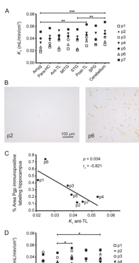
RESULTS Preoperative Pgp activity. In all patients, VPM-K1 values from preoperative VPM-PET scans
were lower in regions pathophysiologically connected to the epileptic focus (amygdala: 0.032 6 0.011; superior temporal gyrus: 0.037 6 0.012) compared with a reference region considered not to be involved in epileptogenic pathways (cerebellum: 0.045 6 0.012) (figure 1A). Moreover, VPM-K1was lower in
amygdala than in posterior TL (0.04360.011). There were no differences between TL VPM-K1 values
ipsilaterally or contralaterally to the seizure focus.
Correlation of preoperative Pgp activity with Pgp expression.Pgp immunoreactivity in surgically resected hippocampal specimens was determined using Pgp-specific antibodies (figure 1B). The higher the Pgp immunoreactivity in surgically resected hippocampal specimens, the lower were the ipsilateral VPM-K1
values for anterior TL (rs 5 20.821, p 5 0.034;
figure 1C) and superior temporal gyrus (rs5 20.857,
p50.024).
Postoperative Pgp activity.Postoperative VPM-K1
val-ues were lower in superior temporal gyrus (0.0336 0.007) compared with cerebellum (0.04160.008) and the posterior TL (0.041 6 0.009; figure 1D). The AED load was reduced in all patients, with 2 patients coming off all AEDs, but this did not affect peripheral metabolism of VPM; i.e., fractions of polar radiolabeled metabolites of VPM in plasma at 10 mi-nutes after injection were similar before (0.140 6 0.039) and after surgery (0.13660.042,p50.938).
Correlation of pre- and postoperative Pgp activity with postoperative outcome. Patients with better outcome had lower mean presurgical VPM-K1values in
ipsilat-eral TL (higher Pgp function) than patients with poorer outcome (rs50.837,p50.024) (figure 2A). Patients
low Pgp immunoreactivity (rs5 20.782,p50.048;
figure 2B). In the 3 patients with best postoperative outcome (patients 1 and 6: seizure-free and without medication; patient 3: seizure-free and with very low AED load), VPM-K1 values increased relative to
preoperative PET scans in all brain VOIs studied, whereas K1 values decreased in patients with worst
outcome (figure 2C).
DISCUSSION In a longitudinal pilot VPM-PET study in patients with drug-resistant TLE before and after TL surgery with a median follow-up period after surgery of 6 years (range 4–7), we detected a
“normalization” of Pgp activity in patients with long-lasting seizure freedom postoperatively. Our preoperative in vivo PET findings are supported by direct correlation with Pgp immunoreactivity in surgical specimens in the same patients. Furthermore, high Pgp activity in epileptogenic pathways preop-eratively was associated with better postoperative outcome. Our longitudinal PET data suggest that Pgp overactivity is dynamic rather than static, and seizure freedom and reduction in AED load are associated with reversal of upregulation.
In line with the transporter hypothesis of drug resistance, we found significantly lower VPM-K1
val-ues in some TL brain regions close to the presumed epileptic focus (amygdala, superior temporal gyrus) as compared with a reference region outside the TL (cerebellum) or with a region more distant from the focus (posterior TL), pointing to Pgp overactivity in epileptogenic brain tissue. Consistent with previous findings, we show that Pgp overactivity was not restricted to the ipsilateral TL, but also extended to the contralateral side involved in epileptogenic pathways.8
After epilepsy surgery, patients with optimal sur-gery outcome defined as seizure freedom and with-drawal of AEDs had global VPM-K1 increases
relative to presurgery PET scans, suggestive of decreased Pgp function. This “return to normal” after successful epilepsy surgery is consistent with our recent report of higher VPM-K1values in
phar-macosensitive compared with drug-refractory pa-tients8 and with findings in postmortem brain
tissue, which showed almost no Pgp overexpression in the sclerotic hippocampus of patients with epi-lepsy who had entered terminal remission before death.11
A previous immunohistochemistry study showed higher Pgp expression in the TL white matter of patients with postoperative seizure relapse compared with postoperatively seizure-free patients,12 in
keeping with our finding of increased Pgp activity postoperatively in patients with poorer outcome. Our preoperative in vivo PET data and ex vivo
Table Clinical, EEG, and MRI data for patients with drug-resistant TLE who underwent surgery a Patient Age, y/ sex Syndrome MRI, EEG Age at onset of epilep sy, y Averag e seizure fre quency per year befor e surgery Interval last seizure to presurgery PET, d Time interval presurgery PET to sur gery, mo Time interval sur gery to posts urgery PET, mo AEDs at presurg ery PET (dose, mg/d) AEDs at postsurgery PET (dose, mg/d) No. of
seizures since surgery
Follow-u p period after surgery , mo Outcom e rank b 1 37/M RmTLE RHA, RTL 3 12 7 3 54 L TG (200) , LEV (1,0 00) None 0 84 1 2 50/M LmTLE LHA, LTL 11 36 15 10 42 OXC (1,8 00) OXC (1,200) 36 (aur as only) 72 5 3 54/F LmTLE LHA, BTL 3 12 17 12 48 CB Z (750) CBZ (300) 0 72 3 4 52/F RmTLE
No atrophy, RTL
34 144 1 37 17 VP A (2,000), L EV (3,000) LEV (2,000) , CB Z (300) 04 8 3 5 33/M RmTLE RHA, BTL 20 72 6 9 45 OXC (2,1 00), L EV (2,000) LEV (1,000) 1 72 6 6 50/M LmTLE LHA, LTL 36 360 4 14 38 P HT (300), LTG (400) None 0 72 1 7 41/M LmTLE LHA, LTL 0 36 2 6 31 L EV (2,500) , CB Z (1,200) LEV (2,000) , CB Z (1,200) 36 0 7 Abbreviations: AED 5 antiepileptic drug; BTL 5 bitemporal; CBZ 5 carbamazepine; LEV 5 levetiracetam; LHA 5 left hippocampal atrophy; LmTLE 5 left mesial temporal lobe epilepsy; LTG 5 lamotrigine; LTL 5 left temporal lobe; OXC 5 oxcarbazepine; PHT 5 phenytoin; RHA 5 right hippocampal atrophy; RmTLE 5 right mesial temporal lobe epilepsy; RTL 5 right temporal lobe; TLE 5 temporal lobe epilepsy; VPA 5 valproic
acid. aPatients
immunohistochemistry, however, suggest that Pgp overactivity in epileptogenic pathways and hippocam-pal overexpression are associated with optimal outcome after TL surgery. This discrepancy in predicting post-operative outcome might be explained by methodologic differences: we quantified hippocampal Pgp immuno-reactivity, whereas Kwan et al.12used semiquantitative
approaches and found differences in Pgp immunoreac-tivity in TL white matter, but also a trend toward higher neuronal Pgp expression in postoperatively seizure-free patients. The latter finding is in keeping with our in vivo PET finding of Pgp overactivity in TL gray matter as a predictor of optimal outcome.
Our pilot study was not designed to assess the pre-dictive ability of VPM-PET as biomarker of surgery outcome; this should be tested prospectively in a larger cohort, and integrated with other known risk factors for less favorable outcome, such as psychiatric comorbidities and incompleteness of resection. More-over, our study cannot distinguish between the effects of seizure cessation from dose decrease or discontinu-ation of AEDs on postoperative Pgp function. Our cohort was too small to statistically compare sub-groups of patients, e.g., seizure-free (n5 4) vs not seizure-free (n53) patients, or seizure-free patients without AEDs (n 5 2) vs seizure-free patients on AEDs (n52), with meaningful results. Alternatively, one could conduct a prospective study to leave the patients on their medication for the first year after surgery and to acquire 2 separate postsurgery PET scans, before and after the change in medication. However, our data suggest that AED exposure also affects Pgp activity, because our outcome ranking mainly based on seizure burden after surgery corre-lates with AED load. Another limitation of this study is that we were not able to obtain VPM-K1values in
hippocampus, which would require the availability of a PET scanner with higher spatial resolution and advanced image reconstruction to overcome the spill-over from the high VPM uptake in the adjacent cho-roid plexus.8
Our study cannot address whether Pgp overac-tivity is the cause of drug resistance in TLE, and thus“surgical”removal of this overactivity contrib-uted to seizure freedom, or whether upregulation is a marker of seizure burden rather than a cause.9In
both scenarios, Pgp overactivity may reduce target concentrations of AEDs and thereby possibly con-tribute to drug resistance. Our study demonstrates that Pgp overactivity is not static, and thus could be therapeutically targeted by drugs that inhibit or downregulate Pgp to possibly prevent development of, or bring about reversal of, drug resistance medi-ated by Pgp.
Our data confirm previous findings that VPM-PET reflects Pgp function in vivo in patients with TLE, and
Figure 1 Pre- and postoperative Pgp activity and correlation with Pgp
expression
(A) VPM-K1values in different brain regions located ipsilateral to the seizure focus of
indi-vidual patients before epilepsy surgery (***p,0.001,**p,0.01; Friedman test with Dunn
post hoc test). (B) Pgp immunopositive labeling in the surgically resected hippocampus of 2 patients. (C) Correlation of percentage area of Pgp immunopositive labeling in the surgically
resected hippocampus with VPM-K1values in ipsilateral anterior TL (rs5Spearman rank
correlation coefficient). (D) VPM-K1values in different brain regions located ipsilateral to the seizure focus of individual patients after epilepsy surgery. Brain regions affected by surgery
are not shown (*p,0.05; Friedman test with Dunn post hoc test). amyg5amygdala;
ant-TL5anterior temporal lobe; MITG5middle and inferior temporal gyrus; p5patient;
para-HC5parahippocampal gyrus; Pgp5P-glycoprotein; post-TL5posterior temporal lobe;
SPG 5 superior parietal gyrus; STG 5superior temporal gyrus; TL 5temporal lobe;
that cerebral Pgp overactivity is associated with drug resistance in TLE.8Our postoperative VPM-PET data
extend previous findings, indicating a“normalization” of Pgp function in those patients who benefited most from surgery. Our findings also suggest that Pgp over-activity in the TL on preoperative VPM-PET might be indicative of optimal postoperative outcome.
AUTHOR CONTRIBUTIONS
Dr. Bauer: study concept or design, acquisition of data, analysis or inter-pretation of data, drafting/revising the manuscript for content. Dr. Karch: analysis or interpretation of data, statistical analysis, drafting/revising the manuscript for content. Dr. Zeitlinger: study concept and design, draft-ing/revising the manuscript for content. Dr. Liu: acquisition of data, analysis or interpretation of data, drafting/revising the manuscript for content. Dr. Koepp, Dr. Asselin, Dr. Sisodiya, and Dr. Hainfellner: anal-ysis or interpretation of data, drafting/revising the manuscript for con-tent. Dr. Wadsak and Dr. Mitterhauser: drafting/revising the manuscript for content, acquisition of data. Dr. Müller and Dr. Pataraia: study concept or design, analysis or interpretation of data, drafting/revis-ing the manuscript for content. Dr. Langer: study concept or design, acquisition of data, analysis or interpretation of data, drafting/revising the manuscript for content.
ACKNOWLEDGMENT
The authors thank the staff of the PET center at the Department of Biomedical Imaging and Image-guided Therapy (Medical University of Vienna) and Research Nurse Edith Lackner (Department of Clinical Pharmacology, Medi-cal University of Vienna) for their support in performing this study.
STUDY FUNDING
Supported by the European Community’s Seventh Framework Program (grant 201380, to M.J.K, S.M.S., M.-C.A., M. Müller, and O.L.), the Austrian Science Fund (FWF) (grant F 3513-B20, to M. Müller and O.L., and KLI 139-B00, to M. Müller and O.L.), and the Department of Health’s National Institute for Health Research Biomedical Research Centres funding scheme (to M.J.K. and S.M.S.).
DISCLOSURE
M. Bauer, R. Karch, M. Zeitlinger, and J. Liu report no disclosures rel-evant to the manuscript. M. Koepp is funded by the European Commun-ity’s Seventh Framework Program (grant 201380, expired). Dr Koepp served on a scientific advisory board of GE Healthcare, and has received honoraria for lectures from UCB Pharma, Eisai, Novartis, and Desitin. M. Asselin is funded by the European Community’s Seventh Framework Program (grant 201380, expired). S. Sisodiya is funded by the European Community’s Seventh Framework Program (grant 201380, expired). Dr. Sisodiya served on a scientific advisory board for UCB Pharma, and has received institutional support or honoraria for lectures from UCB Pharma, GSK, and Eisai. J. Hainfellner, W. Wadsak, and M. Mitterhauser report no disclosures relevant to the manuscript. M. Müller is funded by the European Community’s Seventh Framework Program (grant 201380, expired) and the Austrian Science Fund (FWF) (grants F 3513-B20 and KLI 139-B00). E. Pataraia reports no disclosures relevant to the manuscript. O. Langer is funded by the European Community’s Seventh Framework Program (grant 201380, expired) and the Austrian Science Fund (FWF) (grants F 3513-B20 and KLI 139-B00). Go to Neurology.org for full disclosures.
Received December 20, 2013. Accepted in final form July 11, 2014.
REFERENCES
1. Kwan P, Brodie MJ. Epilepsy after the first drug fails: substitution or add-on? Seizure 2000;9:464–468. 2. Thom M, Mathern GW, Cross JH, Bertram EH. Mesial
temporal lobe epilepsy: how do we improve surgical out-come? Ann Neurol 2010;68:424–434.
Figure 2 Correlation of Pgp activity and Pgp expression with postoperative
outcome
(A) Correlation of mean presurgical VPM-K1values in ipsilateral TL with surgery outcome
rank in individual patients (see the methods section and figure e-1 for definition of surgery
outcome rank order;rs5Spearman rank correlation coefficient). (B) Correlation of
percent-age area of Pgp immunopositive labeling in the surgically resected hippocampus with
sur-gery outcome rank in individual patients. (C) Mean percent change in VPM-K1values across
all studied TL regions located ipsilateral to the seizure focus in postsurgery PET scan relative to presurgery PET scan. Patients 3 and 4 were both assigned rank 3, but the antiepileptic
drug load of patient 3 was lower than that of patient 4. p5patient; Pgp5P-glycoprotein;
3. Löscher W, Luna-Tortós C, Römermann K, Fedrowitz M. Do ATP-binding cassette transporters cause pharmacore-sistance in epilepsy? Problems and approaches in determin-ing which antiepileptic drugs are affected. Curr Pharm Des 2011;17:2808–2828.
4. van Vliet EA, van Schaik R, Edelbroek PM, et al. Inhibi-tion of the multidrug transporter P-glycoprotein improves seizure control in phenytoin-treated chronic epileptic rats. Epilepsia 2006;47:672–680.
5. Brandt C, Bethmann K, Gastens AM, Löscher W. The multidrug transporter hypothesis of drug resistance in epi-lepsy: proof-of-principle in a rat model of temporal lobe epilepsy. Neurobiol Dis 2006;24:202–211.
6. Bauer M, Zeitlinger M, Karch R, et al. Pgp-mediated interaction between (R)-[11C]verapamil and tariquidar at the human blood-brain barrier: a comparison with rat data. Clin Pharmacol Ther 2012;91:227–233.
7. Langer O, Bauer M, Hammers A, et al. Pharmacoresistance in epilepsy: a pilot PET study with the P-glycoprotein sub-strateR-[11C]verapamil. Epilepsia 2007;48:1774–1784.
8. Feldmann M, Asselin MC, Liu J, et al. P-glycoprotein expression and function in patients with temporal lobe epi-lepsy: a case-control study. Lancet Neurol 2013;12:777–785. 9. French JA. P-glycoprotein expression and antiepileptic
drug resistance. Lancet Neurol 2013;12:732–733. 10. Kwan P, Arzimanoglou A, Berg AT, et al. Definition of
drug resistant epilepsy: consensus proposal by the ad hoc Task Force of the ILAE Commission on Therapeutic Strategies. Epilepsia 2010;51:1069–1077.
11. Liu JY, Thom M, Catarino CB, et al. Neuropathology of the blood-brain barrier and pharmaco-resistance in human epilepsy. Brain 2012;135:3115–3133.
12. Kwan P, Li HM, Al-Jufairi E, et al. Association between temporal lobe P-glycoprotein expression and seizure recur-rence after surgery for pharmacoresistant temporal lobe epilepsy. Neurobiol Dis 2010;39:192–197.
13. Wieser HG, Blume WT, Fish D, et al. ILAE Commission Report: proposal for a new classification of outcome with respect to epileptic seizures following epilepsy surgery. Epi-lepsia 2001;42:282–286.
Save These Dates for AAN CME Opportunities!
Mark these dates on your calendar for exciting continuing education conferences by the American Academy of Neurology. Learn more atAAN.com/conferences.AAN Fall Conference
• October 31-November 2, 2014, Las Vegas, NV, The Cosmopolitan of Las Vegas
AAN Annual Meeting
• April 18-25, 2015, Washington, DC, Walter E. Washington Convention Center
BrainPAC
BrainPAC is the American Academy of Neurology’s federal political action committee.
• Since its inception, nearly 3,000 AAN members have contributed $1,500,000 to BrainPAC. • BrainPAC will contribute $600,000 to candidates running for Congress in 2014.
• During the 2012 congressional campaign, 89 percent of candidates supported by BrainPAC won their elections.
DOI 10.1212/WNL.0000000000000858
2014;83;1326-1331 Published Online before print September 3, 2014
Neurology
Martin Bauer, Rudolf Karch, Markus Zeitlinger, et al.
In vivo P-glycoprotein function before and after epilepsy surgery
This information is current as of September 3, 2014
Services
Updated Information &
http://n.neurology.org/content/83/15/1326.full
including high resolution figures, can be found at:
Supplementary Material
858.DC1
http://n.neurology.org/content/suppl/2014/09/03/WNL.0000000000000
Supplementary material can be found at:
References
http://n.neurology.org/content/83/15/1326.full#ref-list-1
This article cites 13 articles, 0 of which you can access for free at:
Subspecialty Collections
http://n.neurology.org/cgi/collection/pet_in_epilepsy
PET in epilepsy
http://n.neurology.org/cgi/collection/pet
PET
http://n.neurology.org/cgi/collection/functional_neuroimaging
Functional neuroimaging
http://n.neurology.org/cgi/collection/epilepsy_surgery_
Epilepsy surgery
http://n.neurology.org/cgi/collection/all_epilepsy_seizures
All Epilepsy/Seizures
following collection(s):
This article, along with others on similar topics, appears in the
Permissions & Licensing
http://www.neurology.org/about/about_the_journal#permissions
its entirety can be found online at:
Information about reproducing this article in parts (figures,tables) or in
Reprints
http://n.neurology.org/subscribers/advertise
Information about ordering reprints can be found online:
rights reserved. Print ISSN: 0028-3878. Online ISSN: 1526-632X.
1951, it is now a weekly with 48 issues per year. Copyright © 2014 American Academy of Neurology. All ® is the official journal of the American Academy of Neurology. Published continuously since