Journal of Pain Research
Dovepress
R e v i e w
open access to scientific and medical research
Open Access Full Text Article
intra-articular hyaluronans: the treatment
of knee pain in osteoarthritis
victor M Goldberg1
Laura Goldberg2
1Department of Orthopaedics, Case
Medical Center, Cleveland, Ohio, USA;
2Cleveland Clinic, Cleveland, Ohio,
USA
Correspondence: victor M Goldberg Department of Orthopaedics, Case Medical Center, 11100 euclid Avenue, Cleveland, Ohio 44106, USA email vmgoldbg@aol.com
Abstract: The etiology of pain in osteoarthritis is multifactoral, and includes mechanical and inflammatory processes. Intra-articular injections of hyaluronans (HAs) are indicated when non-pharmacological and simple analgesics have failed to relieve symptoms. The HAs appear to reduce pain by restoring both mechanical and biomechanical homeostasis in the joint. There are five FDA-approved injectable preparations of HAs: Hyalgan®, Synvisc®, Supartz®, Orthovisc® and Euflexxa®. They all appear to relieve pain from 4 to 14 weeks after injection and may have disease-modification properties. Although several randomized controlled trials have established the efficacy of this treatment modality, additional high quality randomized control studies with appropriate comparison are still required to clearly define the role of intra-articular HA injections in the treatment of osteoarthritis.
Keywords: hyaluronans, knee, pain, osteoarthritis
Introduction
Osteoarthritis (OA) is the most common form of arthritis. It has been reported that 40% of people over the age of 80 will have at least one joint involved and 27 million people in the United States have clinical OA.1,2 OA is a slowly evolving
process, characterized by joint pain, stiffness and loss of range of motion. Weight bearing usually worsens the pain and it is improved with rest. OA results from a complex interaction of biomechanical, biochemical and genetic factors and is characterized by degradation of cartilage and hypertrophy of bone. The etiology of OA remains unknown; however, significant risk factors have been identified with OA development.1,3 These include joint trauma, hypermobility, mal-alignment and
genetic abnormalities. On physical examination patients usually have a swollen joint with warmth, palpable osteophytes, crepitus with movement, tenderness and reduced range of motion. The treatment of OA includes non-pharmacological interventions such as patient education, physical therapy, weight loss and low-impact exercises.4–7
Pharmacological treatment options include acetaminophen, non-steroidal anti-inflammatory drugs (NSAIDs), topical NSAIDs, glucosamine and/or chondrotin sulfate and intra-articular (IA) corticosteroids. Opioid and non-narcotic analgesics may be prescribed in refractory pain patients. IA hyaluronans (HAs) have recently been used for the treatment of painful knee joints with OA.8 Surgical intervention
should be considered only after pharmacological and non-pharmacological treatment have failed.3 The purpose of this review is to address the role of IA HAs treatment
for pain in OA of the knee.
Journal of Pain Research downloaded from https://www.dovepress.com/ by 118.70.13.36 on 24-Aug-2020
For personal use only.
Number of times this article has been viewed
This article was published in the following Dove Press journal: Journal of Pain Research
Dovepress
Goldberg and Goldberg
Pain mechanism in OA
The etiology of pain in the joint with OA is complex but a brief summary of the pathophysiology of pain production is helpful to understand the role of HAs in its treatment.1
Studies have identified the three major types of pain. Acute pain is directly associated with the activation of peripheral nerve receptors. Chronic pain is associated with an ongoing inflammation of peripheral tissues.9 When there is a
sig-nificant chronic pain, damage occurs to the pain pathways either peripherally or centrally, which results in the third type of pain, neuropathic.10 Synovial joints are innervated
by nerves that originate in primary sensory neurons located in the dorsal root ganglion.11 The tissues of synovial joints
that are innervated by nerve endings are the capsule, liga-ments, synovial membrane and subchondral bone. Hyaline cartilage does not have nerve endings. The nerves of the synovial joint are sensitive to the detection of both noxious and non-noxious stimuli. Activation of the nerve endings can begin with any mechanical, chemical, or thermal pro-cess. The pathophysiology of OA involves the release of a large number of inflammatory mediators and these directly act on the nerve endings and reduce their threshold to pain recognition.11 The result is an enhanced discharge of nerve
impulses that are perceived as a painful stimuli. IA HAs have been shown to have a pain-reduction effect through a number of mechanisms which will be discussed later.
Hyaluronans
Hyaluronan or hyaluronic acid (HA) is a complex gly-cosaminoglycan composed of repeated disaccharide units to form a linear polymer. It is widely present in mammalian tissues and has the highest concentration in synovial fluid. Its function in the diarthrodial joint is both mechanical and metabolic. HA provides important viscoelasticity and lubri-cating properties to synovial fluid, thereby reducing articular cartilage wear.10 Further, HA molecules restrict large plasma
protein from entering into the synovial fluid while facilitating the passage of small molecules into the joint for maintenance of nutrition.12 The synthesis of HA in OA is disrupted by
increased levels of pro-inflammatory cytokines, free radi-cals and proteinases, resulting in an HA with a significantly reduced molecular weight, more molecular polydisaccha-rides, and a reduction in synovial fluid viscoelasticity.12 This
abnormal hyaluronic acid increases the potential for articular cartilage wear and accelerates progression of the disease. The progression of OA results from the disruption of the mechanical, biochemical and homeostasis of the diarthro-dial joint, in part provided by ultra-high molecular weight
HA. HA functions in articular cartilage as a supramolecular aggregate to retain proteoglycans, aggrecan and link protein. It therefore acts as a scaffold for these important extracellular matrix molecules. Loss of this normal HA disrupts cartilage matrix stability and perpetuates the destruction of the articu-lar cartilage. Mechanically HA provides viscoelastic proper-ties to the joint allowing for normal fluid flow, lubrication and smooth joint motion. When the ultra-molecular weight HA is enzymatically cleaved by the increased levels of the proteinases observed in OA, the lower-molecular-weight HA no longer can maintain the mechanical integrity of the joint. Further, this lower-weight HA may be proinflamma-tory, further accelerating the disease. The goals of IA HA injections are to improve function, reduce pain and possibly modify disease activity. Potential disease-modifying activi-ties of the HA include promotion of healing and repair by stimulating chondrocyte growth, decreasing apoptosis and stimulating synthesis of cartilage matrix components: col-lagen, proteoglycans and endogenous hyaluronans.12–14 There
are data that suggests a potential inhibition of the synthesis and activity of the chondrodegradating enzymes, eg, metal-loproteinases as well as the inhibition of matrix destructive inflammatory processes.15–17
Exogenous HA with a molecular weight of 500 to 4000 kDa increases synovial fluid viscosity and enhances shock absorption and the lubricating capabilities of synovial fluid.18 HA has been shown to have an effect on reducing pain
by several mechanisms.19 HA binds neuropeptides, creating a
boundary layer around nocireceptors which is one mechanism to reduce pain. Additional mechanisms that potentially can reduce pain in OA include inhibition of inflammatory media-tors, eg, cytokines and prostaglandin. HA has been shown to stimulate endogenous HA synthesis by synovial sites through CD 44 receptor binding.12,20 Exogenous HA downregulates
matrix metalloproteinase-3 expression and inhibit metal-loproteinase synthesis which results in decreased cartilage destruction. Exogenous HA restores metabolic homeostasis thereby enhancing synovial fluid flow and reducting pain. The synergistic effect of exogenous HAs reduces the mechani-cal, chemical or thermal noxious stimuli to the innervated tissues of the synovial joint restoring normal homeostasis and reducing pain and stiffness.
HAs in clinical use
There are currently five FDA-approved injectable prepara-tions of HAs used for the painful knee in OA: Synvisc®,
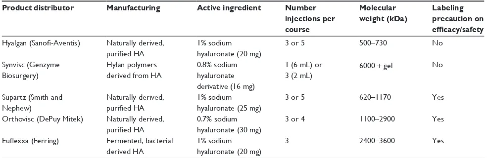
Hyalgan®, Supartz®, Orthovisc® and Euflexxa®. Table 1
summarizes their product characteristics.21–24
Journal of Pain Research downloaded from https://www.dovepress.com/ by 118.70.13.36 on 24-Aug-2020
Dovepress intra-articular hyaluronans for knee pain
The available HAs range in molecular weight from 500 to 6000 kDa. HAs with molecular weights less than 500 kDa have not been effective in either pain relief or improvement of function. The active ingredients are sodium hyaluronate in a concentration of about 20 mg per dose. The recommended number of injections per course range from a single injection for Synvisc to five for Hyalgan or Supartz. The total dose of HA per series varies from 45 mg for Orthovisc to 125 mg for Supartz. There have been no well done randomized controlled head-to-head studies reported, so conclusions on the outcomes of different dosages are difficult to make. Hyalgan, Synvisc, Supartz and Orthovisc are naturally derived products from purified HA extracted from rooster comb. Euflexxa and the recently introduced Supartz are made by a different process in which their HA is derived from an engineering-based process that extracts HA from fermented bacterial sources.
The current guidelines for the treatment of OA of the knee suggest the use of IA HAs for the treatment of pain in patients who have failed to respond adequately to pharmacological therapy as well as simple analgesics.1,3–5,7
IA HAs are contra-indicated if there is known hypersensitiv-ity to HA preparations, if there is a IA infection or a skin problem in the area of the injection site. As a class the HAs are relatively safe with no significant systemic adverse events. There have been no drug interactions reported.25
Five meta-analyses have been published on IA HA treatment in knee OA with the primary outcome being pain relief.25–29 Table 2 summarizes these meta-analyses.
In general the studies support the efficacy of IA HAs in reducing pain; however, the data indicate only a small but clinically important effect. The outcome metrics of most studies centered upon the improvement of pain measures,
eg, WOMAC A or VAS scores; however, in some studies when function was also assessed usually by WOMAC Scores, similar parallel improvements of function and stiffness were seen. The improvement did not last beyond 14 weeks after treatment and the effect size was no more than 0,3. Issues that reduce the value of these meta-analyses include the lack of head-to-head comparisons, the high variability because of different outcome measures and the use of different outcome scales. One study has suggested that IA HA injections provide a longer-term pain benefit than IA corticosteroids.8 The recent
introduction of Synvisc I, a single 6 mm injection of Hyalan G-F 20, was shown to be effective in a randomized, double-blind placebo controlled trial. This is a unique formulation and demonstrated after one injection a significant decrease in WOMAC A pain scores.30
The 2006 Cochrane Review reviewed 76 trials, and is the most comprehensive review to date.7 Many different
HA products were examined for effects from 1 to 52 weeks. In assessing the pooled effect size in comparison to placebo at 1 to 4 weeks after treatment, the reduction in pain was significant and physical function also improved as joint stiffness declined. There is considerable diversity in the outcomes between many of these trials. Previous data had suggested that the higher-molecular-weight products had a greater efficiency, especially in pain relief, but recent studies indicated that the pooled effect size of higher molecular weights were not more effective in relieving pain.14,15,18,19,25,31 Further, the data suggested that pain
reduc-tion diminished with time and was no longer significant after 14 weeks. A number of trials have compared IA HAs to IA corticosteroids.4,32–34 The data indicate that IA corticosteroids
significantly improveed pain during the first 4 weeks after injection but that IA HAs were shown to be more effective
Table 1 US-approved hyaluronan (HA) therapies: summary of product profiles Product distributor Manufacturing Active ingredient Number
injections per course
Molecular weight (kDa)
Labeling precaution on efficacy/safety
Hyalgan (Sanofi-Aventis) Naturally derived, purified HA
1% sodium hyaluronate (20 mg)
3 or 5 500–730 No
Synvisc (Genzyme Biosurgery)
Hylan polymers derived from HA
0.8% sodium hyaluronate derivative (16 mg)
1 (6 mL) or
3 (2 mL) 6000 + gel
No
Supartz (Smith and Nephew)
Naturally derived, purified HA
1% sodium hyaluronate (25 mg)
3 or 5 620–1170 Yes
Orthovisc (DePuy Mitek) Naturally derived, purified HA
0.7% sodium hyaluronate (30 mg)
3 or 4 1100–2900 Yes
Euflexxa (Ferring) Fermented, bacterial derived HA
1% sodium hyaluronate (20 mg)
3 2400–3600 Yes
Notes: Product profiles based on package insert of Hyalgan (2001), Synvisc (2006), Synvisc I (2008), Supartz (2004), Orthovisc (2005), Euflexxa (2005).
Journal of Pain Research downloaded from https://www.dovepress.com/ by 118.70.13.36 on 24-Aug-2020
Dovepress
Goldberg and Goldberg
from 5 to 13 weeks post-injection. Pain relief was greatest following IA corticosteroids at 2 weeks, but not at 4 weeks after injection. By contrast IA HAs demonstrated superior reduction in pain at 8 weeks and continued to be significant until 14 weeks after the injections.
IA HAs have an excellent safety profile with few serious side-effects.25 Systemically there is no differences observed
in gastrointestinal and cardiovascular events between HAs and controls. There have been reported a number of local adverse events such as transient pain and swelling at the injection site and in some circumstances, IA injections of HAs were followed by a greater frequency of pain and swelling when compared to placebo.9,35–37 Pseudoseptic
reactions after injections have been reported primarily with Hylan G-F 20 treatment.38 Pseudosepsis is characterized by
a severe inflammatory response in the joint with significant cellular infiltration and painful effusion occurring within 72 hours after the injection. It usually occurs after a second or third injection in the treatment course and appears similar to an infectious process or a gouty response so that sepsis or pseudogout should be excluded. The synovial fluid may have a high number of mononuclear cells with some neu-rophils, but with an increased number of eosinophils and macrophages.6 It generally is not self limiting and requires
some clinical intervention such as arthrocentesis and an IA steroid injection, with the addition of oral NSAIDs. If there is a concern about infection, while awaiting cultures, one should consider administering prophylactic oral antibiotics. If the condition does continue to deteriorate, intravenous antibiotics may be indicated. If a chronic granuloma develops, surgi-cal intervention may be required. Data from clinisurgi-cal and
pre-clinical studies suggest that there is an immunological basis for this reaction; however, more studies in this area are needed to substantiate this mechanism and their long-term consequences.38
IA HAs are indicated in the US for use in knee OA. However, there have been a number of physician directed uses of HA in other joints. Qvistgaard in a randomized controlled trial of HA compared to isotonic saline in hip OA demonstrated a significant improvement in pain and an overall improvement in clinical activities.39 Another
study utilizing a population of patients who were total hip replacement candidates demonstrated a significant improvement in both pain and Harris Hip Scores.40,41 More
randomized control trials are necessary to establish the risk/benefit of the use of IA HAs in hip OA. Other joints have been studied including the carpal-metacarpal joint, shoulder and ankle.42–44 The results have been variable
and it is difficult to conclude anything from these studies without further randomized control trials. One orthopedic application is the injection of HA in the post-arthroscopy period to enhance pain and accelerate rehabilitation. One recent study evaluated the safety and efficacy of a high-molecular-weight sodium hyaluronate after a knee arthros-copy for treatment of a symptomatic meniscus tear.45 The
study utilized 3 injections after arthroscopy and randomized patients into a group treated with the IA HA compared to a group using only acetaminophen, up to 1000 mg 4 times a day. The patients in the treated groups 3 and 6 months after surgery had significantly less pain and more flexion than the comparative control group. This is an area that is fertile for additional randomized controlled trials.
Table 2 intra-articular hyaluronan therapy in knee osteoarthritis: summary of meta-analyses
Reference Lo27 Wang29 Arrich26 Modawal28 Bellamy25
Total no. of studies
22 20 22 11 76
Efficacy outcome measures
effect size (change from baseline pain vs placebo)
Mean difference, ASPiD vs placebo
wMD in pain during/after exercise (vAS) vs control
vAS wMD in pain on
weight bearing vs placebo
Key pooled results
effect size: 0.32 (P , 0.001)
Mean difference for ASPiD: 13.4%
wMD 10–14 wk: -4.3 mm (P = 0.013) wMD 22–30 wk: -7.1 mm (P = 0.013)
wMD 5–7 wk: 17.6 (95% Ci, +7.5, +28.0) 8–12 wk: 18.1 (95% Ci, +6.3, +29.9) 15–22 wk: 4.4 (95% Ci, -15.3, 24.1)
% Change 5–13 wk: +26% for pain; +23% for function
Conclusion Small treatment effect
Safe and effective Not effective in measured outcomes
Moderately effective at 5–10 wk, not effective at 15–22 wk
effective treatment for pain, function, pt, global assessment at 5–13 wk Abbreviations: ASPiD, adjusted sum of pain intensity differences; wMD, weighted mean difference; vAS, visual analog scale; pt, patient.
Journal of Pain Research downloaded from https://www.dovepress.com/ by 118.70.13.36 on 24-Aug-2020
Dovepress intra-articular hyaluronans for knee pain
In conclusion pain is a central symptom of OA and requires an integrated approach to it’s treatment. Both non-pharmacological and non-pharmacological treatments offer the best chance for pain relief. Pharmacological treatments include NSAIDs, cox-2 inhibitors, opioids, anti-inflammatory creams and IA corticosteroids. IA corticosteroids have been shown to be effective in relieving pain during the first 2 weeks after treatment. IA HA injections have a longer onset of action and longer duration of effect. The exact mechanism of action is still to be delineated, although it is clear that IA HA reduces pain through its effect on peripheral pain receptors as well as an impact on the synovial tissue and its role in enhancing the viscoelastic properties of synovial fluid. Although there have been many studies, there still is a need for additional high quality, randomized control trials with placebos or comparators to clearly delineate the role of IA HA in the treatment of pain in OA. The use of IA HA in other joints requires additional well structured clinical studies.
Disclosures
One or more of the authors is a consultant for Ferring Phar-maceuticals and has/will receive compensation.
References
1. Manek NJ, Lane NE: Osteoarthritis current concepts in diagnosis and management. Am Fam Physician. 2000;61(6):1795–1804.
2. Lawrence RC, Felson DT, Helmick CG, et al. Estimates of the prevalence of arthritis and other rheumatic conditions in the United States. Part II.
Arthritis Rheum. 2008;58(1):26–35.
3. Kelly MA, Goldberg VM, Healy WL, Pagnano MW, Hamburger MI. Osteoarthritis and beyond: a consensus on the past, present, and future of hyaluronans in orthopedics. Orthopedics. 2003;26(10):1064–1079; quiz 1080–1081.
4. Neustadt DH, Altman R: Intra-articular therapy. In: Moskowitz RW, Altman RD, Hochberg, MC, Buckwalter JA, Goldberg VM, editors. Osteoarthritis: Diagnosis and Management/Surgical Management. 4th ed. Philadlphia, PA: Lippincott Williams & Wilkins. 2002; 287–302.
5. Zhang W, Moskowitz RW, Nuki G, et al. OARSI recommendations for the management of hip and knee osteoarthritis, Part II: OARSI evidence-based, expert consensus guidelines. Osteoarthritis Cartilage. 2008;16(2):137–162.
6. Recommendations for the medical management of osteoarthritis of the hip and knee: 2000 update. American College of Rheumatology
Arthritis Rheum. 2000;43(9):1905–1915.
7. Zhang W, Moskowitz RW, Nuki G, et al. OARSI recommendations for the management of hip and knee osteoarthritis, part I: critical appraisal of existing treatment guidelines and systematic review of current research evidence. Osteoarthritis Cartilage. 2007;15(9):981–1000. 8. Divins JG, Zazulak BT, Hewett TE: Viscosupplementation for
knee osteoarthritis: a systematic review. Clin Orthop Relat Res. 2007;445:113–122.
9. Han F, Ishiguron N, Ito T, et al: Effects of sodium hyaluronate on experimental osteoarthritis in rabbit knee joints. Nagoya J Med Sci. 1999;62(3–4):115–126.
10. Balazs EA, Denlinger JL: Viscosupplementation: a new concept in the treatment of osteoarthritis. J Rheumatol Suppl. 1993;39:3–9.
11. Belmonte C: Signal transduction in nocicptors; general principles In: Belmonte C, Cervero F, editors. Neurobiology of Nociceptors. Oxford, England: Oxford Unviersity Press. 1996;241–257.
12. Goldberg VM, Buckwalter JA: Hyaluronans in the treatment of osteoarthritis of the knee: evidence for disease-modifying activity.
Osteoarthritis and Cartilage. 2005;31:216–224.
13. Yoshioka M, Shimizu C, Harwood FL, et al: The effects of hyaluronan during the development of osteoarthritis. Osteoarthritis Cartilage. 1997;5:251–260.
14. Kikuchi T, Yamada H, Shimmei M: Effect of high molecular weight hyaluronan on cartilage degradation in a rabbit model of osteoarthritis.
Osteoarthritis Cartilage. 1996;4:990110.
15. Ghosh P, Guidolin D: Potential mechanism of action of intra-articular hyaluronan therapy in osteoarthritis: are the effects molecular weight dependent? Semin Arthritis Rheum. 2002;32:10–37.
16. Takahashi K, Goomer RS, Harwood F, et al: The effects of hyaluronan on matrix metalloproteinase-3 (MMP-3), interleukin-1β, and tissue inhibitor of metalloproteinase-1 (TIMP-1) gene expression during the develop-ment of osteoarthritis. Osteoarthritis Cartilage. 1999;8:182–190. 17. Takahashi K, Hashimoto S, Kubo T, et al: Effect of hyaluronan on
chondrocyte apoptosis and nitric oxide production in experimentally induced osteoarthritis. J Rheumatol. 2000;27:1713–20.
18. Ghosh P, Kipic B, Smith MM, et al: The viscosity and elasticity of synovial fluids from osteoarthritis joints are improved 5 weeks post treatment by 5 intra-articular injections of hyaluronan with a molecular weight (MW) of 0.5–0.730 a 106 but not by a preparation with MW
of 3.0–6.9 × 10⁶ Da. Annual European Congress of Rheumatology
(EULAR). Stockholm, Sweden, 2002.
19. Goroh S, Onaya J, Abe M, et al: Effects of the molecular weight of hyaluronic acid and its action mechanisms on experimental joint pain in rats. Ann Rheum. Dis. 1993;52(11):817–822.
20. Balazs EA, Watson D, Duff IF, Roseman S. Hyaluronic acid in synovial fluid. I. Molecular parameters of hyaluronic acid in normal and arthritis human fluids. Arthritis Rheum. 1967;10(4):357–376.
21. Orthovisc® (sodium hyaluronate) (prescribing information) Bridgewater,
NJ: Ortho Biotech Products, LP: 2004.
22. Synvisc® (hylan G-F 20) (prescribing information) Philadelphia, PA:
Wyeth-Ayerst, 2003.
23. Hyalgan® (sodium hyaluronate) (prescribing information) New York,
NY: Sanofi Pharmaceuticals, Inc, 2001.
24. Supartz® (sodium hyaluronate) (prescribing information). Tokyo, Japan:
Seikagaku Corp. 2001.
25. Bellamy N, Campbell J, Robinson V, et al: Viscosupplementation for the treatment of osteoarthritis of the knee. Cochrane Database Syst Rev. 2-CD005321, 2006.
26. Arrich J, Piribauer F, Mad P, Schmid D, Klaushofer K, Mullner M. Intra-articular hyaluronic acid for the treatment of osteoarthritis of the knee: systematic review and meta-analysis. CMAJ. 2005;172(8):1039–1043. 27. Lo GH, LaValley M, McAlindon T, Felson DT. Intra-articular
hyaluronic acid in treatment of knee osteoarthritis: a meta-analysis.
JAMA. 2003;290(23):3115–3121.
28. Modawal A, Ferrer M, Choi HK, Castle JA. Hyaluronic acid injections relieve knee pain. J Fam Pract. 2005;54(9):758–767.
29. Wang CT, Lin J, Chang CJ, Lin YT, Hou SM. Therapeutic effects of hyaluronic acid on osteoarthritis of the knee. A meta-analysis of random-ized controlled trials. J Bone Joint Surg Am. 2004;86-A(3):538–545. 30. Chevalier X, Jerosch J, Goupille P, et al. Single, intra-articular
treat-ment with 6 mL of hylan G-F 20 in patients with symptomatic primary osteoarthritis of the knee: A randomized, multi-centre, double-blind, placebo-controlled trial. Ann Rheum Dis. 2009.
31. Dougados M, Nguyen M, Listrat V, et al: High molecular weight sodium hyaluronate (hyalectin) in osteoarthritis of the knee: a 1 year placebo-controlled trial. Osteoarthritis Cartilage. 1993;l:97–103. 32. Bannuru RR, Natov NS, Obadan IE, et al: Therapeutic trajectory of
hyaluronic acid versus corticosteroids in the treatment of knee osteoar-thritis: A systematic review and meta-analysis. Arthritis and Rheum. 2009;Vol 61:1704–1711.
Journal of Pain Research downloaded from https://www.dovepress.com/ by 118.70.13.36 on 24-Aug-2020
Journal of Pain Research
Publish your work in this journal
Submit your manuscript here: http://www.dovepress.com/journal-of-pain-research-journal
The Journal of Pain Research is an international, peer-reviewed, open access, online journal that welcomes laboratory and clinical findings in the fields of pain research and the prevention and management of pain. Original research, reviews, symposium reports, hypoth-esis formation and commentaries are all considered for publication.
The manuscript management system is completely online and includes a very quick and fair peer-review system, which is all easy to use. Visit http://www.dovepress.com/testimonials.php to read real quotes from published authors.
Dovepress
Dovepress
Goldberg and Goldberg
33. Bellamy N, Campbell J, Robinson V, Gee T, Bourne R, Wells G. Intraarticular corticosteroid for treatment of osteoarthritis of the knee.
Cochrane Database Syst Rev. 2006;(2):CD005328.
34. Fuchs S, Monikes R, Wohlmeiner A, Heyse T. Intra-articular hyaluronic acid compared with corticoid injections for the treatment of rhizarthro-sis. Osteoarthritis Cartilage. 2006;14(1):82–88.
35. Altman RD, Moskowitz R: Intraarticular sodium hyaluronate (Hyalgan) in the treatment of patients with osteoarthritis of the knee; a randomized clinical trial. Hyalgan Study Group. J Rheuatol. 1998;25(11):2203–2212.
36. Brandt KD, Block JA, Michalski JP, et al: Efficacy and safety of intraarticular sodium hyaluronate in knee osteoarthritis. ORTHOVISC Study Group. Clinic Ortho. 2001;385:130–143.
37. Lohmander LS, Dalen N, Englund G, et al: Intra-articular hyaluronan injections in the treatment of osteoarthritis of the knee: a randomised, double blind, placebo controlled multicenter trial. Ann Rheum Dis. 1996;55:424–431.
38. Goldberg VM, Coutts RD: Pseudoseptic Reactions to Hylan Visco-supplementation. Clin Orthop. 2004;No 419:130–137.
39. Qvistgaard E, Christensen R, Torp-Pedersen S, Bliddal H. Intra-articular treatment of hip osteoarthritis: a randomized trial of hyaluronic acid, corticosteroid, and isotonic saline. Osteoarthritis Cartilage. 2006;14(2):163–170.
40. van den Bekerom MP, Lamme B, Sermon A, Mulier M. What is the evidence for viscosupplementation in the treatment of patients with hip osteoarthritis? Systematic review of the literature. Arch Orthop Trauma Surg. 2008;128(8):815–823.
41. van den Bekerom MP, Rys B, Mulier M. Viscosupplementation in the hip: evaluation of hyaluronic acid formulations. Arch Orthop Trauma Surg. 2008;128(3):275–280.
42. Roux C, Fontas E, Breuil V, Brocq O, Albert C, Euller-Ziegler L. Injection of intra-articular sodium hyaluronidate (Sinovial) into the carpometacarpal joint of the thumb (CMC1) in osteoarthritis. A prospec-tive evaluation of efficacy. Joint Bone Spine. 2007;74(4):368–372. 43. Blaine T, Moskowitz R, Udell J, et al. Treatment of persistent shoulder
pain with sodium hyaluronate: a randomized, controlled trial. A multi-center study. J Bone Joint Surg Am. 2008;90(5):970–979.
44. Cohen MM, Altman RD, Hollstrom R, Hollstrom C, Sun C, Gipson B. Safety and efficacy of intra-articular sodium hyaluronate (Hyalgan) in a randomized, double-blind study for osteoarthritis of the ankle. Foot Ankle Int. 2008;29(7):657–663.
45. Westrich G, Schaefer S, Walcott-Sapp S, Lyman S, Randomized Prospective Evaluation of Adjuvan Hyaluronic Acid Therapy Admin-istered After Knee Arthroplasty. Am J Orthop. 2009;38(12):612–616.
Journal of Pain Research downloaded from https://www.dovepress.com/ by 118.70.13.36 on 24-Aug-2020