INVESTIGATION
Sequence-Based Detection and Breakpoint
Assembly of Polymorphic Inversions
Russell B. Corbett-Detig,*,1Charis Cardeno,†and Charles H. Langley† *Department of Organismic and Evolutionary Biology, Harvard University, Cambridge, Massachusetts 02138, and†Department of Evolution and Ecology, University of California, Davis, California 95616
ABSTRACTInversion polymorphisms have occupied a privileged place inDrosophilagenetic research since their discovery in the 1920s. Indeed, inversions seem to be nearly ubiquitous, and the majority of species that have been thoroughly surveyed have been found to be polymorphic for one or more chromosomal inversions. Despite enduring interest, however, inversions remain difficult to study because their effects are often cryptic, and few efficient assays have been developed. Even inDrosophila melanogaster, in which inversions can be reliably detected and have received considerable attention, the breakpoints of only three inversions have been characterized molecularly. Hence, inversion detection and assay design remain important unsolved problems. Here, we present a method for identification and localde novoassembly of inversion breakpoints using next-generation paired-end reads derived from D. melanogaster isofemale lines. PCR and cytological confirmations demonstrate that our method can reliably assemble inversion breakpoints, providing tools for future research onD. melanogasterinversions as well as a framework for detection and assay design of inversions and other chromosome aberrations in diverse taxa.
S
TURTEVANT (1917) discovered the first chromosomal inversion as a suppressor of recombination inDrosophila melanogaster. Shortly after his initial finding, Sturtevant produced evidence that inversions, structurally reversed segments of the linear map order of chromosome arms, ac-count for this observation (Sturtevant 1926). A vast body of empirical work has followed this original discovery, yielding several key results that inform our understanding of the genetic, and potential evolutionary, implications of polymor-phic inversions. First, single crossovers within the inverted regions of inversion heterokaryotypes are expected to yield aneuploid gametes, effectively suppressing exchange be-tween arrangements (Sturtevant and Beadle 1936). Inver-sion heterokaryotypy redistributes chiasma elsewhere in the genome, both intrachromosomally in colinear segments and via the interchromosomal effect (Lucchesi and Suzuki 1968).This primary effect—suppression of recombination in the inverted regions of heterokaryotypes, especially near the
breakpoints—is the subject of much of the theoretical pop-ulation genetic research focused on inversions. Generally, interpretations in the literature favor a model in which inversions achieve high frequencies by suppressing recombi-nation between coadapted alleles located near the inversion breakpoints (Dobzhansky 1951), although there are many other possible mechanisms (Kirkpatrick and Barton 2006; Hoffmann and Reiseberg 2008). Empirical research on poly-morphic inversions has been extensive as well, the central result being that chromosomal inversions are pervasive. In-deed, segregating inversions are found in abundance in the majority of organisms that have been examined in depth, including plants, insects, mammals, and humans (Hoffmann and Reiseberg 2008). However, the selective forces that gov-ern the evolution of inversion polymorphisms remain largely obscure, with important exceptions being inversions associ-ated with novel sex chromosomes (Charlesworth et al. 2005), sex ratio distortion (Jaenike 2001), and autosomal segregation distortion (Kusanoet al.2003; Lyon 2003).
D. melanogaster is highly polymorphic for chromosomal inversions. In fact, since the pioneering work of Sturtevant (1931) and Dubinin et al. (1937), .500 segregating inversions have been found in natural populations of D. melanogaster, encompassing a broad-frequency spec-trum ranging from present at low frequency in single Copyright © 2012 by the Genetics Society of America
doi: 10.1534/genetics.112.141622
Manuscript received April 30, 2012; accepted for publication May 21, 2012 Supporting information is available online at http://www.genetics.org/content/ suppl/2012/06/05/genetics.112.141622.DC1.
1Corresponding author: Harvard University, Biological Laboratories 2109, 16 Divinity
populations to present in virtually all populations world-wide (Ashburner and Lemeunier 1976; Aulard et al. 2004). This distribution is conventionally subdivided in-to four classes that correspond in-to the inversions’prevalence in natural populations: unique endemic, recurrent endemic, rare cosmopolitan, and common cosmopolitan (Ashburner and Lemeunier 1976; Krimbas and Powell 1992).
The latter class has received by far the most attention. Common cosmopolitan inversions exhibit frequency clines, diminishing from high frequency in equatorial regions, to nearly absent in higher lattitudes. This pattern is replicated independently on several continents, suggesting that strong selective forces govern the distributions of these inversions (Knibbet al. 1981). This observation has prompted numer-ous attempts to identify the traits that are experiencing se-lection, and several ecologically relevant traits have been associated with common cosmopolitan inversions (Van Delden and Kamping 1991; Frydenberget al.2003; Kennington et al. 2007). However, it remains unknown if the genetic elements that confer these traits are the targets of selection or hitchhiking as a result of reduced recombination and the rel-atively young age of these inversions (Andolfattoet al.1999; Matzkinet al.2005).
Even less is known about the rare cosmopolitan and recurrent endemic inversions, which are comparatively understudied and sometimes not recorded in published frequency assays (Knibb et al. 1981; Krimbas and Powell 1992). The rare cosmopolitans are distributed worldwide, but often entirely absent from populations, while the recur-rent endemic inversions may be at high frequency in one geographic region, but have rarely or never been identified elsewhere (Krimbas and Powell 1992). A detailed under-standing of their limited distributions and selective poten-tials is essential both as a comparison to the more“successful” common cosmopolitan inversions and to a nuanced and com-plete conception of polymorphic inversions inD. melanogaster and the broader topic of genome evolution.
Despite continuing interest in the inversion polymor-phisms ofD. melanogaster, only three inversions, all common cosmopolitans, can be assayed directly using molecular means (Wesley and Eanes 1994; Andolfatto et al. 1999; Matzkinet al. 2005). Others must be identified via the la-borious original method: crossing to a stock with known chromosomal arrangements and examining the banding pat-terns of the giant salivary gland polytene chromsomes in the larval progeny. In fact, all inversion breakpoints that have been characterized molecularly in D. melanogaster were identified using this convenient cytogenetic feature in com-bination with fluorescent in situ hybridization techniques (Wesley and Eanes 1994; Andolfatto et al. 1999; Matzkin et al. 2005). By hybridizing larval polytene chromosomes with DNA fragments of known mapping positions, it is pos-sible to narrow down the breakpoint regions through suc-cessively closer hybridizations (Wesley and Eanes 1994). This method is both time-consuming and, perhaps most problematic, completely impractical for organisms that lack
visible polytene chromosomes. Considering the largely quan-titative goals of population genetic research, a more general and efficient means of inversion detection and assay design is essential to furthering our understanding of inversion poly-morphisms in natural populations.
Genomic techniques have presented two appealing alter-natives for identifying and characterizing structural poly-morphisms segregating within populations. One method, originally developed by Tuzun et al. (2005), is based on sequencing both ends of short DNA fragments with an ap-proximate known distance and orientation with respect to each other. By first mapping paired reads to a reference sequence, and subsequently identifying clusters of read pairs that do not map in the expected orientation or distance relative to one another, it is possible to reliably identify the breakpoints of structural polymorphisms (Tuzun et al. 2005; Kidd et al. 2008; Medvedev et al. 2009). This ap-proach is appealing because it can be used tofine-map struc-tural breakpoint and it has previously been validated as a tool for interrogation of structural polymorphisms in D. melanogaster (Cridland and Thornton 2010). While these methods have high sensitivity for breakpoint detection, it is often not possible based solely on the breakpoints to dis-tinguish inversions from other structural rearrangements, such as duplications that reinsert in inverted orientation (Cridland and Thornton 2010). The relatively short length of inserts that are currently used in the majority of rese-quencing projects, as opposed to the long inserts used in the previous landmark studies (Tuzun et al. 2005; Kidd et al.2008), may exacerbate this issue, as short inserts pro-vide little resolution beyond detected breakpoints.
Alternative approaches, which rely on data from many densely genotyped individuals, use an expected signature of nucleotide variation to detect polymorphic inversions (Bansal et al. 2007; Sindi and Raphael 2010). While these methods have provided valuable insights and many candidate inver-sions, the inherent genomic limitations of SNP genotype data have proved to be a major impediment. To accurately predict inversions, these methods require substantial minor allele fre-quencies of inversion and large sample sizes (Bansal et al. 2007; Sindi and Raphael 2010). Additionally, genotyping approaches offer poor resolution of inversion breakpoints, which may be of interest for population genetic analyses, de-signing PCR assays, and studying the mechanisms of inversion formation and DNA repair.
we identify breakpoints and design molecular assays forfive previously uncharacterized polymorphic inversions. We also improve on existing assays for two previously characterized inversions. Cytology as well as previously developed PCR assays confirm that our method is highly accurate. We ex-pect that these primer pairs will be immediately useful as tools for researchers interested in assaying inversions di-rectly in individualD. melanogasteror existing stocks. Addi-tionally, this general framework could be implemented to detect and design molecular assays for structural polymor-phisms in other species.
Materials and Methods
Details for allfly stocks used in this study can be found at http://www.dpgp.org. In short, all short-read sequences are derived from 76-bp Illumina paired-end reads, separated by 300-bp inserts, and represent numerous African popula-tions and one French population. All genomic regions ana-lyzed are haploid through chromosomal extractions, inbreeding, or haploid embryo extractions (Langley et al. 2011). The average coverage depth is31·(range: 8–47). We aligned short reads for each line to the D. mela-nogasterreference sequence v5.22 (Adamset al.2000) using ELAND v2 as a standard part of the Illumina Casava pipe-line. Alignments were performed over the course of more than a year; as such, several versions of the Casava pipeline were used (see http://www.dpgp.org for details). Eland alignments were ported to MAQ using the “export2maq” utility in the MAQ v0.7.3 software package (Li et al. 2008). We called consensus sequences for each line, requir-ing that each site called have a minimum read depth of 3 and a minimum quality of 30. All other sites were excluded from the resulting assemblies. We identified seven lines showing excess identity by descent as those with regions of little pairwise divergence on more than one chromosome arm; these are obvious in cursory inspections, so this was done by hand and primarily excluded genomes that are drawn from the same isofemale line. From each pair show-ing identity by descent, we discarded the line that was se-quenced to lower depth.
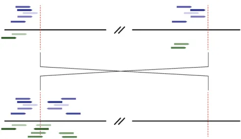
From the alignment files, we discarded read pairs that share identical mapping coordinates with another pair, as well as those for which one read maps to an annotated transposable element. Next, we parsed read pairs for which both reads mapped uniquely to the reference sequence but mapped in parallel orientation (or). We restricted this set of reads to those that mapped to the same chromosome arm. For each line individually, we assigned aberrant read pairs to clusters, requiring that both reads in a pair mapped within 500 bp of another read included in that cluster. Thus, each cluster contains sets of read pairs for which one of the pair maps to one 500-bp region of the genome, and the other read maps to another 500-bp region of the same chro-mosome arm. We further required that all reads in one clus-ter map in the same orientation and that genomic positions
within each cluster be.1 megabase from each other. Clus-ters supported by fewer than five clones were discarded. Finally, we parsed from the “export.txt”file read pairs for which one read maps to the same genomic location and in the same orientation as the clusters identified previously, and the other read is unmapped. The unmapped reads are expected to cross the breakpoint. We then folded these reads into the original cluster (Figure 1).
We compared each line’s set of read clusters to all other lines’sets of read clusters. Overlapping clusters in the same genomic position and orientation were identified as poten-tially confirming the same inversion breakpoint. Due to their unique origins, which results in zero polymorphisms in the inverted population at formation, inversion breakpoints will immediately attain highFSTrelative to the standard arrange-ment population. As genetic exchange is almost completely suppressed near inversion breakpoints (Novitski and Braver 1954; Wesley and Eanes 1994; Andolfatto et al. 1999), genetic differentiation will be maintained between arrange-ments and is an expected signature of all inversion break-points. Provided that an inversion is present in more than two individuals, this expectation suggests an ideal way to sift through identified breakpoints for inversion false pos-itives. For lines sharing identical breakpoints, we compared the consensus sequences in 20-kb windows centered on each potential breakpoint and calculated FST between the lines that share the putative breakpoint and the lines that do not. We retained candidate inversions for which both break-points’ FST was .0.25. We calculated FST as described in Hudson et al. (1992), using only sites that were called in all lines. Importantly, we did not weightFSTby sample size, which enables the detection of low-frequency inversions. Because of this, even immediately after formation, we ex-pect to observe strong genetic differentiation, and inver-sions’FST’s will initially be0.5.
Wede novoassembled the set of reads corresponding to each remaining potential breakpoint using Phrap v1.090518 (Green 1996) All contigs were aligned to the D. mela-nogasterreference sequence v5.22 (Adamset al.2000), us-ing Blast v2.2.25 (Altschul et al. 1990), and contigs with significant alignments to both sides of the expected break-point were retained. Blast alignments with corresponding e-values ,10210and alignment lengths.30 bp were con-sidered significant.
To assist in assigning identities for novel inversions, we compared the cytogenetic positions of the inversions’ break-points with reported breakpoint coordinates. To do this, we downloaded the cytologically predicted positions of inver-sion breakpoints and the map converinver-sion table for cytolog-ical coordinates from FlyBase (http://www.flybase.org).
Inversion breakpoints may take two forms: cut-and-paste and inverted duplication (see Ranzet al.2007 for a descrip-tion of breakpoint structure). After aligning breakpoint-spanning contigs to the reference genome, we inferred the structure on the basis of the following criteria. If both break-point-spanning contigs appear to map in convergent orien-tations to within 50 bp of each other at both ends of the inversion, they are assumed to be cut-and-paste breakpoints. Otherwise, we assume that the sequence between mapping positions is present as a duplication at the other breakpoint. We confirmed these structural predictions via comparisons with the three inversions that have previously been exam-ined (Wesley and Eanes 1994; Andolfattoet al.1999; Matzkin et al.2005) and by comparison with the copy-number varia-tion analysis performed by C. H. Langleyet al. (2012), whose stocks are known to bear many of these inversions.
To confirm breakpoints, we developed a PCR-based inversion assay. We designed primers using Primer3 (http://frodo.wi.mit.edu/primer3/) that would produce an amplicon unique to the standard or inverted chromosomal arrangement on the basis of these putative breakpoints. We extracted genomic DNA from flies using the Quick Fly Genomic DNA Prep provided by the Berkeley Drosophila Genome Project (http://www.fruitfly.org/about/methods/ inverse.pcr.html). Briefly, we ground 30 flies in Buffer A (100 mM Tris–HCl, 100 mM EDTA, 100 mM NaCl, 0.5% SDS) and incubated theflies at 65for 30 min. We added a 1:2.5 solution (1 part 5 M KAc to 2.5 parts 6 M LiCl) to the samples and incubated them on ice for at least 10 min. The DNA was precipitated with isopropanol, washed, and resus-pended in ddH2O.
All of the PCR inversion assays [except for the standard chromosomal arrangement of In(3R)P] used standard PCR reaction conditions: 2.0 mM MgCl2, 0.2 mM each of dNTPs, 0.5 uM each of forward and reverse primers, 1 unit of Taq, and 50 ng of DNA. Specific PCR conditions for each reaction are described inSupporting Information,Table S1. Appro-priately sized amplicons were identified with agarose gels. We used long PCR to assay the In(3R)Pstandard chromo-somal arrangement. This was necessary because the inverted duplications present at each breakpoint are too long for
stan-dard PCR. We followed the manufacturer’s PCR general reac-tion mixture and condireac-tions (TaKaRa LA Taq) with a few exceptions: a final MgCl2 concentration of 1.75 mM (Table S1) and an annealing/elongation time of 5 min. We included the reference strainy;cn bw;spas well as several other stan-dard orientation lines as negative controls for inversion-specific primers and positive controls for standard inversion-specific primers. For inversion-positive controls, we obtained sev-eral lines known by cytology to harbor the putative inversion from the Bloomington Stock Center and C. H. Langleyet al. (2012;Table S1).
For each inversion that had not been detected previously, we sequenced via Sanger PCR at least one breakpoint to further validate Phrap assemblies. Sequences were assem-bled from forward and reverse chromatograms using phred-Phrap, which is distributed as a part of the Consed package. We inspected all assembled PCR fragments by hand in Consed v1.090518 (Gordon et al. 1998). These sequences are available inFile S1.
Results
Across all genomes, we recovered.15,000 breakpoints that map in parallel orientation to a single chromosome arm. After pooling across all samples, we found .200 break-points that were present in more than one line. Finally, after applying the FSTfilter, we found 12 aberrant read clusters whose corresponding consensus sequences showed in-creased FSTaround both breakpoints. HeightenedFSTis an expected signature of nucleotide variation between inverted and standard arrangements owing to the unique origin of inversion and suppressed exchange between arrangements immediately surrounding each breakpoint (Novitski and Braver 1954; Wesley and Eanes 1994; Andolfatto et al. 1999). Importantly, heightened FST at both breakpoints is not expected for breakpoints associated with other re-arrangements that may occur at higher frequencies. This is because only a single breakpoint actually harbors the novel insertion event; the other “breakpoint” reflects reads that map uniquely to the single copy present in the reference sequence, but are not actually linked to this genomic loca-tion. We also surveyed all clusters present in more than one line for breakpoints consistent with cytologically known inversions that have been identified in populations from sub-Saharan Africa (Aulard et al. 2002). We did not find any additional breakpoints that were consistent with these inversions; thus,FSTappears to be a successfulfilter for in-version true positives.
due to the presence offixed repetitive elements immediately adjacent to the proximal breakpoints of each inversion (Andolfattoet al.1999; Matzkinet al.2005).
In a recent study, C. H. Langleyet al.(2012) found a pat-tern of excess long-distance linkage disequilibrium and sig-nificantly decreased nucleotide diversity associated with a large paracentric inversion,In(3R)Mo, in a Raleigh, North Carolina, population of D. melanogaster. Because the se-quence data used in this study were derived from single-end reads, they are not suitable for direct comparison via our method, which relies on independently mapped paired-end reads. We did identify one line, FR310, which shares this pattern of long-distance linkage disequilibrium and that had been sequenced using paired-end reads. Because of this inversion’s unexpected prevalence in this Raleigh, North Carolina, population, we surveyed this line specifically for potential breakpoints, without requiring that the identified read clusters be corroborated by clones derived from an-other line. We found two breakpoints whose positions are consistent with our expectations for this inversion on the basis of the observed pattern of nucleotide diversity and confirmed these breakpoints via PCR in the eight lines iden-tified by C. H. Langleyet al.(2012).
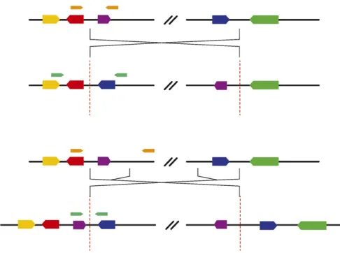
We recovered two classes of breakpoints: simple cut-and-paste breakpoints and staggered break-plus-inverted-duplication breakpoints (Figure 2). We were able to confirm structural predictions for three inversions whose breakpoints were previously characterized (Wesley and Eanes 1994; Andolfatto et al. 1999; Matzkin et al. 2005). In(3R)K, In(3R)Mo,In(1)A, andIn(2R)NSare all present in the sample analyzed by C. H. Langleyet al.(2012), and we were able to support breakpoint structure predictions for each on the basis of comparisons to the copy-number variation analysis included in that work. Five of the eight inversions contain inverted duplications at their breakpoints (Table S2), which is similar to the proportions that were found by a study that focused on inversions thatfix between species in themelanogaster subgroup (17 of 29) (Ranzet al.2007).
We designed assays for each class that amplify a product of unique length for either the standard or the inverted haplotype. Cut-and-paste breakpoints can be assayed easily as described in Andolfatto et al. (1999). Because in stag-gered-break inversion structures there is no single break-point that is unique to the standard arrangement, primers that span a single breakpoint cannot be used to distinguish between inversion heterozygotes and inversion homozy-gotes. Our solution is to design primers that span the dupli-cated regions at either end of the inversion (Figure 2). This produces a PCR product that is unique to the standard ar-rangement and may be more robust than an allele-specific PCR approach (e.g., Andersonet al.2005).
To make use of this advantage, we designed new primers for the standard arrangement of In(3R)P (Table S1), for which only ambiguous or allele-specific primers were pre-viously available. Likely due to the presence of a repetitive element immediately adjacent to the proximal breakpoints
ofIn(3R)P(Matzkinet al.2005), we recovered only a single breakpoint for this inversion. Fortunately, Matzkin et al. (2005) have previously sequenced both breakpoints via long-range PCR forIn(3R)P. We downloaded these sequen-ces from GenBank (acsequen-cession nos. AY886890–AY886892) and used them to design a novel set of primers that yield a unique amplicon for the standard arrangement. All PCR fragments that we sequenced are identical to the Phrap as-semblies, except in the low-quality bases toward the ends of the traces (File S1).
For the three inversions that were previously charac-terized at the molecular level, breakpoint coordinates are known, and we identified these breakpoints directly in our sequence data. We also confirmed our results for these inversions using published primers (Table S3). For In(3L)P, the existing primers did not work reliably. We elected to design new primers (Table S1) and have found these to be more reliable. For four other inversions, all putative inversion identities were confirmed by positive controls (Table S1).
For all previously uncharacterized inversions, the cyto-logically derived mapping positions based on previous surveys were within 100 kb of the breakpoint that we identified. In most previously uncharacterized inversions, it was also possible to test our primers on stocks known via cytology to bear the inversion. This was not possible for one inversion on the X chromosome for which no independent positive controls are available. However, the breakpoint coordinates, geographic distribution, and frequency of this inversion are all consistent withIn(1)Beand do not suggest any other known inversions. Hence, although we cannot be certain of the identity, we refer to this inversion asIn(1)Be (Table S1).
Discussion
While our method was quite successful and has immediate applications to many short-read sequencing projects, it should be noted that there are two important drawbacks, both of which will be ameliorated by imminent advances in sequencing technologies. First, our method requires accu-rate mapping information and a well-characterized refer-ence genome. Already, several species’genomes have been fully assembled using next-generation sequencing technolo-gies (e.g., Liet al.2009); hence, the anticipated availability of many additional reference sequences may make the pro-posed method widely serviceable. Second, the extent to which transposable elements contribute to the formation of chromosomal inversions remains an open question (Mathiopoulos et al. 1998; Cacereset al. 1999). Although this does not appear to be a common mechanism of inver-sion formation in theD. melanogastersubgroup (Ranzet al. 2007), it is possible that transposable elements contribute more to structural polymorphisms in other species. Because of the modest insert lengths used in sequencing, our method has little power to detect inversions that form via ectopic recombination between repetitive elements. However, this limitation will also diminish in importance with the increas-ing availability and quality of larger insert sizes in library preparation, which will be able to span individual repetitive elements. Hence, if anything, the applicability and useful-ness of this approach will increase as sequencing technolo-gies continue to progress.
Despite these potential drawbacks, our method has performed well. A recent survey of African D. melanogaster inversion polymorphisms (Aulardet al.2002) reported gen-erally the same set of polymorphic inversions at moderate frequencies. So, while we cannot estimate a true false-negative rate, this suggests that we have recovered the majority of inversions that are likely to be segregating at frequencies.2 in this sample. Requiring FST calculations means that our method will miss inversions present in only one individual. This drawback is unavoidable, since there are thousands of aberrant read clusters in individual genomes, and it is not always possible to distinguish between inversions and other structural variants solely on the basis of breakpoint coordi-nates. Regardless of the error rates, our method is a vast im-provement over conventional methods, and it allows us to rapidly characterize and develop novel molecular assays for five chromosomal inversions and to improve on two existing assays. This more than doubles the available assays, providing a substantial improvement in the tools available for studies of the polymorphic inversions ofD. melanogaster.
Another advantage of this approach is its broad applica-bility. Cytological methods, beyond being time-consuming, require visible polytene chromosomes, as well as an approx-imate idea of where inversion breakpoints might be expected to occur and in what strains. Our method circumvents these issues, and it allows us to examine numerous individuals simultaneously and to identify polymorphic inversions without
requiring any prior knowledge of the lines or inversion content of the genome. Hence, we expect this will be a useful framework for researchers interested in characterizing and developing molecular assays for polymorphic inversions, especially in developing model systems. Although we ana-lyzed haploid data, this method could feasibly be extended to accommodate diploid samples with sufficient sequencing or sampling depth.
It should also be emphasized that clustering putative breakpoints across samples would allow detection not only of inversions but also of other types of chromosome aberra-tions. These need not necessarily be aberrations transmitted through the germ line. For example, our approach may have applications in the study of rearrangements among somatic or cancer cells where relevant independent sampling can be conducted.
Chromosomal inversion polymorphisms are a ubiquitous evolutionary phenomenon. They are present in virtually all species and may have potent evolutionary effects ranging from resisting geneflow in hybrid zones, to maintaining co-adapted gene complexes, to the long-term maintenance of epistatically interacting segregation distortion systems (Hoffmann and Reiseberg 2008). However, a complete un-derstanding of the selective effects of polymorphic inver-sions is elusive. Even—perhaps especially—in the species in which chromosomal inversions were originally discovered, D. melanogasterinversions remain an enigmatic and intrigu-ing feature of virtually all populations. A central impediment to quantitative studies of these polymorphisms, especially rare cosmopolitan and recurrent endemic inversions, is a lack of low-cost efficient assays. Here, we provide these tools. Although there are certainly numerous interesting popula-tion genetics quespopula-tions that could feasibly be addressed us-ing these data (many of which are subjects of ongous-ing research), our goal with this work is to make these resources available to the community as soon as possible.
Acknowledgments
We thank John Pool, Daniel Hartl, and two anonymous reviewers for helpful comments on this manuscript, as well as Shu Fang for providing cytology information. We also acknowledge funding by National Institutes of Health grant HG02942 (to C.H.L.). R.B.C-D. is supported by a Harvard Prize Fellowship.
Literature Cited
Adams, M. D., S. E. Celniker, R. A. Holt, C. A. Evans, J. D. Gocayne
et al., 2000 The genome sequence ofDrosophila melanogaster. Science 287: 2185–2195.
Altschul, S. F., W. Gish, W. Miller, E. W. Myers, and D. J. Lipman, 1990 Basic local alignment search tool. J. Mol. Biol. 3: 403– 410.
inversion polymorphism has shifted in the last 20 years in Aus-tralianDrosophila melanogasterpopulations. Mol. Ecol. 14: 851– 858.
Andolfatto, P., J. D. Wall, and M. Kreitman, 1999 Unusual hap-lotype structure at the proximal breakpoint ofIn(2L)tin a nat-ural population of Drosophila melanogaster. Genetics 153: 1297–1311.
Ashburner, M., and F. Lemeunier, 1976 Relationships within the
melanogasterspecies subgroup of the genusDrosophila (Sopho-phora). 1. Inversion polymorphisms inDrosophila melanogaster
andDrosophila simulans. Proc. R. Soc. Lond. 193: 137–157. Aulard S., J. R. David, and F. Lemeunier, 2002 Chromosomal
in-version polymorphism in Afrotropical populations ofDrosophila melanogaster. Genet. Res. 79: 49–63.
Aulard, S., L. Monti, N. Chaminade, and F. Lemeunier, 2004 Mitotic and polytene chromosomes: comparisons betweenDrosophila melanogaster and Drosophila simulans. Genetica 120: 137– 150.
Bansal, V., A. Bashir, and V. Bafna, 2007 Evidence for large in-version polymorphisms in the human genome from HapMap data. Genome Res. 17: 219–230.
Caceres, M., J. M. Ranz, A. Barbadilla, M. Long, and A. Ruiz, 1999 Generation of a widespread Drosophila inversion by a transposable element. Science 285: 415–418.
Charlesworth, D., B. Charlesworth, and G. Marais, 2005 Steps in the evolution of heteromorphic sex chromosomes. Heredity 95: 118–128.
Cridland, J., and K. Thornton, 2010 Validation of rearrangement breakpoints identified by paired-end sequencing in natural pop-ulations ofDrosophila melanogaster. Genome Biol. Evol. 2: 83– 101.
Dobzhansky, T., 1951 Genetics and the Origin of Species, Ed. 3. Columbia University Press, New York.
Dubinin, N. P., N. N. Sokolov, and G. G. Tiniakov, 1937 Intraspecific chromosomal variability. Biol Zh. 6: 1007.
Frydenberg, J., A. A. Hoffmann, and V. Loeschcke, 2003 DNA sequence variation and latitudinal associations in hsp23, hsp26 and hsp27 from natural populations ofDrosophila mela-nogaster. Mol. Ecol. 12: 2025–2032.
Gordon, D., C. Abajian, and P. Green, 1998 Consed: a graphical tool for sequencefinishing. Genome Res. 8: 195–202.
Green, P., 1996 Phrap documentation. Available at:http://www. phrap.org/phredphrap/phrap.html
Hoffmann, A. A., and L. H. Rieseberg, 2008 Revisiting the impact of inversions in evolution: From population genetic markers to drivers of adaptive shifts and speciation? Annu. Rev. Ecol. Evol. Syst. 39: 21–42.
Hudson, R. R., M. Slatkin, and W. P. Maddison, 1992 Estimation of levels of geneflow from DNA-sequence data. Genetics 132: 583–589.
Jaenike, J., 2001 Sex chromosome meiotic drive. Annu. Rev. Ecol. Syst. 32: 25–49.
Kennington, W. J., A. A. Hoffmann, and L. Partridge, 2007 Mapping regions within cosmopolitan inversion In(3R)Payne associated with natural variation in body size in Drosophila melanogaster. Genetics 177: 549–556.
Kidd, J. M., G. M. Cooper, W. F. Donahue, H. S. Hayden, N. Sampas
et al., 2008 Mapping and sequencing of structural variation from eight human genomes. Nature 453: 56–64.
Kirkpatrick, M., and N. Barton, 2006 Chromosome inversions, lo-cal adaptation and speciation. Genetics 173: 419–434. Knibb, W. R., J. G. Oakenshot, and J. B. Gibson, 1981 Chromosome
inversion polymorphism inDrosophila melanogaster.I. Latitudinal clines and associations between inversions in Australasian popu-lations. Genetics 98: 833–847.
Krimbas, C. B., and J. R. Powell, 1992 Drosophila Inversion Poly-morphism. CRC Press, Boca Raton, FL.
Kusano, A., C. Staber, H. Y. E. Chan, and B. Ganetzky, 2003 Closing the (Ran)GAP on segregation distortion in Drosophila. Bioessays 25: 108–115.
Langley, C. H., M. Crepeau, C. Cardeno, R. Corbett-Detig, and K. Stevens, 2011 Circumventing heterozygosity: sequencing the amplified genome of a single haploid Drosophila melanogster
embryo. Genetics 188: 239–246.
Langley, C. H., K. Stevens, C. Cardeno, Y. C. G. Lee, D. R. Schrider
et al. 2012 Genomic variation in natural populations of
Drosophila melanogaster. Genetics 192: 533–598.
Li, H., J. Ruan, and R. Durbin, 2008 Mapping short DNA sequenc-ing reads and callsequenc-ing variants ussequenc-ing mappsequenc-ing quality scores. Genome Res. 18: 1851–1858.
Li, R., W. Fan, G. Tian, H. Zhu, L. Heet al., 2009 The sequence andde novoassembly of the giant panda genome. Nature 463: 311–317.
Lucchesi, J. C., and D. T. Suzuki, 1968 The interchromosomal control of recombination. Annu. Rev. Genet. 2: 53–86. Lyon, M. F., 2003 Transmission ratio distortion in mice. Annu.
Rev. Genet. 37: 393–408.
Mathiopoulos, K. D., A. della Torre, V. Predazzi, V. Petrarca, and M. Coluzzi, 1998 Cloning of inversion breakpoints in the Anoph-eles gambiaecomplex traces a transposable element at the in-version junction. Proc. Natl. Acad. Sci. USA 95: 12444–12449. Matzkin, L. M., T. J. S. Merritt, C. T. Zhu, and W. F. Eanes,
2005 The structure and population genetics of the breakpoints associated with the cosmopolitan chromosomal inversionIn(3R) PayneinDrosophila melanogaster. Genetics 170: 1143–1152. Medvedev, P., M. Stanciu, and M. Brudno, 2009 Computational
methods for discovering structural variation with next-generation sequencing. Nat. Methods 6: S13–S20.
Novitski, E., and G. Braver, 1954 An analysis of crossing-over within a heterozygous inversion inDrosophila melanogaster. Ge-netics 39: 197–209.
Ranz, J. M., D. Maurin, Y. S. Chan, M. V. Grotthuss, L. W. Hillier
et al., 2007 Principles of genome evolution in theDrosophila melanogasterspecies group. PLoS Biol. 5: 1366–1381.
Sindi, S. S., and B. J. Raphael, 2010 Identification and frequency estimation of inversion polymorphisms from haplotype data. J. Comput. Biol. 17(3): 517–531.
Sturtevant, A. H., 1917 Genetic factors affecting the strength of linkage inDrosophila. Proc. Natl. Acad. Sci. USA 3: 555–558. Sturtevant, A. H., 1926 A crossover reducer inDrosophila
mela-nogasterdue to inversion of a section of the third chromosome. Biol. Zentralbl. 46: 697–702.
Sturtevant, A. H., 1931 Known and probable inverted sections of the autosomes ofDrosophila melanogaster. Carnegie Inst.Wash-ington Publ. 421: 1–27.
Sturtevant, A. H., and G. W. Beadle, 1936 The relations of inver-sions in the X chromosome ofDrosophila melanogasterto cross-ing over and disjunction. Genetics 21: 544–604.
Tuzun, E., A. J. Sharp, J. A. Bailey, R. Kaul, V. A. Morrisonet al., 2005 Fine-scale structural variation of the human genome. Nat. Genet. 37: 727–732.
Van Delden, W., and A. Kamping, 1991 Changes in relativefitness with temperature among second chromosome arrangements in
Drosophila melanogaster. Genetics 127: 507–514.
Wesley, C. S., and W. F. Eanes, 1994 Isolation and analysis of the breakpoint sequences of chromosome inversionIn(3L)Paynein
Drosophila melanogaster. Proc. Natl. Acad. Sci. USA 91: 3132– 3136.