University of South Carolina
Scholar Commons
Theses and Dissertations
2018
Association Of Objective Measures Of Sleep And
Inflammation Markers On Police Officers: A
Cross-Sectional Analysis
Megan R. Buss
University of South Carolina
Follow this and additional works at:https://scholarcommons.sc.edu/etd Part of theEpidemiology Commons
This Open Access Thesis is brought to you by Scholar Commons. It has been accepted for inclusion in Theses and Dissertations by an authorized administrator of Scholar Commons. For more information, please contactdillarda@mailbox.sc.edu.
Recommended Citation
Buss, M. R.(2018).Association Of Objective Measures Of Sleep And Inflammation Markers On Police Officers: A Cross-Sectional Analysis.
ASSOCIATION OF OBJECTIVE MEASURES OF SLEEP AND INFLAMMATION MARKERS ON POLICE OFFICERS: A CROSS-SECTIONAL ANALYSIS
By
Megan R. Buss
Bachelor of Science
University of Pittsburgh at Johnstown, 2016
Submitted in Partial Fulfillment of the Requirements
For the Degree of Master of Science in Public Health in
Epidemiology
The Norman J. Arnold School of Public Health
University of South Carolina
2018
Accepted By:
Michael D. Wirth, Director of Thesis
James Burch, Reader
Jim Hussey, Reader
ii
iii ABSTRACT
Police officers are a unique occupational group due to the fact that they
have more health problems than many other occupations. These health problems
could be a result of elevated inflammation markers caused by poor sleep. Sleep
influences circadian rhythms, which thereby influences the function of the immune
system. The immune system is responsible for the body’s inflammatory response
using pro-inflammatory cytokines such as IL-6, CRP, Fibrinogen, and TNF-a. These
cytokines can become elevated if disruption of the sleep cycle occurs. Elevated
levels of inflammatory markers are associated with increased risk of cardiovascular
disease. Police officers also work shifts and have a large amount of occupational
stress that may contribute to increased levels of pro-inflammatory markers as well.
This analysis aimed to examine the influence that objective and subjective
measures of sleep have on inflammatory markers among police officers within the
Buffalo Cardio-Metabolic Occupational Police Stress (BCOPS) cohort
cross-sectionally. Body mass index (BMI), shift work, and stress measures were
examined as potential effect modifiers. Subjective measures of sleep were
obtained by the Pittsburgh Sleep Quality Index (PSQI) and objective measures of
sleep were obtained through actigraph data. Police officers wore an Actiwatch for
iv
Sleep latency, quality, duration, efficiency and daytime dysfunction were
used from the PSQI, and wake after sleep onset, sleep onset latency, sleep
duration, and efficiency were used from the actigraph measures. The inflammation
markers were collected from blood samples after a 12 hour fast. Each inflammatory
marker was measured using different assays at the University of Vermont.
General linear models were used to compare adjusted means of categorical
sleep measures and beta coefficients for continuous sleep measures for each
inflammation marker. Analyses were stratified by normal (18.5-24.9 BMI),
overweight (25-29.9 BMI), and obese (≥30 BMI), and then by day and
evening/night shiftwork. Logistic regression was performed on a dichotomous
version of CRP, using a clinial cut point, and odds ratios were obtained for
high-risk CRP. Statistically significant associations were seen between various sleep
measures and inflammation markers. It is seen that as sleep worsens, there is an
v
TABLE OF CONTENTS
ABSTRACT ... iii
LIST OF TABLES ... vii
CHAPTER 1: Introduction ... 1
CHAPTER 2: Background ... 4
2.1 Sleep ... 4
2.2 Inflammation ... 6
2.3 Inflammation and Sleep ... 7
2.4 Potential Effect Modifiers, Sleep, and Inflammation ... 11
CHAPTER 3: Methods ... 15
3.1 Sleep ... 16
3.2 Outcomes ... 18
3.3 Covariates... 19
3.4 Statistical Analysis ... 21
vi
CHAPTER 5: Discussion ... 53
CHAPTER 6: Conclusion ... 66
vii
LIST OF TABLES
Table 3.1 Types of Sleep Parameters Used ... 26
Table 4.1 Characteristics of BCOPS Population by PSQI Global ... 33
Table 4.2 Comparison of BCOPS Population Characteristics by Subjective and Objective Sleep ... 34
Table 4.3 Distribution of PSQI Components ... 35
Table 4.4 Distribution of Actigraph Measures of Sleep ... 36
Table 4.5 Distribution of Actigraph Measures of Sleep by Shiftwork ... 36
Table 4.6 Adjusted Mean Inflammation Markers by PSQI Components ... 37
Table 4.7 Adjusted Inflammation Markers by Actigraph Measures of Sleep ... 39
Table 4.8 Adjusted Mean Inflammation Markers by PSQI Components by BMI Status ... 40
Table 4.9 Adjusted Inflammation Markers by Actigraph Measures of Sleep by BMI Status ... 44
Table 4.11 Adjusted Inflammation Markers by Actigraph Measures of Sleep by Shiftwork Status... 50
CHAPTER 1
Introduction
In the health conscious world we live in today, tracking sleep has become
a new trend among the public (1). Although still largely unstudied, there has been
increased interest in the role sleep plays in health. Underlying this are the circadian
rhythms that control sleep and other bodily systems (2). When sleep is disrupted,
circadian rhythms become misaligned, which in turn affects the other circadian or
clock-controlled bodily functions. One large system controlled by circadian rhythms
is the immune system (3).
The immune system controls the pro- and anti-inflammatory cytokines
found in the blood. The proliferation of these cytokines occurs under a circadian
rhythm; during the light phase anti-inflammatory cytokines are released and
during the dark phase pro-inflammatory cytokines are released (4, 5). If circadian
misalignment occurs, the balance of the cytokine release becomes uneven, and a
higher rate of pro-inflammatory cytokine production happens (6).
Pro-inflammatory cytokines also are released during an infection; this is referred to as
acute inflammation. In acute inflammation, once the injury is healed, the
pro-inflammatory cytokines are broken down. However, if interleukin-6 (IL-6) remains
more pro-inflammatory cytokines, creating chronic inflammation (7-10). A
common form of chronic inflammation is obesity. Obesity is a low-grade chronic
inflammatory state caused by the release of pro-inflammatory cytokines from
adipose tissue due to overflow (11).
Occupational stress also can play a role in the quality and quantity of sleep
a person receives. Of all occupational stressors, the ones that police officers
experience are the most detrimental (12). They are subject to environmental
stimuli that can cause stress like shootings and violence, as well as shift work (13,
14). Shiftwork can cause circadian misalignment because shift workers tend to
work during the dark phase and sleep during the light phase (15). When coupling
stress and shiftwork, police officers are at a higher risk of developing disease, and
more specifically, chronic diseases (16, 17).
Although there has been research on the effect of sleep on inflammation
markers, most previous studies have measured sleep subjectively or performed an
experiment on non-habitual sleep (4, 18-25). Most of the observational studies
that have measured sleep subjectively show no association between poor sleep
and an increase in inflammation markers (19-22). Experimental studies, using
objective measures of sleep, however, do find associations indicating a difference
between subjective and objective measures of sleep (4, 22-25). This study
attempts to bridge this gap by using an objective measure of sleep, actigraphy,
proven to be similar to the polysomnography (PSG) measure of sleep used in
Cardio-Metabolic Occupational Police Stress (BCOPS) study, and involves collection of
blood samples, objective and subjective measures of sleep, past records of
shiftwork, a range of psychosocial metrics, and occupational stress measures. This
will allow for a more objectively-measured investigation of the relationship
between sleep and inflammation markers.
The specific aims of this study are to:
1. Determine if sleep affects levels of inflammatory markers c-reactive
protein (CRP), interleukin-6 (IL-6), tumor necrosis factor alpha
(TNF-α), and fibrinogen by addressing the following hypotheses:
a. A poorer sleep profile (shorter sleep duration, lower sleep
efficiency, longer sleep latency, and higher wake after sleep onset
[WASO]) is associated with higher levels of inflammation markers
b. Those with scores indicating poorer sleep on the Pittsburgh Sleep
Quality Index will have higher levels of inflammation
2. There will be a difference in the relationship between sleep and
inflammation by BMI levels after:
a. Stratification by body mass index (BMI)
b. Stratification by shiftwork
CHAPTER 2
Background
2.1 Sleep
Sleep is an essential part of everyone’s lives, but the concept of sleep and
what is does for the body is sometimes not understood (27). Sleep deprivation or
poor sleep is now becoming a larger issue because we are beginning to understand
some of the mechanisms that sleep controls (28). Studies have shown that it is
not only the amount of sleep we get, but the quality that puts people at higher
risk for disease (27, 29, 30).
Sleep is separated into two stages: non-rapid-eye-movement-sleep
(NREMS) and rapid-eye-movement sleep (REMS)(2, 31-33). The first stage of sleep
is further separated into 4 sections, all having distinct functions. Stage 1 is believed
to be the transition between wakefulness and sleep and is referred to as light
sleep. Stage 2 shows an increase in higher-frequency brain waves and is where
greater depth of sleep begins. Stages 3 and 4 are characterized by slow-wave
sleep and then followed by REMS (31). In humans, sleep is typically a 90-minute
cycle of NREMS to REMS, which can repeat 5-6 times a night (34). The length of
each cycle, however, can vary drastically depending on the type of sleep a person
Good sleep can be defined as optimal length of each sleep stage with proper
distribution of sleep stages and low arousal from sleep. Good sleep is comprised
of approximately 80% NREMS (31). A person receiving good sleep should
experience no fatigue upon full wakefulness. Sleep also plays a role in memory
consolidation, the transition of newly learned tasks or materials into memories
(35). Poor sleep is characterized as deviant sleep patterns in either quantity and/or
quality. With deviant sleep patterns comes an increased risk of physical and
psychological problems (19, 36-39). A few of these problems include risk of
diabetes (36), hypertension (37), coronary heart disease (38), occupational
functioning, mood disturbance (39), depression, and anxiety (19).
The regulation of sleep, however, is dependent upon a homeostatic need
and circadian rhythms (2). The circadian rhythm is controlled by a natural clock
within the suprachiasmatic nucleus (SCN) of the brain that runs on a 24-hour cycle
that tends to follow the 24-hour light-dark cycle of the environment, but can be
active even in the absence of light-cues. Circadian clock mechanisms are present
in many cell types and organs. Cells related to the immune system are an example
of these types of cells (40, 41). Within the central clock located in the SCN are
three proteins that have a large impact on the immune function. These proteins
are circadian locomotor output cycles kaput (CLOCK), brain and muscle aryl
hydrocarbon receptor nuclear translocator-like 1 (BMAL1), three period regulators
(PER1, PER2, PER3), and reverse-Erb alpha (REV-ERBα) (3, 42-45). It has been
in limiting inflammation(3). PER 3 can have varying lengths that have an effect on
morning preference, cognitive performance, and circulating concentrations on
IL-6 (44).
2.2 Inflammation
The immune system can be monitored by measuring the production of
inflammation biomarkers through measurements of blood cytokines. Cytokines can
be categorized as pro-inflammatory (type 1) and anti-inflammatory (type 2) (5,
46, 47). Maximum production of pro-inflammatory cytokines, including type-1,
IL-2, IL-6, IL-1IL-2, TNF-α, and interferon (IFN)-gamma, occurs during the dark phase
or nocturnal sleep. The production of IL-12 and TNF-α are completely dependent
on sleep, whereas production of anti-inflammatory cytokines are dependent on
wakefulness (48-51). However, a study on sleep deprivation showed a shift from
type 1-type 2 cytokine balance to type 1 cytokine production, indicating an
elevation of pro-inflammatory cytokines (4, 6).
Inflammation can be beneficial; in its acute phase, it aids in fighting
infections. However, chronic inflammation leads to tissue damage and disease.
Acute inflammation is defined by the recruitment of neutrophils and then
monocytic cells to damaged tissue by the immune response. Chronic inflammation
is associated with a large presence of macrophages and lymphocytes (7-10). The
switch from acute to chronic inflammation can be linked through IL-6. IL-6 acts as
a mediator during acute inflammation, but when IL-6 remains after the infection
another immune response is activated, causing mononuclear cell accumulation and
chronic inflammation proliferation (53). This then creates a cycle of chronic
inflammation because of the increase in IL-6 re-triggering the immune response.
2.3 Inflammation and Sleep
The association between sleep and inflammation has been studied, but
previous results are conflicting and founded on subjective measures of sleep,
creating the potential for information bias. In an experimental study assessing
sleep deprivation and activation of morning levels inflammation markers, 30
healthy adults spent 4 days in the National Institute of Health General Clinical
Research Center. The first 3 days they were permitted to sleep between 11 pm
and 7 am, for baseline information, and on the 4th day sleep was permitted from
3 am to 7 am, for the sleep deprivation information. Blood samples were taken on
each day at 8am, 12pm, 4pm, 8pm, and 11pm. The study results showed that
after partial sleep deprivation, IL-6 and TNF-α showed a significant increase (t107=
-2.3, P<.05) compared to the baseline sleep duration in the morning. The cytokine
levels approached baseline ranges as the day progressed (22).
Vgontzas et al. and Meier-Ewert et al. showed the same inverse association
with IL-6 and CRP, respectively, in an experimental study design (23, 24). In
another experimental study by Vgontzas et al., the effect of modest sleep
deprivation (a loss of 2 hours compared to the normal 8 hours) on levels of
inflammatory markers was assessed. Once again, this study showed that on the
than when individuals received all 8 hours of sleep (4). Another laboratory study
assigned participants to 12 days of sleeping 8 hours a night or 4 hours a night to
compare the effect of sleep restriction on inflammatory markers (54). Haack et.
al. found elevated levels of IL-6 in people sleeping 4 hours a night compared to 8
hours a night (p<0.05) but no significant increase in CRP (54).
Patel et al. used individuals from the Cleveland Family Study to look at the
association between sleep duration and biomarkers of inflammation. The sleep
measure was based on self-reported habitual sleep time and a separate PSG study.
For the observational and the experimental study, the inflammatory markers CRP,
IL-6, TNF-α, IL-1, and IL-10 were collected between 7 am and 8 am after the PSG
and an overnight fast. The experimental section of this study used an overnight
PSG to measure sleep duration. The observational study found a positive linear
relationship between sleep duration and CRP and IL-6. Conversely, the
experimental study found an inverse association between sleep duration and
TNF-α (25). The different findings suggest that the self-reported sleep data is modeling
a different relationship than the PSG.
The findings from the NSDA study, however, contradict the laboratory
performed studies showing the difference between experimental and real-world
associations (19). A study on sleep duration, insomnia and markers of systemic
inflammation was conducted within the Netherlands Study of Depression and
Anxiety (NSDA). Sleep was measured through a questionnaire completed after an
at baseline from fasting blood samples collected between 8am-9am (19). They
found that longer sleep durations were associated with significantly higher levels
of CRP (p-value=0.005) and IL-6 (p-value<0.001) compared to short sleep
duration and when comparing normal sleep to short sleep duration levels of CRP
(p-value=0.575) and IL-6 (p-value=0.916). This study failed to see a significant
association between short sleep duration and inflammatory markers (19). These
results also were found in a study of sleep duration and quality among a Taiwanese
population and a cross-sectional study performed within the 2007-2008 cycle of
NHANES (20, 21).
There also are observational studies that have examined at the association
between poor sleep quality and inflammation markers. In the Heart and Soul
Study, a prospective cohort of men and women with established coronary heart
disease, a cross-sectional analysis was done on self-reported sleep quality and
biomarkers of systematic inflammation (18). The self-reported sleep measure was
from the Pittsburgh Sleep Quality Index (PSQI), and asked participants at baseline
and 5 years later, “During the past month, how would you rate your sleep?”
Secondary sleep variables also were included. Inflammation markers were
collected at baseline and at the 5-year follow up after a 12-hour fast. The
inflammation markers collected were CRP, IL-6, and fibrinogen. After analysis
there was no evidence that self-reported sleep quality was associated with
cross-sectional or 5-year difference in levels of IL-6, CRP, and fibrinogen. Prather et al.
in IL-6 (p=0.003) , CRP (p=0.02), and fibrinogen (p=0.02) after a 5-year increase
(18). This study suggests that gender has an effect on the association between
sleep and inflammation markers.
Another cross-sectional study performed within the 2005-2006 US National
Health and Nutrition Examination Survey (NHANES) cycle looked at the association
between self-reported sleep quality, using two questions from the Sleep Disorders
Questionnaire, and mediators of cardio-metabolic health, one being CRP (28). The
findings concluded that, although above clinical reference range, there is a
J-shaped relationship between sleep quality and CRP levels. On this J-J-shaped curve,
there is a steep increase in CRP from fair to very poor sleep quality, with the
association between very poor sleep quality and CRP being statistically significant
(28).
A study examining the link between sleep, exaggerated inflammatory
response and adverse health outcomes focused on gender-specific responses. In
women, poor sleep quality was associated with higher CRP levels but there were
no relationships of note between PSQI scores and IL-6 or TNF-a (55). A cohort
made of western Australian men also looked at the association between
inflammation and poor sleep. They found a significant association between
difficulty falling asleep and higher levels of CRP(56).
This relationship has been examined extensively in individuals with
obstructive sleep apnea (OSA). After comparing 15 studies, it is seen that on
difference increased significantly when individuals were obese (57). These studies
show that poor sleep can affect inflammation.
All of these previous cross-sectional studies have used self-reported sleep
measures, and four of them demonstrated a positive linear association between
sleep and inflammation markers. One study showed an inverse relationship when
stratified by gender for all inflammation markers, and the other showed an inverse
relationship for just CRP. However, all of the experimental studies showed an
inverse relationship between sleep duration/quality and levels of inflammation
markers. The next step in this field of research is to combine the experimental
research findings using PSG with an observational study. This can be done by
performing a cross-sectional analysis using objective measures of sleep.
2.4 Potential Effect Modifiers, Sleep, and Inflammation
Obesity is one of the most burdensome diseases in the world, and is a result
of excessive energy intake (58). Obesity is characterized not only by high BMI, but
is also an inflammatory state (58, 59). Obesity has an impact on immune function
just like malnutrition, because it is a form of malnutrition caused by excess dietary
intake (59). The link between immune function and obesity is the adipose tissue
where fat is stored. When obesity persists, the pro-inflammatory cytokines
localized within the adipose tissue are pushed into systemic circulation creating a
As sleep duration and sleep quality have been decreasing over the past
decades, obesity has been increasing (27, 61). When studying obesity, body mass
index (BMI, kg/m2) is the standard measurement used, because it incorporates
height and weight into the relationship. Overweight is defined as 25 kg/m2 ≥ BMI
£ 30 kg/m2, and obesity is defined as a BMI ≥ 30 kg/m2 (62). The link between
short sleep duration and obesity has been observed and shows positive
associations for children and adults. The nature of the relationship, however,
remains a mystery (63). Some indicate a linear inverse association, showing that
as sleep duration decreases, BMI increases. Other studies indicate more of a
U-shaped association, showing that short and long sleep duration are associated with
high BMIs (64). The U-shaped associations signal that the relationship does differ
by age category but it is still present (63).
Occupational stress is a major element of physical and mental health. Law
enforcement and more specifically police officers experience some of the highest
levels of stress related to work (13, 14). Stress is the strain placed on an individual
by environmental stimuli (65). A normal day of work for a police officer can entail
duties such as crime scene violence, involvement in shootings, seeing and handling
dead bodies, injury on the job, and negative news coverage. Police officers are
exposed to these environmental stimuli which can contribute to greater stress than
other occupational stress stimuli. Therefore, police officers are at a higher risk for
Hypothalamic-pituitary-adrenal axis (HPA) and the autonomic nervous
system show the highest response to stress. These systems are therefore used to
look at the impact stress has on the body (66). Events that occur on the job can
cause a wide range of diseases. Acute Post-Traumatic Stress Disorder (PTSD) is
caused by occupational stress. PTSD in police officers can become long-term
because of the cycle of stimuli re-occurring. Long-term effects can lead to an
increase in behavioral dysfunction (17, 67, 68). Psychological stress also can be
shown to play a role in the development of heart disease, like atherosclerosis and
coronary heart disease (69-71).
The development of a disease due to stress can in part be explained by
cortisol secretion. Constant challenges to the HPA axis can create abnormal cortisol
secretion patterns. These patterns could change so that cortisol is not being
secreted upon awakening or is failing to return to normative values after several
hours. Another way to explain the development of risk factors for cardiovascular
disease, type II diabetes, and stroke can be low variability in pathological HPA axis
(72-74).
In addition to occupational stress caused by events, police officers also are
exposed to shift work, another occupational stressor. Shift work is when an
individual’s work schedule will change regularly in terms of number of shifts
worked and time of day those shifts start (75). This type of work is common among
police officers and plays a significant role in their health (76, 77). When timing of
Individuals working the night shift or switching between night and day shifts
experience circadian stress resulting in sleep deprivation and stress reaction (78).
Distribution of circadian timing of food intake shows weight gain among shift
workers. Excess weight gain can lead to obesity and an increase in
pro-inflammatory cytokines (15, 79). Circadian misalignment causes dysregulation of
the immune system, meaning an increased risk of chronic disease is also
associated with shiftwork (16, 80-83).
Shiftwork and other occupational stressors that police officers experience
affect quality and quantity of sleep. Sleep disorders experienced by police officers
include obstructive sleep apnea, insomnia, restless legs syndrome, and narcolepsy.
Among shift workers, excessive wake-time sleepiness, insomnia, and wake-time
drowsiness are found (84).
Previous studies have examined the association between sleep and
inflammation but show a lack in information. Observational studies used only a
subjective measure of sleep and were inconsistent with respect to poor sleep’s
effect on inflammation levels. There is not a lot of associations that hold true across
the different types of studies. This could be due to the different cohorts used or
differences in measures of sleep. Experimental studies show similar results across
each study indicating that subjective measures of sleep may not be capturing
actual sleep quality. Therefore, this study will employ both objective measures of
CHAPTER 3
Methods
The study population consisted of officers within The Buffalo
Cardio-Metabolic Occupational Police Stress (BCOPS) study (n=464). BCOPS is a
retrospective cohort starting in 1998-1999, looking back to 1994, and a prospective
cohort starting in 1998-1999. Data for this cross-sectional analysis was from visit
3, which occurred between 2009 (most clinic visits occurred between
2004-2005) and derived from a single examination (17). Visit 1 and visit 2 were pilot
studies with only selected officers.
The BCOPS study provided a cohort to examine biological processes
associated with police work and its influence on health outcomes. The protocol
includes characterization of basic demographics, anthropometric information, a
blood draw, questionnaire data, stress biomarkers, psychosocial factors, shiftwork
from electronic payroll records from 1994 to the date of the officer’s examination,
sleep, markers of adverse health outcomes (17, 77). All officers provided written
informed consent prior to examination. The BCOPS study received Institutional
Review Board approval from The State University of New York at Buffalo and the
3.1 Sleep
The primary exposures for this cross-sectional analysis were sleep quality
and quantity. For the objective measures of sleep quality and quantity, actigraphy
was used. Actigraphy correlates with PSG, but allows for continuous recording of
data, eliminating the need of overnight stays in the laboratory (26). However,
actigraphy cannot be used to detect specific sleep disorders. The Actiwatch used
was the Octagonal Motionlogger Sleep Watch #26.100 with an Octagonal
Motionlogger Computer Interface with ACT #25.111PS and ACTION analysis
software 21.123 (85).
Actigraph assessment spanned a 15-day cycle of: four days on shift, four
days off-duty, four days back on shift, and three days off-duty. Officers were
instructed to only remove watches when they were going to be exposed to water.
Determination of sleep-wake cycle for each participant was processed through a
variety of sleep scoring algorithms to show if a person was awake or asleep at any
given moment. Sleep parameters were then developed based on this sleep score.
We used four of the sleep parameters available: sleep duration, sleep efficiency,
sleep onset latency (SOL), and wake after sleep onset (WASO). Napping
information was not available for this analysis. Sleep duration is defined as the
number of hours spent asleep. Sleep efficiency is described as the hours spent
asleep divided by hours in bed; this gives the ratio of time actually sleeping versus
time just lying in bed. SOL is the interval of time between the participant starting
time of periods of wakefulness that occur after the participant actually falls asleep
(86).
Subjective measures of sleep were analyzed as a secondary exposure. The
Pittsburgh Sleep Quality Index (PSQI) was used to assess self-reported sleep
quality. It has been shown that the PSQI has high homogeneity, reliability, and
validity (87-89). This self-administered questionnaire contains 11 questions that
can measure sleep quality and disturbances over the past month. Among the 11
questions, there are multiple parts that address habitual bed time, time spent
falling asleep, habitual waking time, habitual hours slept per night, various forms
of sleep disturbances, sleep quality, use of sleep medication, day time sleepiness,
lack of enthusiasm, sharing room or bed, and symptoms of sleep disordered
breathing. Responses range from 0-3 with a different meaning per question and
adjustment for reverse coding. The codes were created to give scores for the
following components: sleep quality, sleep latency, hours of actual sleep, sleep
efficiency, sleep problems, sleep medication, and daytime dysfunction. When
added together, these create a global quality sleep score.
Sleep quality ranges from very good to very bad with 0 denoting very good.
This measure refers to participants’ opnions of how well they are sleeping. Sleep
duration has the following response range: ³7 hours is 0, 6-7 hours is 1, 5-6 hours
is 2, and £ 5 hours is 3. Sleep efficiency in the PSQI is defined the same as it was
for the actigraph measures (89). The responses are categorized as ³ 85% as 0,
asleep and was calculated by combining different questions. Time increases from
level 0 to 3 for sleep latency. Daytime dysfunction is a composite of how often
participants have trouble staying awake while performing activities and how much
of a problem they have had with keeping up enough enthusiasm to get things
done. Sleep problems and sleep medication were not used in the analysis because
they had a distribution of responses that made it impossible to examine them as
exposures, more than one group had less than 10% of the population within them.
Sleep quality was distributed uniformly, but sleep latency, sleep duration, and day
time dysfunction did not have more than 10% of the population in at least one of
the four levels. For all components, we combined levels 2 and 3 together except
for sleep quality which did not need to be recoded. Global sleep was given as a
contious variable.
3.2 Outcomes
The primary outcomes were inflammation markers found in the blood. A
staff phlebotomist obtained blood from an officer, in the morning, who had fasted
for 12 hours. The blood was then centrifuged to separate and remove serum and
was frozen. To allow for quality control checks and future measurements, an
adequate amount of blood must be collected. Samples were stored at -80 C with
only an identifying number at the UB biological specimen bank. The biological
specimen bank was created as part of the baseline activates of the Western New
York Health Study at the Center for Health Research in the Department of Social
analytical determinates to avoid exposure to thawing and re-freezing cycles.
Quality control for lab analytes included 5% blind replicate assay.
The inflammation markers analyzed were CRP (produced in the liver in
response to inflammation); fibrinogen (protein used in blood clot formation); IL-6
(regulates the immune system which have pro- and anti- inflammatory
components); and TNF- α (signaling protein involved in systemic inflammation)
(23, 90, 91). The assays for the four inflammation markers were performed by
laboratory personnel from the University of Vermont. High-sensitivity CRP was
measured on serum, heparin-, or EDTA- anticoagulated plasma using BNII
nephelometer from Dade Behring utilizing a particle-enhanced
immunonephelometric assay. Fibrinogen was measured by using the BNII
nephelometer (92). IL-6 was measured by an ultra-sensitive ELISA technique (93).
TNF-α was measured using the Human Serum Adipokine Panel B LINCOplex Kit
(94).
3.3 Covariates
Basic demographic information, including sex, age, race/ethnicity were
viewed as potential covariates. Body mass index (BMI) was calculated from
measurements taken by staff who were trained and certified specifically for
anthropometric measurements. Height and weight were measured with shoes
removed. Height was recorded to the nearest half of a centimeter. Weight was
including physical activity, smoking status, and drinks per week were also reviewed
as potential covariates.
Shift work was developed as an objective measure through payroll records.
Day-to-day accounts of shift work and overtime were compiled for each officer
over from beginning of their police career or 1994, whichever came last, to the
date of the exam. Shifts were categorized as day shift, start time between 0400
and 1100 hours; afternoon shift, start time between 1200 and 1900 hours; and
midnight shift, start time of 2000 and 0300 hours. Officers also were classified into
one of those three shifts based on which shift had the largest percentage of hours
worked.
The following stress measures were used in the analysis: Spielberger Police
Stress Survey (SPSS), Perceived Stress Scale (PSS-14), and Impact of
Events-Revised (IES-R). The SPSS consist of 60 items used to report self-reported stress
rating and frequency of occurrence. Each item describes an event or condition and
is given a stress rating of 0 to 100 and check boxes for the frequency that has
occurred within the past month and year. Total stress score is calculated by
multiplying the subjective stress rating by the frequency and then adding together
all 60 items (95).
The PSS-14 is a measure of global stress levels. It is a 14-item self-reported
inventory used to measure the degree to which situations, during the past month,
are appraised as stressful on a 5-point scale. The summary score was calculated
the resulting scores for the 14 items. The PSS-14 is internally consistent and
recommended when assessing non-specific stress in relation to disease outcomes
or behavioral disorders (96, 97).
The IES-R is widely used and noted for providing continuous measures of
PTSD symptoms. It consists of 22 items describing the subjective impact of a
traumatic event. These are related to three subscales: Intrusion, Avoidance, and
Hyperarousal. Each item has a 5-point response measuring how much participants
were bothered by these “difficulties” in the past 7 days. Subscales are obtained by
calculating the mean of the appropriate items. The overall IES-R is obtained by
summing all 22 items (98).
Depressive symptoms are measured using the Center for Epidemiological
Studies Depression scale (CESD). This is a 20-item questionnaire with a 4-point
scale for each response (99). The scale represents how often each symptom
occurred over the past 7 days with the highest score being most of the time. The
test is scored by reverse coding appropriate items and then adding together all
scores. This scale has been shown to correlate with other measures of depression
and shows similar psychometric properties across different populations (100).
3.4 Statistical Analysis
All analyses were performed using SAS® version 9.4 (Cary, North Carolina,
USA). The exposure variables were: sleep duration (numeric), sleep efficiency
scores (numeric and categorical). The outcome variables were: IL-6 (continuous),
TNF-α (numeric), fibrinogen (numeric), and CRP (numeric and categorical). Effect
Modifiers were: stress measures (numeric), BMI (categorical), and shift work
(categorical). Gender, age, race, ethnicity, education, rank, years of service, work
status, smoking status, drinks per week, physical activity score, metabolic
syndrome, systolic and diastolic blood pressure, HDL, triglyceride, glucose, insulin,
adipose, HBA1C, and leptin were analyzed as potential confounders.
All outcomes and exposures were assessed to verify no more than 10% of
people in the sample were missing these values. Subjectively measured
sleep-related analyses had a total 457 officers available for analysis and 149 individuals
did not have actigraph data. Correlations were performed on the descriptive
variables, outcomes, sleep parameters, and stress measures. BMI and waist
circumference were highly correlated (0.87), so only BMI was examined as an
effect modifier because of the well-known cut points. None of the stress measures
showed strong correlations to each other. Hence, all were examined as potential
effect modifiers. Correlations for the actigraph measures indicated that time in bed
and sleep duration were highly correlated (0.85). We decided to use just sleep
duration. Wake after sleep onset and sleep efficiency also were highly correlated
(0.92). Both were used in the analysis because WASO measures time they woke
up during the night and sleep efficiency is the ratio of time spent asleep and time
(0.75) and the rest of the measures had low correlations. Neither the inflammation
markers nor the PSQI measures were highly correlated with each other.
A descriptive table was created using means and standard deviations with
test of significance based on t-tests for the continuous variables. For categorical
variables, frequencies and percentages were determined and chi-square tests were
used for significance. PSQI global has a cut point of 5 and was used to classify
participants as either having good sleep (PSQI global <5) or bad sleep (PSQI global
≥5) (89). Characteristics were compared between individuals with good sleep and
bad sleep. After creating the descriptive table, we compared the descriptive
statistics of objective sleep measures to subjective sleep measures since the
number of observations for objective measures was 149 less than for subjective
measures of sleep. The same procedures were used for this comparison defined
for the descriptive table. The difference in shiftwork among the actigraph
individuals was also assessed by looking at the distribution between day and
evening/night shift workers.
Variable selection was performed for various outcomes and exposures of
interest. Possible covariates were added into separate models (e.g., CRP= sleep
duration + gender) and any potential covariate with a p value of <0.20 were added
to a full model. After the full model was produced, a backward confounder
reduction process to remove covariates one at a time was applied. This was
performed starting with variables that had p-values >0.05. Once removed, if the
retained in the model. Any statistically significant covariates remained in the model
as well.
When the final model was made for each immune marker and sleep
parameter, the assumptions of linear regression were assessed using the model’s
residuals. For CRP and IL-6, a cut point of 10 was assigned because CRP mg/L and
IL-6 pg/mL levels above 10 are indicative of acute infections. IL-6 then had six
more observations coded as missing with the following IL-6 levels: 7.32, 7.33,
8.45, 7.49, 6.55, and 7.04 because these values had high studentized residuals
leading to non-normal model residuals. After applying these limitations to CRP and
IL-6, the residual graphs showed no violation of the assumptions of linear
regression. This held true for TNF-a and fibrinogen.
General linear models were used to conduct the main analysis. GLM allows
for calculation of least squares means and 95% confidence intervals for each
inflammation markers according to the sleep measure used. Linear regression was
performed on all sleep measures (sleep duration, sleep efficiency, sleep latency,
WASO, and the PSQI) by each inflammation marker (CRP, IL-6, TNF-α, and
fibrinogen) using the final model created during variable selection. Table 3.1 shows
the exposures and their data format (i.e., numeric vs. categorical). Sleep duration
was categorized into three groups ( ³ 7 hours, 7-6 hours, and £ 6 hours) for the
actigraph data because just as not enough sleep is bad so is too much sleep. The
middle level of sleep duration for the PSQI components was also used as the
It was decided a priori that the analysis would be stratified by shiftwork.
Adjusted means and 95% confidence intervals for each outcome were obtained
for the categorical measures of sleep and beta coefficients with standard errors
for the continuous measures by shiftwork category. A similar approach was used
for the BMI categories. Interactions between the sleep parameters and stress
measures were then assessed to see if stress acts as an effect modifier in the
relationship between sleep and inflammation markers. The interactions between
the exposures and CESD and PSS were examined. Given the limited number of
significant interactions, this information is not tabulated. However, the significant
interactions are described in the results in greater detail.
Logistic regression was used for analysis of CRP when it was dichotomized
at its standard of 3.0 mg/L (101). Logistic regression was performed for each sleep
measure with greater than 3.0 mg/L being the outcome of interest for CRP. Using
our logistic model, we obtained the odds ratios and 95% confidence interval for
Table 3.1 Types of Sleep Parameters Used
Objective Subjective Categorical Numeric Sleep Efficiency Yes Yes Sleep Duration Yes Yes Sleep onset Latency Yes Yes Wake After Sleep Onset Yes Yes PSQI Sleep Quality Yes Yes
PSQI Sleep Latency Yes Yes PSQI Daytime Dysfunction Yes Yes PSQI Sleep Duration Yes Yes
PSQI Global Yes Yes Yes
CHAPTER 4
Results
There was a total of 464 officers in BCOPS during the 2004-2005 data
collection time point. However, 7 had missing data for either for all exposures or
for all outcomes. The final sample size was 457 with 233 people in the good sleep
category and 224 in the bad sleep category as defined by the PSQI. Table 4.1
shows the general characteristics of the participants by good and bad sleep
according to the PSQI. Overall the population was primarily white, middle-aged
men ranked as police officers. The population also was primarily overweight with
a mean BMI of 29.28 ± 4.75 kg/m2. There was a statistically significant difference
between good and bad sleepers for systolic blood pressure (mean= 120.13 vs
122.61 mmHG, respectively, p-value=0.03) and drinks per week (mean= 4.68 vs
6.45, respectively p-value=<0.01), with bad sleep having higher mean values. No
other statistically significant differences were seen.
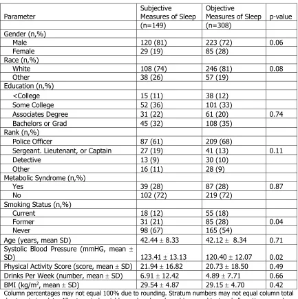
The objective measures of sleep were missing 149 observations due to
missing data. Table 4.2 compares the differences in the general characteristics
between those with and without objective measures of sleep. Small statistically
significant differences were observed for smoking status where without objective
objective meaures (67% vs. 54%, respectively, p-value=0.04) and systolic blood
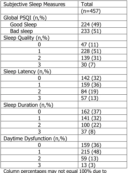
pressure (mean= 120.40 vs 123.41 mmHG, respectively, p-value=0.02). Table 4.3
shows that the distribution of observations between good and bad sleep were
almost identical with 233 individuals in the good sleep category and 224 in the bad
sleep. Sleep duration distribution shows that more officers were sleeping ³ 6 hours
on average. This table also shows that more than 50% of officers were have a
poor sleep quality. The population had an average 6.42 global PSQI score,
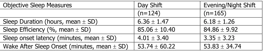
categorized as bad sleep. A descriptive analysis on the objective measures of sleep,
presented in Table 4.4, found the average sleep duration was 6.2 ± 1.4 hours with
average sleep efficiency being 84.8 ± 10.1%. Wake after sleep onset had an
average of 54.2 ± 46.6 minutes. The average minutes of sleep onset latency for
all observations in objective sleep measures was 3.6 ± 3.3. After looking at the
distribution of actigraph measures by their shift type there was no significant
difference between day and evening/night workers (Table 4.5).
The models that were used for the rest of the analysis are as follows. For
all inflammation markers and all PSQI components, models were adjusted for age,
systolic blood pressure, and total drinks per week.For all actigraphy metrics,
models were adjusted for metabolic syndrome and age. Additionally models with
actigraph sleep duration as the exposure were adjusted for systolic blood pressure,
total physical activity score, and rank. For models meauresing TNF-a, race and
gender were adjusted for as well. For models measuring CRP, additional
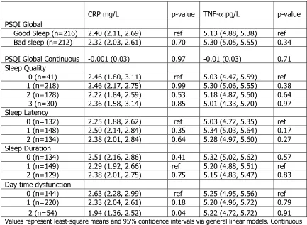
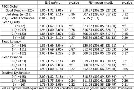
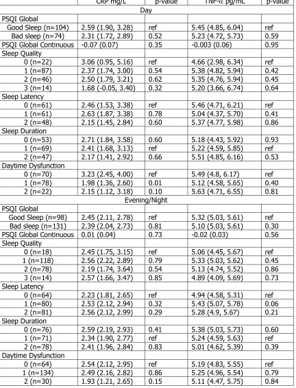
The results from the linear regression of the inflammation markers with
the subjective exposure are in Table 4.6. PSQI global, as a numeric exposure, was
significantly associated with fibrinogen (beta=-2.15, p-value=0.04). The mean of
CRP among those in the highest level of daytime dysfunction (worst category) was
significantly higher compared to the best level (mean=1.94 vs. 2.63 mg/L,
respectively, p-value=0.04). These were the only statistically significant
associations found between inflammation markers and subjective measures of
sleep.
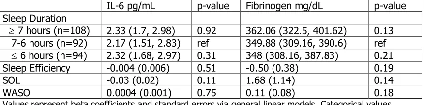
The linear regression with the objective measures of sleep as the exposure
are found in Table 4.7. Sleep efficiency was significantly associated with CRP
(beta=-0.03, p-value=0.02). Sleep onset latency (beta=0.07, p-value=0.05) and
wake after sleep onset (beta=0.01, p-value=<0.01) also were significantly
associated with CRP. No other associations were statistically significant.
The interactions between the stress and depression measures (i.e., PSS and
CESD) and sleep paramters were examined. It was found that the association
between CRP and sleep onset latency was modified by CESD (p-value=0.01). The
association between TNF-a and PSQI sleep duration also was modified by the PSS
(p-value=0.03). When IL-6 was the outcome, interactions were found between
the CESD and PSQI daytime dysfunction component (p-value=0.02). There was
an interaction for wake after sleep onset and CESD when modeling fibrinogen
(p-value=0.01). CESD and PSS were categorized with cut points (CESD³16, PSS³25)
CRP and sleep onset latency was statistically significant for people with a high
CESD score (beta=0.75, p-value=<0.01), but it was not among those with a low
CESD score (beta=0.07, p-value=0.06). The relationship between fibrinogen and
wake after sleep onset was statistically significant for people with a high CESD
score (beta=1.01, p-value=0.01) but it was not among those with a low CESD
score (beta=1.06, p-vlaue=0.44). TNF-a was statistical significantly associated
with the middle sleep duration category (5-6 hour) for people with a high PSS
score compared to the referent category, (³ 7 hours, means=5.95 vs. 4.71 pg/mL,
respectively, p-value=0.02), but not among people with a low PSS score
(means=5.01 vs. 5.43 pg/mL, respectively, p-vlaue=0.08). After categorization,
there was no statistically significant association found between IL-6 and PSQI
daytime dysfunction for CESD.
Among obese officers, the highest level of sleep latency (worst category)
was found to be significantly associated with fibrinogen (mean=306.49 vs. 336.26
mg/dL, respectively, p-value=0.03) compared to the best category of sleep
latency. Again, in those who are obese, the highest level of daytime dysfunction
(worst category) was significantly associated with fibrinogen (means=339.11 vs.
305.23 mg/dL, respectively, p-value=0.05) compared to the best category of
daytime dysfunction. There were numerous significant interactions which can be
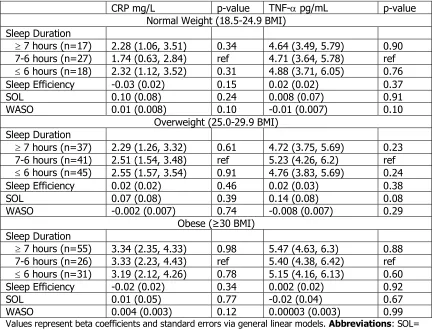
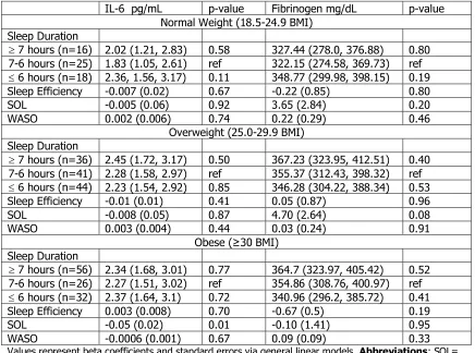
found in Table 4.8. Table 4.9 presents results for stratification by BMI for objective
measures of sleep. It was found that sleep onset latency was statistically
p-value=0.01). These relationships were not found for normal weight and
overweight. No other statistically significant associations for the interaction with
BMI for all other outcomes were found.
Lastly, analyses were stratified by shiftwork, with results present in Table
4.10. Daytime dysfunction for the middle category (level 2) was significantly
associated with CRP among people working dayshifts (mean=1.98 vs. 3.23 mg/L,
respectively, p-value=0.03) compared to the best category of daytime dysfunction.
Sleep duration also was found to be significantly associated with IL-6 for day shift
workers in the ³ 7 hours category (means=2.43 vs. 1.81 pg/mL, respectively,
p-value=0.03) compared to the middle sleep duration level (6-7 hours). For the worst
sleep duration category (£ 5), there also was a significant association with IL-6 for
day shift workers (means= 2.37 vs. 1.81 pg/mL, respectively, p-value=0.04)
compared to the middle sleep duration level (6-7 hours).
The stratification by shiftwork for the objective measures showed
statistically significant associations for CRP and fibrinogen, presented in Table
4.11. Sleep duration was found to be significantly associated with CRP for day shift
workers in the ³ 7 hours category (means=3.19 vs. 2.34 mg/L, respectively,
p-value=0.02) compared to the middle sleep duration level (6-7 hours). Sleep onset
latency (beta=0.20, p-value=0.04) was significantly associated with CRP for day
shift workers while the evening/night shift showed no statically significant
associations for CRp. Wake after sleep onset (beta=0.006, p-value=0.01) was
was not observed among day shift workers. Sleep duration was found to be
significantly associated with fibrinogen for eveing/night shift workers in the ³ 7
hours category (means=377.89 vs. 351.27 pg/mL, respectively, p-value=0.04)
compared to the middle sleep duration level (6-7 hours).
CRP has a recognized cut point of ³ 3.0mg/L (101). The results of logistic
regression with CRP as a categorical outcome with all of the exposures can be
found in Table 4.12. It was found that the odds ratio for a one-unit increase in
sleep efficiency was 0.97 (0.95, 0.99) for a CRP ³ 3.0. Logistic regression was
repeated for a 5 and a 10 unit increase for the continuous actigraph measures.
The 5 unit increases results showed the odds for sleep efficiency was 0.86 (0.76,
0.97), sleep onset latency was 1.47 (1.00, 2.17), and wake after sleep onset was
1.06 (1.02, 1.10) for high-risk CRP. The 10 unit increased the odds for sleep
efficiency was 0.74 (0.58, 0.95), sleep onset latency was 2.17 (1.00, 4.71), and
Table 4.1 Characteristics of BCOPS Population by PSQI Global
Parameter Total Good Sleep Bad Sleep p-value (n=457) (n=233) (n=224)
Gender (n,%)
Male 343 (75) 180 (39) 163 (36) 0.27 Female 114 (25) 53 (12) 61 (13)
Race (n,%)
White 354 (79) 178 (40) 176 (39) 0.56 Other 95 (21) 51 (11) 44 (10)
Education (n,%)
<College 60 (13) 36 (8) 24 (5)
Some College 154 (33) 81 (17) 73 (16) 0.28 Associates Degree 95 (20) 42 (9) 53 (11)
Bachelors or Grad 155 (33) 81 (17) 74 (16) Rank (n,%)
Police Officer 302 (65) 155 (33) 147 (32)
Sergeant. Lieutenant, or Captain 75 (16) 37 (8) 38 (8) 0.87 Detective 43 (9) 23 (5) 20 (4)
Other 44 (9) 25 (5.39) 19 (4.09) Metabolic Syndrome (n,%)
Yes 126 (28) 66 (15) 60 (13) 0.55 No 321 (72) 158 (35) 163 (36)
Smoking Status (n, %)
Current 73 (16) 35 (8) 38 (8)
Former 116 (26) 58 (13) 58 (13) 0.87 Never 263 (58) 135 (30) 128 (28)
Age (years, mean ± SD) 42.23 8.60 ± 42.32 ± 8.99 42.13 ± 8.18 0.81 Systolic Blood Pressure (mmHG, mean
± SD) 121.35 12.48 ± 120.13 12.13 ± 122.61 12.74 ± 0.03 Physical Activity Score (score, mean ±
SD) 21.11 17.97 ± 19.62 16.23 ± 22.65 19.53 ± 0.08 Drinks Per Week (number, mean ±
SD) 5.55 ± 9.53 4.68 ± 8.70 6.45 ± 10.25 <0.01 BMI (kg/m2, mean ± SD) 29.28 ±
4.75 29.24 ± 4.39 29.32 ± 5.11 0.86
Column percentages may not equal 100% due to rounding. Stratum numbers may not equal column total due to missing data. All categorical variable p-values based on chi-squared test and all continues p-values are based on t-tests or Wilcoxon rank sums test. Abbreviations: BCOPS=The Buffalo Cardio-Metabolic Occupational Police Stress, PSQI=Pittsburgh Sleep Quality Index, BMI=Body Mass Index. Cut points:
Table 4.2 Comparison of BCOPS Population Characteristics by Subjective and Objective Sleep
Parameter
Subjective
Measures of Sleep
Objective
Measures of Sleep p-value (n=149) (n=308)
Gender (n,%)
Male 120 (81) 223 (72) 0.06 Female 29 (19) 85 (28)
Race (n,%)
White 108 (74) 246 (81) 0.08 Other 38 (26) 57 (19)
Education (n,%)
<College 15 (11) 38 (12) Some College 52 (36) 101 (33)
Associates Degree 31 (22) 61 (20) 0.74 Bachelors or Grad 45 (32) 108 (35)
Rank (n,%)
Police Officer 87 (61) 209 (68)
Sergeant. Lieutenant, or Captain 27 (19) 41 (13) 0.11 Detective 13 (9) 30 (10)
Other 16 (11) 28 (9) Metabolic Syndrome (n,%)
Yes 39 (28) 87 (28) 0.87 No 102 (72) 219 (72)
Smoking Status (n,%)
Current 18 (12) 55 (18)
Former 31 (21) 85 (28) 0.04 Never 98 (67) 165 (54)
Age (years, mean SD) 42.44 ± 8.33 42.12 ± 8.34 0.71 Systolic Blood Pressure (mmHG, mean ±
SD) 123.41 ± 13.13 120.40 ± 12.07 0.02 Physical Activity Score (score, mean ± SD) 21.94 ± 16.82 20.73 ± 18.50 0.49 Drinks Per Week (number, mean ± SD) 6.91 ± 12.42 4.89 ± 7.71 0.66 BMI (kg/m2, mean ± SD) 29.54 ± 4.87 29.15 ± 4.70 0.42
Table 4.3 Distribution of PSQI Components
Subjective Sleep Measures Total (n=457) Global PSQI (n,%)
Good Sleep 224 (49) Bad sleep 233 (51) Sleep Quality (n,%)
0 47 (11) 1 228 (51) 2 139 (31) 3 30 (7) Sleep Latency (n,%)
0 142 (32) 1 159 (36) 2 84 (19) 3 57 (13) Sleep Duration (n,%)
0 162 (37) 1 141 (32) 2 100 (22) 3 37 (8) Daytime Dysfunction (n,%)
0 159 (36) 1 215 (48) 2 59 (13) 3 13 (3)
Table 4.4 Distribution of Actigraph Measures of Sleep
Objective Sleep Measures Total (n=308) Sleep Duration (hours, mean ± SD) 6.25 ± 1.38 Sleep Efficiency (%, mean ± SD) 84.85 ± 10.06 Sleep onset latency (minutes, mean ± SD) 3.57 ± 3.25 Wake After Sleep Onset (minutes, mean ± SD) 54.19 ± 46.65
Table 4.5 Distribution of Actigraph Measures of Sleep by Shiftwork
Objective Sleep Measures Day Shift Evening/Night Shift (n=124) (n=165)
Table 4.6. Adjusted Mean Inflammation Markers by PSQI Components
CRP mg/L p-value TNF-a pg/L p-value PSQI Global
Good Sleep (n=216) 2.40 (2.11, 2.69) ref 5.13 (4.88, 5.38) ref Bad sleep (n=212) 2.32 (2.03, 2.61) 0.70 5.30 (5.05, 5.55) 0.34
PSQI Global Continuous -0.001 (0.03) 0.97 -0.01 (0.03) 0.71 Sleep Quality
0 (n=41) 2.46 (1.80, 3.11) ref 5.03 (4.47, 5.59) ref 1 (n=218) 2.46 (2.17, 2.75) 0.99 5.30 (5.06, 5.55) 0.38 2 (n=128) 2.22 (1.84, 2.59) 0.53 5.18 (4.87, 5.50) 0.64 3 (n=30) 2.36 (1.58, 3.14) 0.85 5.01 (4.33, 5.70) 0.97 Sleep Latency
0 (n=132) 2.25 (1.88, 2.62) ref 5.03 (4.72, 5.35) ref 1 (n=148) 2.50 (2.14, 2.84) 0.35 5.34 (5.03, 5.64) 0.17 2 (n=134) 2.38 (2.01, 2.84) 0.64 5.28 (4.97, 5.60) 0.27 Sleep Duration
0 (n=134) 2.51 (2.16, 2.86) 0.41 5.32 (5.02, 5.62) 0.57 1 (n=149) 2.29 (1.92, 2.66) ref 5.20 (4.88, 5.51) ref 2 (n=129) 2.38 (2.01, 2.75) 0.75 5.15 (4.83, 5.47) 0.83 Day time dysfunction
0 (n=144) 2.63 (2.28, 2.99) ref 5.25 (4.95, 5.56) ref 1 (n=220) 2.33 (2.04, 2.61) 0.18 5.20 (4.96, 5.72) 0.79
2 (n=54) 1.94 (1.36, 2.52) 0.04 5.22 (4.72, 5.72) 0.91
Table 4.6. (Continued) Adjusted Mean Inflammation Markers by PSQI Components
IL-6 pg/mL p-value Fibrinogen mg/dL p-value PSQI Global
Good Sleep (n=220) 1.86 (1.72, 2.01) ref 318.37 (309.20, 327.53) ref Bad sleep (n=211) 1.96 (1.81, 2.11) 0.36 307.92 (298.63, 317.22) 0.12 PSQI Global Continuous 0.01 (0.02) 0.59 -2.15 (1.03) 0.04 Sleep Quality
0 (n=42) 2.00 (1.67, 2.33) ref 323.32 (302.85, 343.80) ref 1 (n=217) 1.94 (1.80, 2.09) 0.76 316.83 (307.65, 326.01) 0.57 2 (n=133) 1.88 (1.69, 2.07) 0.53 306.20 (294.59, 317.81) 0.15 3 (n=28) 1.76 (1.34, 2.17) 0.37 305.89 (280.67, 331.12) 0.29 Sleep Latency
0 (n=134) 1.85 (1.66, 2.04) ref 320.30 (308.68, 331.91) ref 1 (n=151) 1.87 (1.69, 2.05) 0.87 312.40 (301.17, 323.63) 0.34 2 (n=132) 2.03 (1.84, 2.22) 0.19 308.65 (297.05, 320.25) 0.17 Sleep Duration
0 (n=133) 1.93 (1.75, 2.11) 0.49 319.23 (308.03, 330.42) 0.21 1 (n=151) 1.84 (1.65, 2.03) ref 308.80 (297.17, 320.44) ref 2 (n=131) 1.99 (1.80, 2.18) 0.27 312.62 (300.66, 324.58) 0.65 Daytime Dysfunction
0 (n=145) 2.00 (1.82, 2.18) ref 318.32 (307.09, 329.54) ref 1 (n=220) 1.89 (1.75, 2.04) 0.34 311.52 (302.41, 320.64) 0.36
2 (n=56) 1.79 (1.50, 2.08) 0.22 311.08 (292.58, 329.49) 0.51
Table 4.7. Adjusted Inflammation Markers by Actigraph Measures of Sleep
CRP mg/L p-value TNF-a pg/mL p-value Sleep Duration
³ 7 hours (n=109) 2.71 (1.8, 3.63) 0.98 5.16 (4.37, 5.94) 0.58 7-6 hours (n=94) 2.54 (1.64, 3.43) ref 5.25 (4.40, 6.09) ref
£ 6 hours (n=94) 2.72 (1.82, 3.62) 0.53 5.01 (4.19, 5.83) 0.73 Sleep Efficiency -0.03 (0.01) 0.02 0.002 (0.01) 0.87 SOL 0.07 (0.03) 0.05 0.03 (0.03) 0.37 WASO 0.006 (0.002) <0.01 -0.0004 (0.002) 0.87
Values represent beta coefficients and standard errors via general linear models. Categorical values represent least-square means and 95% confidence intervals via general linear models. Abbreviations: SOL= Sleep onset latency, WASO=Wake after sleep onset. Adjustments: All models were adjusted for metabolic syndrome and age. Sleep Duration models additional adjusted for systolic blood pressure, total physical activity score, and rank. Models with TNF-a additional adjusted for race and gender. Models with CRP additional adjusted for rank.
Table 4.7. (Continued) Adjusted Inflammation Markers by Actigraph Measures of Sleep
IL-6 pg/mL p-value Fibrinogen mg/dL p-value Sleep Duration
³ 7 hours (n=108) 2.33 (1.7, 2.98) 0.92 362.06 (322.5, 401.62) 0.13 7-6 hours (n=92) 2.17 (1.51, 2.83) ref 349.88 (309.16, 390.6) ref
£ 6 hours (n=94) 2.32 (1.68, 2.97) 0.31 348 (308.16, 387.83) 0.21 Sleep Efficiency -0.004 (0.006) 0.51 -0.50 (0.38) 0.19 SOL -0.03 (0.02) 0.11 1.68 (1.14) 0.14 WASO 0.0004 (0.001) 0.75 0.11 (0.08) 0.18
Table 4.8. Adjusted Mean Inflammation Markers by PSQI Components Stratified by BMI Status
CRP mg/L p-value TNF-a pg/mL p-value Normal Weight (18.5-24.9 BMI)
PSQI Global
Good Sleep (n=35) 1.86 (1.15, 2.57) ref 4.53 (3.92, 5.15) ref Bad sleep (n=48) 1.71 (1.11, 2.31) 0.75 4.54 (4.00, 5.07) 0.99 PSQI Global Continuous -0.07 (0.06) 0.25 -0.0003 (0.06) 0.99 Sleep Quality
0 (n=13) 2.61 (1.46, 3.75) ref 5.89 (4.90, 6.87) ref 1 (n=28) 1.77 (0.97, 2.56) 0.24 4.02 (3.34, 4.70) <0.01 2 (n=32) 1.52 (0.77, 2.26) 0.12 4.57 (3.93, 5.21) 0.03
3 (n=8) 1.75 (0.29, 3.20) 0.36 4.74 (3.48, 5.99) 0.16 Sleep Latency
0 (n=21) 1.73 (0.83, 2.64) ref 4.58 (3.80, 5.36) ref 1 (n=34) 1.77 (1.04, 2.50) 0.96 4.73 (4.08, 5.38) 0.77 2 (n=26) 1.91 (1.10, 2.72) 0.77 4.50 (3.79, 5.20) 0.88 Sleep Duration
0 (n=23) 2.34 (1.56, 3.12) 0.07 4.76 (4.07, 5.45) 0.55 1 (n=29) 1.25 (0.37, 2.13) ref 4.45 (3.68, 5.21) ref 2 (n=28) 1.68 (0.91, 2.46) 0.46 4.58 (3.88, 5.28) 0.80 Daytime Dysfunction
0 (n=24) 2.41 (1.54, 3.28) ref 4.78 (4.01, 5.55) ref 1 (n=42) 1.64 (1.01, 2.28) 0.16 4.26 (3.70, 4.82) 0.28 2 (n=15) 1.32 (0.27, 2.38) 0.12 5.29 (4.35, 6.22) 0.41
Overweight (25.0-29.9 BMI) PSQI Global
Good Sleep (n=98) 2.10 (1.68, 2.52) ref 4.98 (4.62, 5.34) Bad sleep (n=80) 2.05 (1.58, 2.51) 0.86 5.16 (4.77, 5.56) 0.5 PSQI Global Continuous 0.02 (0.05) 0.64 0.05 (0.04) 0.25 Sleep Quality
0 (n=17) 1.66 (0.66, 2.66) ref 3.64 (2.82, 4.45) ref 1 (n=96) 2.14 (1.71, 2.56) 0.39 5.29 (4.92, 5.65) <0.01 2 (n=49) 1.98 (1.39, 2.57) 0.59 5.23 (4.74, 5.72) <0.01 3 (n=12) 2.42 (1.17, 3.66) 0.35 4.90 (3.84, 5.97) 0.06 Sleep Latency
0 (n=65) 2.13 (1.62, 2.65) ref 4.82 (4.38, 5.27) ref 1 (n=55) 2.02 (1.46, 2.58) 0.77 4.95 (4.47, 5.44) 0.7 2 (n=52) 2.01 (1.43, 2.58) 0.74 5.50 (5.01, 5.99) 0.05 Sleep Duration
0 (n=54) 2.13 (1.63, 2.64) 0.34 5.10 (4.67, 5.54) 0.63 1 (n=68) 1.77 (1.21, 2.33) ref 4.94 (4.45, 5.43) ref 2 (n=50) 2.34 (1.76, 2.93) 0.16 5.14 (4.63, 5.65) 0.58 Daytime Dysfunction
0 (n=65) 2.34 (1.84, 2.85) ref 5.03 (4.60, 5.46) ref 1 (n=91) 1.93 (1.50, 2.36) 0.22 5.06 (4.68, 5.43) 0.94 2 (n=18) 1.64 (0.67, 2.61) 0.21 5.23 (4.37, 6.09) 0.69
PSQI Global
Good Sleep (n=83) 2.95 (2.50, 3.41) ref 5.96 (5.57, 6.35) ref Bad sleep (n=84) 2.97 (2.51, 3.43) 0.96 5.44 (5.04, 5.83) 0.06 PSQI Global Continuous 0.02 (0.06) 0.74 -0.07 (0.05) 0.13 Sleep Quality
0 (n=11) 3.43 (2.19, 4.67) ref 6.23 (5.20, 7.25) ref 1 (n=94) 3.01 (2.57, 3.44) 0.53 5.69 (5.32, 6.06) 0.33 2 (n=47) 2.92 (2.32, 3.52) 0.47 5.50 (5.01, 5.99) 0.21 3 (n=10) 2.92 (1.60, 4.23) 0.58 5.42 (4.28, 6.55) 0.29 Sleep Latency
0 (n=46) 2.61 (1.99, 3.22) ref 5.48 (4.97, 5.99) ref 1 (n=59) 3.34 (2.80, 3.89) 0.08 6.00 (5.53, 6.46) 0.14 2 (n=56) 2.98 (2.42, 3.53) 0.38 5.46 (4.98, 5.95) 0.96 Sleep Duration
0 (n=57) 3.04 (2.46, 3.61) 0.64 5.88 (5.38, 6.38) 0.66 1 (n=52) 3.22 (2.67, 3.78) ref 5.72 (5.25, 6.20) ref 2 (n=51) 2.84 (2.26, 3.42) 0.35 5.47 (4.97, 5.97) 0.46 Daytime Dysfunction
0 (n=55) 3.11 (2.55, 3.67) ref 5.72 (5.24, 6.20) ref 1 (n=87) 3.08 (2.63, 3.52) 0.93 5.80 (5.41, 6.18) 0.81 2 (n=21) 2.56 (1.65, 3.47) 0.32 5.08 (4.31, 5.84) 0.16
Table 4.8. (Continued) Adjusted Mean Inflammation Markers by PSQI Components Stratified by BMI Status
IL-6 pg/mL p-value Fibrinogen mg/dL p-value Normal Weight (18.5-24.9 BMI)
PSQI Global
Good Sleep (n=36) 1.49 (1.13, 1.86) ref 303.12 (279.90, 326.35) ref Bad sleep (n=45) 1.84 (1.52, 2.16) 0.16 290.98 (270.87, 311.09) 0.43
PSQI Global
Continuous 0.01 (0.04) 0.71 -2.10 (2.08) 0.32 Sleep Quality
0 (n=13) 1.83 (1.23, 2.42) ref 316.90 (279.18, 1354.62) ref 1 (n=27) 1.71 (1.28, 2.13) 0.74 285.63 (259.55, 311.71) 0.18 2 (n=31) 1.80 (1.41, 2.20) 0.94 300.23 (275.74, 324.73) 0.46 3 (n=8) 1.22 (0.46, 1.98) 0.22 291.90 (244.08, 339.71) 0.42 Sleep Latency
0 (n=21) 1.49 (1.02, 1.96) ref 312.96 (283.78, 342.14) ref 1 (n=33) 1.41 (1.02, 1.79) 0.79 279.72 (255.34, 304.10) 0.08 2 (n=25) 2.27 (1.84, 2.69) 0.02 303.15 (276.64, 329.65) 0.62 Sleep Duration
0 (n=24) 1.64 (1.22, 2.05) 0.74 308.37 (282.41, 334.33) 0.18 1 (n=28) 1.74 (1.29, 2.19) ref 282.34 (253.64, 311.05) ref 2 (n=26) 1.77 (1.35, 2.19) 0.92 295.25 (268.87, 321.63) 0.51 Daytime Dysfunction
0 (n=23) 2.04 (1.58, 2.51) ref 327.49 (298.93, 356.05) ref 1 (n=41) 1.66 (1.33, 2.00) 0.19 284.78 (263.90, 305.65) 0.02 2 (n=15) 1.37 (0.82, 1.92) 0.07 284.36 (249.68, 319.04) 0.06
Overweight (25.0-29.9 BMI) PSQI Global
Good Sleep (n=98) 1.80 (1.59, 2.02) ref 318.83 (305.7, 332.49) ref Bad sleep (n=81) 1.97 (1.73, 2.21) 0.31 307.07 (292.02, 322.12) 0.26
PSQI Global
Continuous 0.01 (0.03) 0.71 -3.21 (1.64) 0.05 Sleep Quality
0 (n=17) 2.11 (1.59, 2.63) ref 318.85 (287.75, 349.95) ref 1 (n=95) 1.86 (1.63, 2.08) 0.38 320.81 (306.91, 334.71) 0.91 2 (n=52) 1.88 (1.59, 2.18) 0.45 303.04 (284.29, 321.82) 0.39 3 (n=12) 1.75 (1.10, 2.40) 0.39 292.18 (251.36, 332.99) 0.13 Sleep Latency
0 (n=65) 1.64 (1.22, 2.05) ref 310.02 (293.34, 326.69) ref 1 (n=56) 1.74 (1.29, 2.19) 0.44 314.26 (296.07, 332.45) 0.74 2 (n=53) 1.77 (1.35, 2.19) 0.40 314.45 (296.19, 332.71) 0.73 Sleep Duration
0 (n=52) 1.92 (1.66, 2.18) 0.57 322.81 (306.38, 339.24) 0.13 1 (n=71) 1.80 (1.50, 2.10) ref 303.99 (285.67, 322.30) ref 2 (n=51) 1.94 (1.64, 2.25) 0.52 310.28 (290.97, 329.60) 0.64 Daytime Dysfunction