Can Fetuin-A Level, CRP, and WBC be a Predictive
1
Value in the Diagnosis of Acute Appendicitis in
2
Children with Abdominal Pain?
3
Author Names:, 1*Abuzer Coskun, MD, 2Cengiz Güney, Assoc. Prof.
4
Author Affiliations: 1Sivas Numune Hospital, Department of Emergency, Sivas, Turkey
5
2Cumhuriyet University Medical Faculty, Department of Pediatric Surgery, Sivas, Turkey
6
Corrsponding: *Abuzer Coskun, MD
7
Sivas Numune Hospital, Department of Emergency
8
Yesilyurt Mah. Sifa Street Sivas, Turkey
9
Telephone: + 90 444 44 58 Fax: + 90 346 223 95 30 Mobile: +90 532 157 79 12
10
E-mail: [email protected] or [email protected]
11
Abstract
: Background: Acute appendicitis (AA) is the most common cause of emergency surgery.12
Perforation is more common than adults. Early diagnosis and new markers are needed. The aim of
13
this study was to investigate the effects of plasma Fetuin-A (FA) levels in patients with the acute
14
abdomen (AB). Material and Method: This prospective study included 107 patients younger than
15
16 years of age who were admitted to the emergency department for abdominal pain between
16
January 2018 and December 2018. The patients who presented to abdominal pain were divided into
17
two groups as AA and other causes (OC) of AB. T Patients with acute appendicitis; intraperitoneal,
18
retrocolic / retrocecal and appendicitis were divided into three groups. Also, the AA group was
19
divided into two groups as perforated appendicitis and non-perforated appendicitis. Serum FA
20
levels of the patients were evaluated in the emergency department. Results: In the AA group,
C-21
reactive protein (CRP) and white blood cell (WBC) levels were higher, and FA levels were
22
significantly lower than in the AB group. Intraperitoneal localization was 95.2% and perforation
23
was frequent. When significant values in the univariate regression analysis for acute abdomen and
24
perforation were compared in the multivariate regression analysis, CRP, WBC, and FA levels were
25
found to be prognostic. Also, decreased FA levels were associated with AA while too much
26
decreased FA levels were associated with the risk of perforation. Conclusion: While trying to
27
diagnose AA in children, the FA level, CRP and WBC may be predictive values to identify risk
28
factors.
29
Keywords: emergency department; pediatric acute appendicitis; perforatio; fetuin-A level
30
31
1. Introduction
32
Appendicitis, the most common cause of surgical abdominal pain in children, is inflammation
33
of appendix vermiformis1,2. In 95% of the cases, the appendicitis was intraperitoneal (in 65%, dorsal
34
to the cecum; in 30%, pelvis), while in 5%, it was retrocolic and retrocecal3.
35
The most significant cause of appendicitis in children is lumen obstruction due to an increase in
36
lymphoid tissue. The second most common decade is in the 10-12 age range. Appendicitis is more
37
common in boys. The possibility of perforated appendicitis in children is higher than in adults. It is
38
thought that this is due to the lack of clear physical examination findings in young children and the
39
lack of their communication skills4.
40
A cheap, reliable, easy and rapidly available biochemical marker with high specificity and
41
sensitivity is not yet available in the diagnosis of acute appendicitis. Fetuin-A (FA) is one of the
42
molecules investigated in various fields. Fetuin-A is an insulin-dependent endogenous tyrosine
43
kinase receptor inhibitor, which is mainly synthesized in the liver5. Furthermore, it has been found
44
that it directly affects the cells of the animal and human adipose tissue, causing subclinical
45
inflammation and cytokine release6. It may be extrahepatically synthesized in the kidneys, choroid
46
plexus and all vital organs during fetal development. Fetuin-A is seen in the α2 band of serum
47
electrophoresis. The serum concentration is in the range of 140-297mg/L7. Fetuin-A was first noted as
48
a negative acute phase reactant similar to albumin in cases of acute inflammation. Factors affecting
49
FA secretion in humans have been reported such as severe liver damage, cirrhosis, acute viral
50
hepatitis and cancer8.
51
Many studies have examined serum FA levels. However, to the best of our knowledge, no study
52
has been conducted to investigate pediatric acute appendicitis. In the present study, we planned to
53
demonstrate the contribution of serum FA levels to the diagnosis of AA.
54
2. Materials and methods
55
Study design and population
56
This prospective cross-sectional study included 107 patients younger than 16 years of age who
57
were admitted to our hospital due to abdominal pain between January 2018 and December 2018. The
58
exclusion criteria were as follows: having known heart and heart valve diseases, drug use due to
59
cardiac causes, having metabolic diseases, chronic liver diseases, chronic renal failure, receiving
60
dialysis treatment, having known inflammatory bowel diseases, malignancies, having known
61
hematological diseases, and receiving erythrocyte suspension over the past six months.
62
The patients who were admitted to the emergency department with abdominal pain were
63
divided into two groups according to clinical and physical examination and laboratory and imaging
64
results: acute abdomen (AB) and other causes (OC) (urinary tract infections, acute gastroenteritis,
65
renal colic, constipation, etc.). The diagnosis of the acute abdomen was made jointly by emergency
66
medicine specialist, pediatric surgeon and radiologist according to the results of the clinical
67
examination, physical examination findings, laboratory results, and radiological imaging results. The
68
patients diagnosed with acute abdomen were divided into three groups: those without appendicitis,
69
intraperitoneal appendicitis, and retrocolic/retrocecal appendicitis. Acute appendicitis was divided
70
into two groups as perforated and not perforated.
71
To determine serum Fetuin-A levels, 5ml of venous blood was collected from the patients
72
presenting with abdominal pain. The blood was centrifuged at 4000 rpm for 5 minutes. Serums were
73
kept in Eppendorf tubes and kept at -80 o C. Fetuin-A levels were analyzed by Human FETUA
74
(Fetuin-A) Sandwich Enzyme-Linked Immunosorbent Assay (ELISA) kit (96-Fine Test, EH0218,
75
Wuhan Fine Biotechnology, China). Range is 140-297mg/L and sensitivity is <0,469ng/ml.
76
Hemogram was measured using a Beckman Coulter Automated CBC Analyzer (Beckman
77
Coulter, Inc., Fullerton, CA, USA).
78
Biochemistry blood was analyzed with the Cobas 6000 (C6000-Core, Cobas c-501 series, Hitachi,
79
Roche, USA).
80
3. Statistical Analysis
81
The data obtained from this study were analyzed by SPSS 15.0 (SPSS Inc., Chicago, IL, USA)
82
software package. While determining the normality of the variables, Shapiro Wilk's was used because
83
of the number of units. When analyzing the differences between the groups, Mann Whitney U Tests
84
were used because the variables did not show normal distribution. The chi-square analysis was
85
performed to examine the relationships between the groups of nominal variables. In the cases where
86
the expected values in the 2x2 tables did not have sufficient volume, Fisher tabs Exact Test was used,
87
and in the RxC tables Spearman Correlation analysis was performed with the help of Monte Carlo
88
Simulation. Besides, linear regression analysis was used for univariate and multivariate analysis of
89
variables. We used univariate analysis to measure the relationship of variables in patient and control
90
groups. The variables that were found to be statistically significant in univariate analysis were used
91
independent prognostic factor. When interpreting the results, the significance level was set at 0.05
93
and P values less than 0.05 were considered as statistically significant.
94
4. Results
95
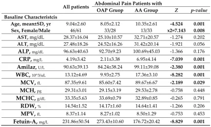
The clinical and demographic characteristics of the patients are listed in Table 1. According to
96
the Mann-Witney U test, significant differences were found between the groups in terms of age, sex,
97
C-reactive protein (CRP), White Blood Cell (WBC), and Fetuin-A (FA) level. CRP and WBC were
98
significantly higher while the FA level was considerably lower in the AA group than in the OC group.
99
Chi-square analysis of AA and OC groups revealed a significant difference between the groups
100
in terms of sex, location of AA, radiological image and perforation (Table 2).
101
Chi-square analysis of the location of AA revealed a significant difference between the groups
102
in terms of sex, presence of AA, radiological image and perforation (Table 3).
103
Chi-square analysis of perforated appendicitis revealed a significant difference between the
104
groups in terms of sex, the location of AA, radiological image and other causes of abdominal pain
105
(Table 4).
106
The univariate regression analysis of groups of abdominal pain revealed no significant
107
differences in terms of alanine aminotransferase (ALT), alkaline phosphatase (ALP) and amylase
108
while significant differences in terms of CRP, WBC, FA, AA, perforation, age, sex, aspartate
109
aminotransferase (AST) and radiological imaging. However, after the adjustment of the variables that
110
are statistically significant in univariate analysis in the multivariate linear regression analysis with
111
advanced stage method, FA, CRP and WBC increased and remained associated with the risk of acute
112
abdomen (Table 5).
113
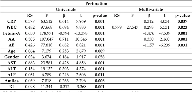
Univariate regression analysis with perforated appendicitis revealed no significant difference in
114
terms of sex while significant differences in terms of CRP, WBC, FA, age, AST, ALT, ALP, amylase,
115
and radiological imaging. However, after the adjustment of the variables that are statistically
116
significant in univariate analysis in the multivariate linear regression analysis with advanced stage
117
method, FA, CRP and WBC increased and remained associated with the risk of perforation (Table 6).
118
There was a statistically significant difference between the acute abdomen and perforated
119
appendicitis in terms of age, FA, CRP, WBC, and sex. Fetuin-A had a strong negative correlation
120
while the other variables strong positive correlation.
121
5. Discussion
122
The key to successful treatment in acute appendicitis is early and accurate diagnosis9. However,
123
the correct diagnosis rate is 72-94%. This rate indicates that the diagnosis is still complicated10,11. We
124
aimed to determine the relationship between serum FA levels and disease activity and inflammatory
125
parameters for early diagnosis and prognosis in pediatric acute appendicitis patients. Our study is
126
the first to report that decreased FA levels are independent predictors of disease activity in patients
127
with AA and that serum FA levels are negatively correlated with CRP concentrations and WBC count.
128
This suggests that Fetuin-A may have a possible inflammatory function in AA and can potentially be
129
used as a biological marker.
130
The most common cause of obstruction in the child appendix lumen is lymphoid tissue
131
hyperplasia. There is a positive correlation between the severity of the inflammatory event in the
132
appendix and the likelihood that the lumen will become obstructed. Due to the secretion of the
133
appendix mucosa, fluid accumulation and distension develop rapidly in the cavity. The appendix
134
mucosa may continue secretion even when the pressure in the lumen is high. Due to these reasons,
135
appendix first gets gangrene, and then it gets perforated. Besides, the proliferation of bacteria living
136
in the appendix due to closed space contributes to this process12-14.
137
In our study, we detected appendicitis in 46 (42.9%) patients. Of these patients, 39 (36.4%) had
138
intraperitoneal, and 7 (6.5%) had retrocecal and retrocolic appendicitis. These patients underwent a
139
standard appendectomy. Appendiceal perforation was observed in 26 (37.7%) patients. These
140
patients underwent standard appendectomy followed by drainage. Fecalith was detected in 11
141
Fetuin-A is a glycoprotein15-17, also known as 2-Heremans Schmid. Its molecular weight is about
143
60 kDa18. It consists of a long chain A and a short B chain connected by a short peptide. Before FA,
144
synthesized in a single chain, becomes mature with its two-chain form in circulation, it undergoes
145
posttranslational modification processes such as proteolysis, glycosylation, and phosphorylation. It
146
is high in serum (140-297mg/L)16-18. Fetuin-A is a negative acute phase reactant15-17. Fetuin-A levels
147
were found to be low in acute alcoholic hepatitis, acute drug-associated hepatitis, chronic
148
autoimmune hepatitis, fatty liver patients, alcoholic and primary cerebrospinal cirrhosis and
149
hepatocellular cancer patients19. Serum fetuin-A levels were found to be low in patients with
end-150
stage renal disease who commonly develop cardiovascular calcification20. Also, Fetuin-A has been
151
shown to be a predictor of mortality in long-term dialysis patients8.
152
Manolakis et al.21 demonstrated that the decrease in FA showed a close association with the acute
153
phase and that chronic inflammation in both Crohn's and ulcerative colitis might be a potential
154
diagnostic and perhaps predictive value molecule. In another study, it was reported that serum levels
155
decrease in response to infection and/or inflammation, play a role as an anti-inflammatory mediator
156
and have protective effects against lipopolysaccharide-associated shock22.
157
In our study, we found a significant decrease in serum FA levels in AA patients compared to the
158
OC group. The mean serum FA level was 273 mg/dl in the OC group and 176 mg/dl in the AA group.
159
Fetuin-A value was 223 mg/dl in the group without perforation and 161 mg/dl in the group with
160
perforation. These values were significant and predictive. This suggests that FA may also play a
161
role in the pathophysiology of AA as a negative inflammatory mediator. In our study, we found a
162
significant relationship between serum FA levels in the AA group and perforated appendicitis group.
163
This relationship was as meaningful as the WBC count and CRP. There was a strong negative
164
correlation between serum FA levels and CRP and WBC in the acute appendicitis group. FA was
165
found to be significant in both univariate and multivariate regression analysis of both AA and
166
perforation. In the perforated appendicitis group where inflammation was more frequent, serum FA
167
levels were found to be lower than the non-perforated AA group. These results indicate that serum
168
FA level can be used as an essential marker in the pathology of appendicitis. Therefore, its clinical
169
significance should be interpreted with caution.
170
Increased levels of CRP determine the presence and severity of inflammation. Wang et al.23
171
found an inverse relationship between CRP levels and Fetuin-A and reported that this inverse
172
relationship was present between FA and inflammation. Ketteler et al.24 reported that the low FA
173
level in patients with chronic renal failure who underwent stable hemodialysis was inversely related
174
to CRP, an indicator of inflammation.
175
The negative relationship between serum FA levels and CRP levels in our study was consistent
176
with the literature. Also, we demonstrated that serum FA levels in patients with AA correlate
177
negatively and strongly with CRP concentrations and WBC count. While all of these values (Table 6)
178
were significant in univariate regression analysis of perforated appendicitis group, only WBC, CRP
179
and FA were significant in multiple linear regression analysis. Therefore, acute phase reactants were
180
high in AA patients with high inflammation, while the negative phase reactant FA was low. Acute
181
phase reactant is a sensitive marker of CRP and tissue damage and systemic inflammation. The
182
degree of inflammation in the acute appendicitis was low with high WBC and CRP levels, and it
183
correlated with serum FA concentration. Serum FA levels were significantly lower in patients with
184
AA compared to perforated appendicitis. In pediatric abdominal pains, low serum FA levels, high
185
CRP and WBC, physical examination and radiological imaging increase the accuracy rate in the
186
diagnosis of acute appendicitis.
187
As a result, the decrease in serum FA levels was associated with the disease in AA and
188
perforation patients. Further research may examine the use of Fetuin-A concentration measurements
189
as markers of disease activity in OC patients.
190
6. Study limitations
191
The most significant limitation of the study was that it was single-centered and had a low
192
participate in the study by some patients, the cost of the FA kit, and the lack of adequate financial
194
support were other challenges of the study.
195
7. Conclusion
196
In acute appendicitis and perforated appendicitis groups, negative acute phase reactant was
197
found to have a significant relationship with serum FA level and other inflammatory parameters.
198
This finding is consistent with other studies that detected serum FA as a negative acute phase
199
reactant. Measurement of serum FA levels in pediatric patients with abdominal pain can be used as
200
a test for the diagnosis of appendicitis. Prospective randomized trials to be conducted with more
201
patient groups are needed on this issue.
202
References
203
1. Karabulut R, Sonmez K, Turkyilmaz Z, Demirogullari B, Ozen IO, Demirtola A, et al.
204
Negative appendectomy experience in children. Ir J Med Sci 2011;180:55-8.
205
2. Deng Y, Chang DC, Zhang Y, Webb J, Gabre-Kidan A, Abdullah F. Seasonal and day of the
206
week variations of perforated appendicitis in US children. Pediatr Surg Int 2010;26:691-6.
207
3. Addiss DG, Sheffer N, Fouler BS, Tauxe RV. The epidemiology of appendicitis and
208
appendectomy in the United States. Am J Epididemiol 1990;132:910-925.
209
4. Newman K, Ponsky T, Kittle K, Dyk L, Throop C, Gieseker K et al. Appendicitis 2000:
210
variability in practice, outcomes and resource utilization at thirty pediatric hospitals. J
211
Pediatr Surg 2003;38:372-379.
212
5. Weikert C, Stefan N, Schulze MB, et al. Plasma Fetuin-A Levels and the Risk of Myocardial
213
Infarction and Ischemic Stroke Circulation 2008; 118:2555-62.
214
6. Fisher E, Stefan N, Saar K, et al. Association of AHSG Gene Polymorphisms With Fetuin-A
215
Plasma Levels and Cardiovascular Diseases in the EPIC-Potsdam Study. Circ Cardiovasc
216
Genet. 2009; 2:607-13
217
7. Westenfeld R, Jahnen-Dechent W, Ketteler M. Vascular calcification and fetuin-A deficiency
218
in chronic kidney disease. Trends Cardiovasc Med 2007;17(4):124-8.
219
8. Stefan N, Hennige AM, Staiger H, Machann J, Schick F, Kröber SM, et al.
Alpha2-Heremans-220
Schmid glycoprotein/fetuin-A is associated with insulin resistance and fat accumulation in
221
the liver in humans. Diabetes Care 2006;29(4):853-7.
222
9. Andersen BR, Kallehave FL, Andersen HK. Antibiotics versus placebo for prevention of
223
postoperative infection after appendectomy. Cochrane Database Syst Rev 2003;CD001439.
224
10. Ates M, Coban S, Sevil S, Terzi A. The efficacy of laparoscopic surgery in patients with
225
peritonitis. Surg Laparosc Endosc Percutan Tech 2008;18:453-6.
226
11. Pearl RH, Hale DA, Molloy M, Schutt DC, Jaques DP. Pediatric appendectomy. J Pediatr Surg
227
1995;30:173-81.
228
12. Gwynn LK. The diagnosis of acute appendicitis: clinical assessment versus computed
229
tomography evaluation. J Emerg Med 2001;21:119-23.
230
13. Wangensteen OH, Buirge RE, Dennis C, Ritchie WP. Studies in the etiology of acute
231
appendicitis: The significance of the structure and function of the vermiform appendix in the
232
genesis of appendicitis. Ann Surg 1937:106:910-42.
233
14. Wangensteen OH, Dennis C. Experimental prof of the obstructive origin of the appendicitis
234
in man. Ann Surg 1939;110:629-47.
235
15. Marhaug G, Shah V, Shroff R, Varsani H, Wedderburn LR, Pilkington CA, Brogan PA.
Age-236
dependent inhibition of ectopic calcification: a possible role for fetuin-A and osteopontin in
237
patients with juvenile dermatomyositis with calcinosis. Rheumatology (Oxford).
238
2008;47(7):1031-7.
239
16. Ix JH, Chertow GM, Shlipak MG, Brandenburg VM, Ketteler M, Whooley MA. Association
240
of Fetuin-A with mitral annular calcification and aortic stenosis among persons with
241
coronary heart disease: Data From the Heart and Soul Study. Circulation.
2007;115(19):2533-242
17. Mori K, Emoto M, Araki T, Yokoyama H, Teramura M, Lee E et al. Association of serum
244
fetuin-A with carotid arterial stiffness. Clin Endocrinol (Oxf). 2007;66(2):246-50.
245
18. Bláha V, Mistrík E, Dusilová-Sulková S, Kalousová M, Andrýs C, Bláha M, Sobotka L.
246
Circulating fetuin-A predicts early mortality in chronic hemodialysis patients. Clin Biochem.
247
2009;42(10-11):996-1000.
248
19. Kalabay L, Gráf L, Vörös K, Jakab L, Benko Z, Telegdy L et al. Human serum fetuin
249
A/alpha2HS-glycoprotein level is associated with long-term survival in patients with
250
alcoholic liver cirrhosis, comparison with the Child-Pugh and MELD scores. BMC
251
Gastroenterol. 2007;7(1):15.
252
20. Kaden JJ, Reinöhl JO, Blesch B, Brueckmann M, Haghi D, Borggrefe M et al. Systemic and
253
local levels of fetuin-A in calcific aortic valve stenosis. Int J Mol Med. 2007;20(2):193-7.
254
21. Manolakis, Anastassios C et al. “α2-Heremans-Schmid Glycoprotein (Fetuin-A)
255
Downregulation and Its Utility in Inflammatory Bowel Disease.” World Journal of
256
Gastroenterology. 2017; 23(3): 437–446.
257
22. Karamessinis PM, Malamitsi-Puchner A, Boutsikou T, Makridakis M, Vougas K,
258
Fountoulakis M., et al. Marked defects in the expression and glycosylation of alpha2-HS
259
glycoprotein/fetuin-A in plasma from neonates with intrauterine growth restriction:
260
proteomics screening and potential clinical implications. Mol Cell Proteomics.
2008;7(3):591-261
9.
262
23. Wang AY, Woo J et al. Associations of serum Fetuin-A with malnutrition, inflammation,
263
atherosclerosis and valvular calcification syndrome and outcome in peritoneal dialysis
264
patients. Nephrol Dial Transplant. 2005;20(8):1676-85.
265
24. Ketteler M. Fetuin-A and extraosseous calcification in uremia. Curr Opin Nephrol Hypertens.
266
2005;14:337-42.
267
268
Table 1. Baseline characteristics of study patients.
269
All patients Abdominal Pain Patients with
OAP Gruop AA Group Z p-value
Basaline Characteristcis Age, mean±SD, yr
Sex, Female/Male 9.04±2.60 46/61 8.05±2.12 33/28 10.35±2.61 13/33 x2=7.143-4.524 0.001 0.008 AST, mg/dL 28.37±16.04 25.10±10.57 32.71±20.57 -1.274 0.202
ALT, mg/dL 27.48±18.26 24.52±16.26 31.42±20.14 -1.921 0.056
ALP,
mg/dL 96.63±40.63 92.70±9.23 100.69±45.03 -1.366 0.176CRP,
mg/L 4.19±3.42 2.11±3.38 6.95±4.14 -7.039 0.001Amilaz
, U/L 90.63±39.13 84.24±38.24 99.11±39.08 -2.380 0.001WBC,
10^3/uL 13.12±4.69 9.93±2.75 17.36±3.10 -8.282 0.001MCV,
fL 87.35±9.61 85.60±7.42 89.67±6.67 -2.189 0.029MCH,
pg 29.31±3.01 29.15±3.19 29.53±2.78 -0.758 0.448MCHC,
g/dL 33.35±5.63 33.69±0.79 32.89±0.85 -0.265 0.791RDW,
% 14.54±1.52 14.17±1.60 14.64±1.41 -1.266 0.206MPV,
fL 8.37±1.14 8.27±1.02 8.50±1.29 -0.753 0.453Fetuin-A,
mg/L 231.86±50.54 273.43±10.60 176.72±20.42 -8.829 0.001OAP: Other abdominal pain, AA: Acute appendicitis, AST:Asptartate aminotransferase; ALT:
270
Alanine aminotransferase;ALP: Alkaline phosphatase; CRP: C reactive protein, WBC: White blood
271
cell, MCV; Mean Corpuscular Volume; MCH: Mean Corpuscular Hemoglobine; MCHC:Mean
272
Corpuscular Hemoglobin Concentration; RDW: Red cell distrubition width; MPV:Mean Platelate
273
Volum; * P<0.05.
Table 2. Chi-Square test results relating to the difference between variables of abdominal pain.
275
Abdominal Pain OAP Group
n(%) AA Group n(%) X2 p-value Gender Female 33(30.8) 13(12.2) 7.143 0.008
Male 28(26.2) 33(30.8)
Radiological Imaging USG 24(22.4) 33(30.8) 11.056 0.001 Abdominal CT 37(34.6) 13(12.1)
Acute Abdomen No 61(57.0) 0(0) 107.00 0.001 Intraperitoneal 0(0) 39(36.4)
Retrocolic/retrocecal 0(0) 7(6.5)
Ferforation No 61(57.0) 20(13.1) 24.39 0.001 Yes 0(0) 26(37.7)
USG: Ultrasonography, CT: Computed Tomography *p<0.05.
276
Table 3. Chi-Square test results relating to the difference between variables of acute abdomen.
277
Abdominal Pain
No
n(%)
IP
n(%)
RC/Rc
n(%) X2 p-valueGender FemaleMale 33(30.8) 28(26.2) 31(29.0) 8(7.5) 5(4.8) 2(1.8) 13.421 0.008 Radiological
Imaging USG 24(22.4)
27(25.2) 6(5.6) 11.703 0.001
Abdominal CT 37(34.6) 12(11.2) 1(0.9)
Abdominal
Pain
OAP 61(57.0) 0(0) 0(0) 107.00 0.001 AA 0(0) 39(36.4) 7(5.6)
Ferforation No 61(57.0) 20(13.1) 0(0) 50.030 0.001 Yes 0(0) 19(17.8) 7(5.6)
IP:Intraperitoneal, RC/Rc: Retrocolic/Retrocecal *P<0.05.
278
Table 4. Chi-Square test results relating to the difference between variables of perforation.
279
Abdominal Pain OAP Group
n(%) AA Group n(%) X2 p-value Gender FemaleMale 39(36.5) 42(39.2) 19(17.8) 7(6.5) 3.618 0.070 Radiological Imaging USG 36(33.6) 21(19.6) 11.056 0.001
Abdominal CT 45(42.1) 5(4.7)
Acute Abdomen No 61(57.0) 0(0) 107.00 0.001 Intraperitoneal 0(0) 19(17.8)
Retrocolic/retrocecal 0(0) 7(6.5)
Abdominal Pain
OAP Yes 61(57.0) 20(18.7) 26(24.3) 0(0) 24.39 0.001Table 5. Univariate and multivariate linear regression analyses for predicting the development of
280
Acute Abdomen.
281
Acute Abdomen
Univariate Multivariate
CRP 0.297 44.316 0.545 6.657 0.001 0.428 3.741 0.041 WBC 0.619 170.816 0.787 13.07 0.001 0.952 153.921 0.542 4.814 0.013 Fetuin-A 0.906 1009.443 -0.952 -31.772 0.001 -0.848 -11.124 0.001
AA 0.854 615.920 0.924 24.818 0.001 0.265 3.972 0.004
Perforation 0.426 77.818 0.652 8.821 0.001 -0.253 -6.239 0.001 Age 0.194 25.203 0.440 5.020 0.001
Gender 0.067 7.511 0.258 2.741 0.007 AST 0.056 6.198 0.236 2.489 0.014 ALT 0.035 3.839 0.188 1.959 0.053 ALP 0.010 1.014 0.098 1.007 0.316 Amilaz 0.036 3.887 0.189 1.971 0.051 RI 0.103 12.009 -0.321 -3.478 0.001 RS; R Sequare, RI;radiological Imaging p<0.05.
282
Table 6. Univariate and multivariate linear regression analyses for predicting the development of
283
perforation.
284
Perforation
Univariate Multivariate
RS F β t p-value RS F β t p-value
CRP 0.377 63.512 0.614 7.969 0.001 0.312 4.034 0.037
WBC 0.482 97.668 0.694 9.883 0.001 0.779 27.547 0.298 5.531 0.023 Fetuin-A 0.630 178.971 -0.794 -13.378 0.001 -1.476 -7.539 0.001
AA 0.505 107.047 0.711 10.346 0.001 0.330 2.160 0.001
AB 0.426 77.818 0.652 8.821 0.001 -1.157 -6.239 0.031
Age 0.064 7.179 0.253 2.679 0.009 Gender 0.034 3.674 0.184 1.917 0.058
AST 0.883 23.581 0.428 4.856 0.001 ALT 0.154 19.132 0.393 4.374 0.001 ALP 0.061 6.789 0.246 2.606 0.011 Amilaz 0.069 7.818 0.263 2.796 0.006 RI 0.098 11.344 -0.312 -3.368 0.001
RS; R Sequare, RI;radiological Imaging, AB; acute abdomen p<0.05.
285
Ethical Statement: All subjects gave their informed consent for inclusion before they participated in
286
the study. The study was conducted in accordance with the Declaration of Helsinki, and the protocol
287
289
Author Contributions: Study conceptualization involved, C.G. and A.C; Methodology, A.C;
290
Validation, C.G. and A.C; Formal analysis, C.G; Investigation, All authors.; Resources, C.G, and A.C;
291
Data curation, C.G.;Writing—original draft preparation, C.G and A.C; Writing—review and editing,
292
All authors; Visualization, C.G and A.C; Supervision, C.G and A.C; Project administration, C.G and
293
B.K.; Funding acquisition, C.G.,
294
Funding: None declared.
295
Acknowledgments: None declared.