Identification of cis-elements regulating expression of Fgf10
during limb development
HIDEMI SASAKI
1, 2,
TAKASHI YAMAOKA
4, HIDEYO OHUCHI
1, AKIHIRO YASUE
3, TSUTOMU NOHNO
5,
HIROTAKA KAWANO
6, SHIGEAKI KATO
6, MITSUO ITAKURA
4, MASARU NAGAYAMA
2and SUMIHARE NOJI*
,11Department of Biological Science and Technology, Faculty of Engineering, The University of Tokushima, Japan, 2Department of Oral and Maxillofacial Surgery I, 3Department of Orthodontics, The University of Tokushima Graduate school of Dentistry, Japan, 4Division of Genetic Information, Institute for Genome Research, Tokushima University, Japan, 5Department of Molecular Biology, Kawasaki Medical
School, Matsushima, Kurashiki City, Japan and 6Institute for Molecular Cellular Biosciences, University of Tokyo, Japan
ABSTRACT Fibroblast growth factor 10 (FGF10) is known to be expressed in limb mesenchymal cells and to function as a mesenchymal signaling factor involved in epithelial-mesenchymal interactions during limb development. To elucidate regulation of Fgf10 expression, we isolated the promoter region of Fgf10 containing its 2.0 kb upstream 5’-fragment from the initiation codon and its 0.9 kb downstream fragment. Transcriptional activity of the fragment was examined with transgenic mice, using a lacZ-reporter system. Although no significant expression of the reporter gene was observed for the 0.2 kb 5’-fragment, expression was detected in the apical ectodermal ridge of the limb bud and developing cartilage of the limb for the 2.0 kb and 0.7 kb 5’-fragments, respectively. From comparison of the mouse sequences of the 2.0 kb fragment with corresponding sequences of human and chicken Fgf10, we identified 17 conserved putative enhancer motifs for AER expression and other unidentified expressions. For limb cartilage expression, we found putative enhancer sequences conserved among the three species in the 0.7 kb 5’-fragment. In the fragment, three DNA binding motifs were identified in the mouse and human sequence, although they are not conserved in the corresponding chicken sequence.
KEY WORDS:
Fgf10, enhancer analysis, mouse, transgenic mouse, developing cartilage
0214-6282/2002/$25.00
© UBC Press Printed in Spain www.ijdb.ehu.es
*Address correspondence to: Dr. Sumihare Noji. The Department of Biological Science and Technology, Faculty of Engineering, The University of Tokushima, 2-1 Minami-Jyosanjima-cho, Tokushima City 770-8506, Japan. Tel: +81-88-656-7528. Fax: +81-88- 656-9074. e-mail:[email protected]
Abbreviations used in this paper: AER, apical ectodermal ridge; fgf, fibroblast
growth factor; lacZ, β galactosidase gene.
Introduction
Limb morphogenesis proceeds upon the regulation of growth and differentiation mediated by epithelial-mesenchymal interac-tion (Reviews: Ohuchi et al., 1999, Dahn and Fallon, 1999, Martin, 2001, Tickle and Munsterberg, 2001, Capdevila and Izpisua Belmonte, 2001). Especially, interaction between apical ectoder-mal ridge (AER) and mesenchyectoder-mal cells underneath the AER is essential for limb development. Both the AER and mesenchymal cells produce various signaling factors including fibroblast growth factors (FGFs) (Martin, 1998, Lewandoski et al., 2000), Wnts, bone morphogenetic proteins (BMPs), etc (References are cited in the above reviews.). In these factors, FGF10 is known to be one of mesenchymal signaling factors expressed in limb mesenchy-mal cells (Ohuchi et al., 1997, Yonei-Tamura et al., 1999). It has been reported that FGF10 can induce formation of an additional limb ectopically between the fore and hind limbs in chick embryos (Ohuchi et al., 1997, For other FGF, Cohn et al., 1995, Ohuchi et al., 1995, Crossley et al., 1996, Vogel et al., 1996), while the
mouse sequences of the 2.0 kb fragment with corresponding sequences of the human and chicken Fgf10, we identified 17 conserved putative enhancer motifs for the AER expression and other unidentified expressions. For the limb cartilage expression, we found putative enhancer sequences conserved among the three species in 0.7 kb 5’-fragment. In the fragment, three DNA binding motifs were identified in the mouse and human sequence, although they are not conserved in the corresponding chicken sequence.
Results and Discussion
Sequence Analyses and Localization of Conserved Regions in the Fgf10 Promoter Regions among Mouse, Chicken and Hu-man
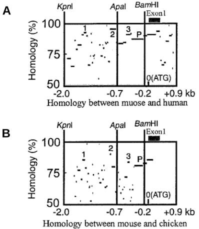
To identify transcriptional regulatory regions of the verte-brate Fgf10, we compared sequences of the mouse 2.0-kb 5’-fragment of the Fgf10 promoter region with the corresponding regions of the human and chicken Fgf10, using the percent identity plot (pip) obtained with PipMaker, which shows both the position in one sequence and the degree of similarity for each aligning segment between the two sequences (Schwartz et al., 2000). The results are shown in Fig. 1 A,B, where several highly conserved sequences (more than 75%) among the three spe-cies are identified (Fig. 1 A,B). There are three regions where mouse sequences are highly homologous to the corresponding human and chicken ones, as indicated by the number 1 to 3, while a region indicated by P may contain promoter sequences. The three regions conserved among human, mouse and chicken may contain some enhancer motifs, regulating expression of Fgf10.
Fig. 1. Homology analysis of 5’-fragment of Fgf10 between mouse and human (A)and between mouse and chicken (B), illustrated by PipMakerA (Schwartz et al., 2000). There are three conserved regions indicated by the number in the fragment. P indicates the promoter region of Fgf10.
Fig. 2. Schematic illustration of the constructs for generating transgenic mice. The 5’- fragment (a KpnI fragment) was digested by restriction enzymes into two smaller fragments with (A) ApaI (0.7 kb) and (B) BamHI (0.2 kb) restriction enzymes. Each fragment was inserted in a lacZ reporter vector containing a promoter of the heat shock protein 68 (hsp68) and Shine-Dalgarno-Kozak (SDK) sequence. PCR primers generated in the lacZ cassette are indicated by arrows.
Identification of Mouse Cis-Control Elements of Fgf10 for Transgene Expression in the Developing Limb
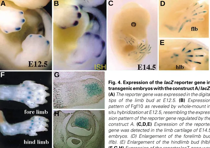
In order to identify the mouse cis-control elements regulating Fgf10 expression in the developing limb, we generated transgenic mice which had a lacZ reporter gene under the control of 2.0 (designated as construct K), 0.7 (A), or 0.2 (B) kb 5’-fragment of Fgf10 (Fig. 2). In embryos (generation 0 (F0)) injected with the construct K (K-transgenic embryos), the transient transgene ex-pression was detected at the apical ectodermal ridge (AER) of the limb bud in E11.5 embryos (n = 2 out of 9)(Fig. 3 A,B). So far, we have not generated any stable transgenic line with the construct K. In the transient transgenic E10 embryos with the construct A (A-transgenic embryos), the transgene was not expressed in the limb bud (Fig. 3 C,D), while the expression was detected clearly in the developing somites (Fig. 3 C,D). However, with the progress of limb development, the expression was observed in the distal tips of the limb bud (E12.5) and then in limb cartilage (from E13.0 to neonate at least). In order to confirm the results obtained in the A-transient F0 mice, we generated two stable transgenic lines with the construct A and analyzed expression patterns more precisely. The transgene was expressed in the distal tip of the limb buds as spots in E12.5 (Fig. 4A). This spotty expression was similar to the expression pattern of Fgf10 in the limb bud of E12.5 embryos (Fig. 4B). Then, the transgene expression was observed in cartilage of the developing limb. In E14.5 embryos, the transgene expression was localized in cartilage of the zoygopod (Fig. 4 C,D). In the limb of neonates, X-gal stained regions were observed in the epiphy-seal cartilage of the autopod. Their sections revealed that the stained cells are localized in the part of the proliferating cartilage cells of the epiphyseal growth plate. The expression in somites, brain, midgut loop, and cardiac vesicle remained unchanged in the A-transgenic mice, as observed in the K-transgenic mice. These results indicated that the enhancers for expression of the epiphy-seal growth plate are involved in the 0.7 kb 5’-fragment.
On the other hand, in the transgenic mice with the shortest construct including 0.2 kb Fgf10 5’-fragment (B-transgenic mice), significant transient expression of the transgene was not detected except for the posterior narrow region of the limb bud and somites (Fig. 3 E,F). This result suggested that the 0.2 kb 5’-region which is a direct upstream of the first codon of Fgf10 and 0.9 kb downstream region do not contain strong regulatory elements for expression in the limb bud. These results indicated that the
A
B
A
B
C
D
E
F
G
H
Fig. 3. Expression of the lacZ reporter gene in E11.5 transgenic embryos with the construct K/lacZ (A,B), A/lacZ (C,D) and B/lacZ (E,F). The expression of the transgene was detected at the apical ectodermal ridge (AER) in transgenic mice with the K construct, but not in mice with the A and B constructs, indicating that there are some enhancers at least for the AER in the KpnI/ ApaI fragment. Weak transgene expression was observed in the posterior region of the limb bud of the transgenic mice with the B construct.
Fig. 4. Expression of the lacZ reporter gene in transgenic embryos with the construct A/lacZ. (A) The reporter gene was expressed in the digital tips of the limb bud at E12.5. (B) Expression pattern of Fgf10 as revealed by whole-mount in situ hybridization at E12.5, resembling the expres-sion pattern of the reporter gene regulated by the construct A. (C,D,E) Expression of the reporter gene was detected in the limb cartilage of E14.5 embryos. (D) Enlargement of the forelimb bud (flb). (E) Enlargement of the hindlimb bud (hlb). (F,G,H) Expression of the reporter lacZ gene was observed in the epiphyseal growth plate of the limb cartilage of the new born limb. Semisagittal and transverse sections of the stained limb were shown in G and H, respectively.
enhancers for expression of limb carti-lage are involved in the 500 bp-fragment localized between - 0.7 and - 0.2 kb 5’ fragment of mouse Fgf10.
Conservation of Putative Regulatory Regions in the Mouse Cis-Element, regulating Expression of Fgf10 in the Developing Limb
To identify DNA-binding motifs in the 2.0 kb sequences of Fgf10, we used MatInd and MatInspector as tools for detection of consensus matches in nucle-otide sequence data, proposed by Quandt et al. (1995). The nucleotide sequences
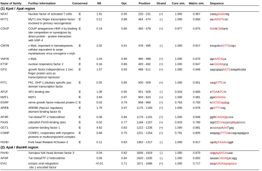
of the identified regulatory elements re-veal several DNA-binding motifs of tran-scription factors which are highly con-served among mouse, human, and chicken, as shown in Fig. 5. There are potential enhancer sequences in the -2.0 – -0.7 kb (KpnI-ApaI) region, containing NFAT, MYT1, COUP, CMYB, VMYB, ETSF, GFI1, PIT1, AP1F, MZF1, EGRF,
AREB, AP4R, PAX5, OCT1, COMP, and FKHD, which are all conserved in the mouse, human, and chicken sequences. De-tailed information is listed in Table 1. Although Fgf10 is a mesen-chymal factor and not expressed in the AER of developing normal limb bud, the transgene expression under the regulation of the construct K (2.0 kb upstream of the first codon of Fgf10) was detected in the AER. On the other hand, this AER expression was not observed in the A-transgenic mice, indicating there are
enhancers for the AER in the 1.3 kb KpnI/ApaI 5’-fragment of Fgf10. This result suggested that the enhancer activity for the AER in this fragment is suppressed somehow in normal develop-ment. In the 1.3 kb KpnI/ApaI fragment, a candidate region for enhancers including the AER enhancer should be a high homol-ogy region conserved among mouse, human and chicken. In this region, there are 17 DNA-binding motifs as listed in Table 1.
For the AER enhancer, Liu et al. (1994) found that 439 bp of 5' flanking sequence of the Msx2 homeobox gene contains regulatory elements for its exclusive expression in the AER of the developing limb. Recently, Pan et al. (2002) found that the B-TAAT site of the four potential homeodomain binding TAAT sites in a 348-bp fragment of the chicken Msx2 gene is critical for AER enhancer activity. However, there is not such se-quence in the KpnI/ApaI fragment. Thus, we can not determine which is the AER enhancer in the conserved 17 motifs. More precise experiment with the short fragment containing the several motifs is necessary to identify enhancer motifs in this region.
The 500-bp ApaI/BamHI murine frag-ment responsible for the cartilage expres-sion of Fgf10 contains a 100 bp sequence conserved highly among mouse, human, and chicken (Fig. 5), suggesting that the sequence may act as cis-element regulat-ing cartilage-specific expression of the Fgf10 gene in developing limb. The 100-bp mouse and human sequences contain three conserved DNA-binding motifs: RREB, GKLF, IRFF, and EVI1 (see Table
A
B
C
D
TABLE 1
CONSERVED ELEMENTS IN THE 5’ REGION OF THE FGF10 PROMOTER BETWEEN MOUSE, HUMAN AND CHICKEN SEQUENCE
Name of family Further Information Conserved RE Opt. Position Strand Core sim. Matrix sim. Sequence
(1) KpnI / ApaI region
NFAT Nuclear factor of activated T-cells C 1.91 0.95 220 - 231 (+) 1.000 0.967 caaagGAAAttg
MYT1 MyT1 zinc finger transcription factor C 0.21 0.88 464 - 474 (-) 1.000 0.894 aaaAAGTtcac
involved in primary neurogenesis
COUP COUP antagonizes HNF-4 by binding C 0.19 0.80 465 - 478 (+) 0.977 0.875 tGAACtttttactt
site competition or synergizes by direct protein - protein interaction with HNF-4
CMYB c-Myb, important in hematopoesis, C 2.02 0.91 478 - 495 (-) 1.000 0.917 tctcgcttccGTTGctga
cellular equivalent to avian myoblastosis virus oncogene v-myb
VMYB v-Myb C 1.04 0.89 480 - 488 (+) 1.000 0.970 agcAACGga
ETSF nuclear respiratory factor 2 C 0.16 0.86 483 - 492 (+) 1.000 0.947 aacGGAAgcg
GFI1 growth factor independence 1 zinc C 1.57 0.93 488 - 511 (+) 1.000 0.946 aagcgagaAATCaatgattcatta
finger protein acts as transcriptional repressor
PIT1 Pit1, GHF-1 pituitary specific pou C 0.55 0.85 500 - 509 (+) 1.000 0.951 aatgATTCat
domain transcription factor
AP1F AP1 binding site C 1.08 0.95 501 - 509 (-) 0.934 0.960 aTGAATCAt
MZF1 MZF1 C 3.84 0.97 903 - 910 (+) 1.000 0.981 ggaGGGGa
EGRF nerve growth factor-induced protein C C 0.03 0.79 958 - 969 (+) 0.763 0.793 tcGCGCtggtgg
AREB AREB6 (Atp1a1 regulatory C 1.76 0.97 1175 - 1183 (+) 1.000 0.976 ggGTTTcag
element binding factor 6)
AP4R Tal-1beta/ITF-2 heterodimer C 0.06 0.84 1176 - 1191 (+) 1.000 0.846 ggtttCAGAtgtccca
PAX5 zebrafish PAX9 binding sites C 0.02 0.77 1184 - 1207 (+) 0.933 0.780 atgtCCCAccgccgtttgaccccc
OCT1 octamer-binding factor 1 C 4.82 0.83 1222 - 1236 (+) 1.000 0.881 acccctgcAAATgag
COMP COMP1, cooperates with myogenic C 0.66 0.75 1231 - 1254 (+) 0.791 0.805 aatgaggTTTGaccagcagaggca
proteins in multicomponent complex
FKHD Fork head Related ACtivator-2 C 0.11 0.83 1302 - 1317 (-) 1.000 0.917 cgcttgTAAAcaggtt
(2) ApaI / BamHI region
FKHD Xenopus fork head domain factor 3 0.04 0.82 1606 - 1619 (-) 1.000 0.870 acggtgAACAaaac
AP4R Tal-1beta/ITF-2 heterodimer 0.06 0.84 1620 - 1635 (-) 1.000 0.892 aaaaaCAGAtgacagg
EVI1 ectopic viral integration <0.01 0.71 1671 - 1686 (+) 1.000 0.717 agtgAAGAagagaaca
site 1 encoded factor
In Conserved column, letter C represents conserved elements among mouse, human and chicken.
Position is shown by the number of nucleotide in the mouse sequence starting from the first nucleotide of the KpnI site.
RE (random expectation): The RE-value for each individual matrix gives an expectation value for the number of matches per 1000 bps of random DNA sequence.
Opt. (optimized matrix threshold): This matrix similarity is the optimized value defined in a way that at most 3 matches are found in 10000 bps of non-regulatory test sequences.
Core sim. (core similarity): The “core sequence” of a matrix is defined as the (usually 4) consecutive highest conserved positions of the matrix. The core similarity is calculated as described in the MatInspector paper. The maximum core similarity of 1.0 is only reached when the highest conserved bases of a matrix match exactly in the sequence.
Matrix sim. (matrix similarity): The matrix similarity is calculated as described in the MatInspector paper. A perfect match to the matrix gets a score of 1.00 (each sequence position corresponds to the highest conserved nucleotide at that position in the matrix), a “good” match to the matrix usually has a similarity of >0.80.
Basepairs marked red are important, i.e. they appear in a position where the matrix exhibits a high conservation profile (ci-value > 60). Basepairs in capital letters denote the core sequence used by MatInspector.
1). Since all of them is not exactly conserved in the corresponding chicken sequence, we can not exclude a possibility that unknown trans-acting factors may bind to these motifs.
Materials and Methods
Isolation of the 5’-Flanking Region of Fgf10
Mouse Fgf10 genomic clones were isolated from a TT2 ES cell genomic library by plaque hybridization using the full- length rat Fgf10 cDNA as a probe(Sekine et al., 1998). Chicken Fgf10 genomic clones were obtained by hybridization screening of the genomic library in lambda phage (Clontech, USA) and used as a template for sequencing after subcloning in pBluescript.
Constructs and Generation of Transgenic Mice
We isolated a mouse genomic clone containing a 3.2 kb 5’upstream fragment of Fgf10. The 3.2 kb fragment was digested with KpnI, ApaI, and BamH I to 2.0 (K), 0.7 (A), 0.3 (B) kb fragments (Fig. 2). To construct transgenes, each fragment was inserted in a vector phsp68lacZpA containing the promoter of the heat shock protein 68 and lacZ reporter gene (Fig. 2) (Sasaki and Hogan, 1996). After excision of a transgene from the vector, the concentration of the transgenes was adjusted to 500 Fig. 5. Position of DNA binding motifs conserved among mouse, human
molecules/pl and the solution was microinjected into the male pronuclei of fertilized eggs derived from superovulated BDF1(C57BL/6 X DBA2 F1) female mice crossed with males of the same strain. Oviducts implantation of surviving injected embryos into pseudopregnant MCH/ICR female mice were carried out according to the standard protocol (Yamaoka et al., 1995). Pregnant mice after implantation were sacrificed and embryos from Embryonic day10 (E10) to E14.5 were treated by 2% paraphormaldehyde for 30 minutes. Reporter activity was analyzed by X-gal staining for over night in a CO2 incubator at 37oC. The integration of the transgene into the mouse genome was detected by PCR, using primers established at the sequence of lacZ cassette and DNA extracted from tail snips of 3-week live offspring by the proteinase K/SDS method (Yamaoka et al., 1995). With the construct A, two stable transgenic lines were obtained. Newborn limbs were stained with X-gal after desqua-mated to enhance permeation of the X-gal, as described previously (Sasaki and Hogan, 1996).
Histology
X-gal stained newborn limbs were fixed in 2% paraphormaldehyde, dehydrated, placed in xylene, and paraffin embedded. The sections with 30µm thickness were prepared to analyze the signals at cellular level.
Acknowledgments
We would like to thank S. Nishimatsu and C. Komaguchi for their technical assistance. This work was supported by grants from the Human Frontier Science Program Organization and from the Ministry of Education, Sports, Culture, Science and Technology to S.N., T.N., and H.O.
References
CAPDEVILA, J. and IZPISUA-BELMONTE, J.C. (2001). Patterning mechanisms controlling vertebrate limb development. Annu Rev Cell Dev Biol. 17:87-132. Review.
COHN, M.J., IZPISUA-BELMONTE, J.C., ABUD, H., HEATH, J.K. and TICKLE, C. (1995). Fibroblast growth factors induce additional limb development from the flank of chick embryos. Cell 80:739-46.
CROSSLEY, P.H., MINOWADA, G., MACARTHUR, C.A. and MARTIN, G.R. (1996). Roles for FGF8 in the induction, initiation, and maintenance of chick limb development. Cell 84, 127-136.
DAHN, R.D. and FALLON, J.F. (1999). Limbiting outgrowth: BMPs as negative regulators in limb development. Bioessays 21:721-725. Review.
ISAAC, A., COHN, M.J., ASHBY, P., ATALIOTIS, P., SPICER, D.B., COOKE, J. and TICKLE, C. (2000). FGF and genes encoding transcription factors in early limb specification. Mech. Dev. 93: 4-8
KAWAKAMI, Y., CAPDEVILA, J., BUSHER, D., ITOH, T., CONCEPCION, R.E. and BELMONTE, J.C. (2001). WNT signals control FGF-dependent limb initiation and AER induction in the chick embryo. Cell 104. 891-900.
KATO, S. and SEKINE, K. (1999) FGF-FGFR signaling in vertebrate organogenesis. Cell Mol. Biol. (Noisy-le-grand) 45: 631-638. Review.
LEWANDOSKI, M., SUN, X. and MARTIN, G.R. (2000). FGF8 signaling from the AER is essential for normal limb development. Nat. Genet. 26: 460-463.
LIU, Y.H., MA. L., WU, L.Y., LUO, W., KUNDU, R., SANGIORGI, F., SNEAD, M.L. and MAXSON, R. (1994). Regulation of the Msx2 homeobox gene during mouse embryogenesis: a transgene with 439 bp of 5' flanking sequence is expressed exclusively in the apical ectodermal ridge of the developing limb. Mech Dev. 48: 187-197.
MARTIN, G.R. (1998). The roles of FGFs in the early development vertebrate limbs. Genes. Dev. 12: 1571-1586.
MARTIN, G. (2001). Making a vertebrate limb: New players enter from the wings. Bioessays 23: 865-858.
MAKARENKOVA, H.P., ITO, M., GOVINDARAJAN, V., FABER, S.C., SUN, L., MCMAHON, G., OVERBEEK, P.A. and LANG, R.A. (2000). FGF10 is an inducer and Pax6 a competence factor for lacrimal gland development. Development 127: 2563-2572.
MIN, H., DANILENCO, D.M., SCULLY, S.A., BOLON, B., RING, B.D., TARPLEY, J.E., DEROSE, M. and SIMONET, W.S. (1998). FGF10 is required for both limb and lung development and exhibits striking functional similarity to Drosophila branch-less. Genes. Dev. 12: 3156-3161.
OHUCHI, H., NAKAGAWA, T., YAMAUCHI, M., OHTA, T., YOSHIOKA, H., KUWANA, T., MIMA, T., MIKAWA, T., NOHNO, T. and NOJI, S. (1995). An additional limb can be induced from the flank of the chick embryo by FGF4. Biochem. Biophys. Res. Commun. 209: 809-816.
OHUCHI, H., NAKAGAWA, T., YAMAMOTO, A., ARAGA, A., OHTA, T., ISHIMARU, Y., YOSHIOKA, H., KUWANA, T., NOHNO, T., YAMASAKI, M. et al (1997). The mesenchymal factor, FGF10, initiates and maintains the outgrowth of the chick limb bud through interaction with FGF8, an apical ectodermal factor. Development 124: 2235-2244.
OHUCHI, H. and NOJI, S. (1999). Fibroblast-growth-factor-induced additional limbs in the study of initiation of limb formation, limb identity, myogenesis, and innerva-tion. Cell Tissue Res. 296: 45-56. Review.
OHUCHI, H., HORI, Y., YAMASAKI, H., SEKINE, K., KATO, S. and ITOH, N. (2000). FGF10 acts as a major ligand for FGF receptor 2 IIIb in mouse multi-organ development. Biochem. Biophys. Res. Commun. 277: 643-649.
PAN, Z.Z., KRONENBERG, M.S., HUANG, D.Y., SUMOY, L., ROGINA, B., LICHTLER, A.C. and UPHOLT, W.B. (2002). MSX2 expression in the apical ectoderm ridge is regulated by an MSX2 and dlx5 binding site. Biochem. Biophys. Res. Commun. 290: 955-961.
QUANDT, K. FRECH, K., KARAS, H., WINGENDER, E. and WERNER, T. (1995). MatInd and MatInspector - New fast and versatile tools for detection of consensus matches in nucleotide sequence data. Nucleic Acids Research 23: 4878-4884. (http://www.genomatix.de/)
SASAKI, H. and HOGAN, B.L. (1996). Enhancer analysis of the mouse HNF-3 beta gene: regulatory elements for node/notochord and floor plate are independent and consist of multiple sub-elements. Genes Cells 1: 59-72
SEKINE, K., OHUCHI, H., FUJIWARA, M., YAMASAKI, M., YOSHIZAWA, T., SATO, T., YAGISHITA, N., MATSUI, D., KOGA, Y., ITOH, N. and KATO, S. (1999). Fgf10 is essential for limb and lung formation Nat Genet. 21: 138-141.
SCHWARTZ, S., ZHANG, Z., FRAZER, K.A., SMIT, A., RIEMER, C., BOUCK, J., GIBBS, R., HARDISON, R. and MILLER, W. (2000). PipMakerA Web Server for Aligning two genomic DNA sequences. Genome Research 10: 577-586. (http:// bio.cse.psu.edu).
SUZUKI, K., YAMANISHI, K., MORI, O., KAMIKAWA, M., ANDERSEN, B., KATO, S., TOIDA, T. and YAMADA, G. (2000). Defective terminal differen-tiation and hypoplasia of the epidermis in mice lacking the Fgf10 gene. FEBS Lett. 481: 53-56.
TICKLE, C. and MUNSTERBERG, A. (2001) Vertebrate limb development—the early stages in chick and mouse. Curr. Opin. Genet. Dev. 11:476-481. Review.
VOGEL, A., RODORIGUEZ, C. and IZPISUA-BELMONTE, J.C. (1996). Involvement of FGF8 in initiation, outgrowth and patterning of the vertebrate limb. Development 122: 1737-1750.
YAMAOKA, T., NISHIMURA, C., YAMASHITA, K., YAMADA, T., TOMONARI, S., MORITANI, M., II, S., YOSHIMOTO, K., HATA, J. and ITAKURA, M. (1995). Acute onset of diabetic pathological changes in transgenic mice with human aldose reductase cDNA. Diabetologia 38: 255-261.