Acknowledgements
To begin, I would like to thank my supervisors. First of all, Rosa Beddington, her insight contributed greatly to this work and her passionate nature was an invaluable source of inspiration. Second, Steve Harrison, who provided thoughtful and thorough feedback that significantly improved this thesis. More than this, he made himself available to become my principle supervisor and was on hand throughout the main body of this work to answer my numerous questions. Finally, Sohaila Rastan and Vassilis Pachnis for their support and useful discussions.
There are also many people I wish to thank for their invaluable contributions to the work. Special thanks to Evelyn Grau for carrying out the blastocyst injections. Graham Duddy for help and advice in tissue culture and our numerous technical discussions in and out of the lab. Davina Gale for invaluable support during the RDA, and Dave Michaelovich for help and advice on bioinformatics. Also, Juan Pedro Martinez Barbera and Alastair Morrison for reading my thesis and giving incisive feedback. Finally, everyone in the division of Mammalian Development and Comparative Genomics for their advice and support.
I'd like to thank some wonderful friends for providing an important source of distraction. Whether it was driving across the country with me to jump in the sea or just getting me out of the lab to the relative sanity of the nearest pub, thanks for
being there!
The isolation and
characterisation o f the novel
gene C53
Ross Kettleborough
National Institute for Medical Research
ProQuest Number: U641885
All rights reserved
INFORMATION TO ALL USERS
The quality of this reproduction is dependent upon the quality of the copy submitted.
In the unlikely event that the author did not send a complete manuscript and there are missing pages, these will be noted. Also, if material had to be removed,
a note will indicate the deletion.
uest.
ProQuest U641885
Published by ProQuest LLC(2015). Copyright of the Dissertation is held by the Author.
All rights reserved.
This work is protected against unauthorized copying under Title 17, United States Code. Microform Edition © ProQuest LLC.
ProQuest LLC
789 East Eisenhower Parkway P.O. Box 1346
Abstract
The study of axis formation is crucial if we are to understand the formation of a functional body plan. In mouse, the first morphological marker of A-P pattern, the primitive streak, forms at the posterior of the embryo and drives a complex process called gastrulation, during which new germ layers are formed and a general body plan is generated that serves as a scaffold for the subsequent morphogenesis of the embryo.
In order to isolate tissue specific genes that are involved in these symmetry breaking events a cDNA library derived from the endoderm of the 7.5dpc mouse embryo was produced. A screen was then performed that used sequence and expression
information to further analyse the endoderm specific library. cDNA clones representing genes that are expressed in tissues intimately involved in the early patterning of the embryo (e.g. the visceral endoderm, the definitive endoderm and the node) were identified.
This study focuses on C53, a novel gene that is expressed in the visceral endoderm, the node, and hematopoietic lineages of the early mouse embryo. A targeted
Contents
CHAPTER 1: GENERAL INTRODUCTION... 10
1.1 Pre and post implantation mouse development and the embryonic axes... 10
1.1.1 Early signs of asymmetry...10
1.1.2 A-P axis formation...13
1.1.3 The VE is also implicated in A-P pattern formation... 15
1.2 Mouse haematopoiesis... 18
1.2.1 Primitive haematopoiesis begins in the yolk sac... 18
1.2.2 Definitive haematopoiesis is mediated by intra-embryonic sites... 19
1.3 Isolation and characterisation of novel genes involved in early patterning events...21
1.3.1 Strategies for the isolation of genes involved in early patterning...21
1.3.2 ES cell technology in the study of gene function...24
CHAPTER 2: MATERIALS AND METHODS... 29
2.1 Screening of the endoderm cDNA library...29
2.1.1 Comparison of sequencing data from the endoderm library with sequence databases... 29
2.1.2 Dissection of peri-implantation mouse embryos...29
2.1.3 Whole mount in situ hybridisation using Digoxigenin-UTP (DIG-UTP) labelled probes... 30
2.1.4 Wax embedding and sectioning of WISH embryos...31
2.2 C53 Full length cDNA Cloning... 31
2.3 Generation of the C53 knockout mouse...32
2.3.1 Isolation of genomic DNA using the Lambda FixII library...32
2.3.2 Isolation of genomic DNA from BAG library (pBeloBACl 1 vector - Research Genetics)...32
2.3.3 Mapping of genomic DNA fragments...33
2.3.4 Production of targeting vectors and targeting constucts...33
2.3.5 General tissue culture techniques...35
2.3.6 Transfection and screening for targeted clones...35
2.3.7 Blastocyst injection and breeding...37
2.3.8 Genotyping techniques...37
2.3.9 Reporter detection...38
2.4 In-Vitro differentiation of null ES cells...39
2.4.1 Generation and in vitro differentiation of ES cell lines...39
2.4.2 cDNA synthesis and reverse transcription polymerase chain reaction (RT- PCR)... 39
2.5 Representational Difference Analysis (RDA) of RNA samples from C53 null yolk sac and liver...40
2.5.1 Isolation of RNA samples from embryonic yolk sac and liver and first strand cDNA synthesis...40
2.5.2 Production of Difference Product (DP) 1 and 2... 40
2.5.3 Cloning and sequencing of Difference Products... 41
CHAPTER 3: SCREENING OF THE BEDDINGTON DISSECTED
ENDODERM CDNA LIBRARY... 44
3.1 Introduction... 44
3.1.1 The endoderm library screen... 44
3.2 R esults... 46
3.2.1 Comparison of sequence from the endoderm library to sequence databases 46 3.2.2 Whole mount RNA in situ hybridisation of prioritised clones...46
3.3 Discussion... 51
3.3.1 The endoderm library screen for novel genes implicated in early patterning. ...51
3.3.2 Expression o f ‘restricted’ mRNAs from this study... 53
CHAPTER 4: C53 CDNA CLONING, BIOINFORMATICS AND EXPRESSION ANALYSIS...56
4.1 Introduction... 56
4.1.1 Full length cDNA cloning by rapid amplification of cDNA ends (RACE) PC R... 56
4.1.2 The use of bioinformatics to assign function to unknown proteins...57
4.1.3 Expression analysis... 61
4.2 R esults... 62
4.2.1 Cloning of the full length mouse C53 cDNA...62
4.2.2 Bio-informatics gives clues about C53 structure and function...64
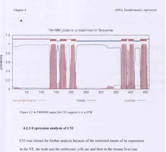
4.2.3 Expression analysis of C53...69
4.3 Discussion... 73
4.3.1 C53 is a 6TM domain protein and may be involved in metal ion transport..73
CHAPTER 5: 053 TARGETED MUTATION... 80
5.1 Introduction... 80
5.1.1 Gene targeting in mouse using homologous recombination in embryonic stem (ES) cells...80
5.1.2 Construct design and consideration for targeting vectors... 82
5.1.3 Selection for targeted clones...85
5.1.4 Knock-in strategies...87
5.1.5 Electroporation of ES cells and screening for targeting events... 89
5.1.6 Blastocyst injection and general breeding regimes... 91
5.2 Results...93
5.2.1 Cloning of the C53 genomic locus...94
5.2.2 Production of targeting vectors... 105
5.2.3 C53 targeted ES cells passed efficiently through the germline... 110
5.2.4 C53 null is embryonic lethal...112
5.3 Discussion... 119
5.3.1 C53 has a complex genomic locus... 119
5.3.2 The C53 targeting Strategy...120
5.3.3 C53 null is embryonic lethal... 124
CHAPTER 6 - IN-VITRO DIFFERENTIATION OF 053 NULL ES CELLS. 128 6.1 Introduction... 128
6.1.1 In vitro differentiation of ES cells... 128
6.1.2 Differentiation of haematopoietic lineages... 129
6.2 R esults...131
6.2.1 Creation of C53 null ES cells... 131
6.2.2 Differentiation of C53 null ES cells in methyl-cellulose medium... 133
6.3 Discussion...142
6.3.1 Induction of haematopoietic genes in control ES cells... 142
CHAPTER 7 - REPRESENTATIONAL DIFFERENCE ANALYSIS (RDA) OF CDNA FROM C53 - I - AND + / + EMBRYONIC YOLK SAC AND LIVER 148
7.1 Introduction...148
7.1.1 Identfifying differences in mRNA expression by representational difference analysis of cDNA... 148
7.1.2 cDNA-RDA of genes expressed in C53 -/- and +/+ embryonic yolk sac and liver... 150
7.2 R esults... 152
7.2.1 RDA with C53 +/+ and -/- embryonic yolk sac and liver...152
7.2.2 Sequence analysis of subtraction products and confirmation of differential expression by quantitative PCR... 154
7.2.3 Genes up regulated and down regulated in C53 null tissues... 159
7.3 Discussion...160
7.3.1 The use of RDA to successfully identify differentially expressed cDNAs in C53 -/- tissues as compared to +/+... 160
CHAPTER 8: GENERAL DISCUSSION... 166
8.1 Screening of the Beddington dissected endoderm cDNA library using a combination of sequencing and expression analysis... 166
8.2 Cloning and Characterisation of C53... 168
8.2.1 Bioinformatics gives clues about C53 structure and function... 169
8.2.2 Expression suggests roles in early axes formation and haematopoietic development... 170
8.3 The creation of the C53 targeted mutation...172
8.3.1 Construct design and targeting of ES cells... 172
8.3.2 The C53 null phenotype... 174
8.4 In vitro differentiation of C53 null ES cells... 176
8.5 RDA analysis of mRNA from C53 null embryonic yolk sac and liver... 178
REFERENCES... ...183
Chapter 1
Chapter 1 General Introduction
Chapter 1: General Introduction
1.1 Pre and post implantation mouse development and the embryonic axes
The formation of the embryonic axes is a crucial aspect of pattern formation as it forms the basis for a functional body plan. The anterior-posterior (A-P) axis of avian and mammalian embryos is of particular interest because the first morphological marker of A-P pattern, the primitive streak, forms at the posterior of the embryo. The primitive streak drives a complex process called gastrulation and is a source of new tissue layers. During gastrulation the basic body plan is generated that serves as a scaffold around which the subsequent morphogenesis of the embryo occurs.
1.1.1 Early signs of asymmetry
It has long been known that in Xenopus laevis localised determinants in the egg seem to play a crucial role in establishing the early axes, and disruption of this early
organisation can prevent the development of a normal embryo. However, the mouse embryo is very tolerant to physical manipulation at early stages, with even removal (Zemicka-Goetz, 1998) or aggregation (Gardner and Mclaren, 1974) of blastomeres leading to normal development, suggesting the origins of embryonic axis formation do not lie within the egg or the blastomeres. However, more recent work tracing polar bodies relative to the sperm entry point suggests the axis of bilateral symmetry in the blastocyst can be traced back to the animal-vegetal axes of the fertilised egg (Piotrowska and Zemicka-Goetz, 2001). With evidence that the polarity of the
Chapter 1 General Introduction
blastocyst influences the Proximal-Distal (P-D) axis in the early post-implantation mouse embryo (Weber et a l, 1999), which in turn influences the induction of the other embryonic axes (reviewed in Lu et al., 2000), it seems the origins of axis formation in mammals may be present much earlier during development than was once thought.
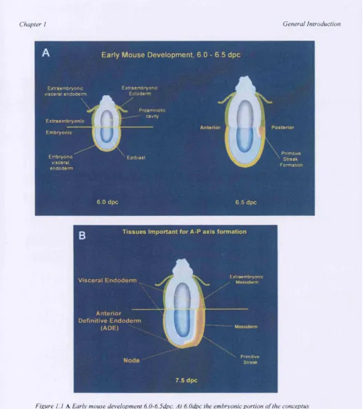
During the immediate post-implantation period (5-6 days post coitum [dpc]), the mouse embryo changes dramatically in size, with the inner cell mass (ICM) and its associated trophectoderm growing into the blastocoelic cavity. A mixture of
apoptotic and survival signals from the surrounding visceral endoderm (VE) creates a cavity in the center of the ICM and results in the embryo aquiring a cup shape made up of two cell layers, the ICM (now termed the epiblast) and the outer VE (see figure
1.1 A). At this stage the conceptus has well delineated embryonic and extraembryonic regions and the prospective dorso-ventral (D-V) axes becomes apparent, with the proamniotic surface of the epiblast corresponding to the dorsal side of the embryo and the outer surface of the visceral endoderm corresponding to the ventral side.
It is now widely accepted that the extraembryonic tissues play an important role in laying down the early axes (reviewed in Beddington and Robertson, 1999). Complex interactions between the extraembryonic ectoderm and the epiblast give the first signs of posterior polarity and define the point at which the primitive streak will form. Both Bone Morphogenic Protein 4 (BMP-4), a member of the TGpp
Chapter 1 General Introduction
epiblast and have been shown to be required for posterior patterning (Lawson et al,
1999; Russ et al, 2000). Furthermore, in embryos lacking nodal (another member of the TGpp superfamily that is normally found in the epiblast at early post
implantation stages) the expression of BMP-4 and eomes are not maintained and genes that are normally expressed in the proximal-posterior region, such as the mesoderm marker Brachyury (T), fail to be expressed (presumably because the extraembryonic ectoderm fails to signal to the epiblast) (Brennan et al, 2001).
These complex interactions between extraembryonic and embryonic tissues
ultimately result in the initiation of gastrulation at 6.5dpc, morphologically marking the posterior aspect of the embryo (see figure 1.1 A). Cells from the embryonic ectoderm (the epiblast) undergo an epithelial to mesenchymal transition and delaminate and ingress through the primitive streak and its specialised anterior component, the node, to produce mesoderm and definitive gut endoderm. The new tissues emerge from the streak and move laterally and anteriorly beneath non- ingressed epiblast. This complex set of morphogenic movements coupled with cell proliferation and differentiation converts the initially two-layered structure into an embryo with all three germ layers. Thus, the formation of the primitive streak at the posterior of the embryo is a symmetry-breaking event, defining the A-P axes and also the left-right axes (L-R).
Chapter 1 General Introduction
1.1.2 A-P axis formation
The tissues that are implicated in A-P pattern formation are illustrated in figure 1 .IB. Where the primitive streak forms is critical in the orientation of the A-P axis. The mouse node is the specialised anterior component of the primitive streak that forms as it elongates towards the distal tip of the egg cylinder. It has many properties in common with the classical 'organiser' of Xenopus, to which anterior patterning is ascribed in the frog (reviewed in Moon and Kimelman, 1998). The node expresses homologs of many 'organiser' genes (Beddington and Smith, 1993), gives rise to a similar repertoire of tissues (Beddington, 1981 and 1994), and can induce a
secondary axis when transplanted (Beddington, 1994; Tam et al, 1997). However, there are important differences, such as its inability to induce a complete secondary axis upon transplantation. Such ectopic axes invariably have anterior truncations (e.g. lacking forebrain) suggesting a further level of regulation is required for anterior pattern formation.
Chapter 1 General Introduction
Early Mouse Development. 6.0 - 6.5 dpc
Extraem bryonic visceral en d o d erm
E xtraem bryonic Em bryonic
Embryonic visceral en d o d erm
Extraem bryonic E ctoderm
Proam niotic - cavity
Epiblast ' S treak Primitive
Form ation
6 0 dpc 6.5 dpc
T issu es Im portant for A-P axis form ation
Visceral Endoderm
A nterior Definitive E ndoderm
(ADE) '
Extraembryonic
Mesoderm
P r im itiv e
Streak
7.5 dpc
Figure I .l A Early mouse development 6.0-6.5dpc. At 6.0dpc the embryonic portion o f the conceptus is made up o f the cup shaped epiblast (Blue) surrounded by a thin layer o f extraembryonic visceral endoderm (yellow). At 6.5 dpc gastrulation is initiated in the posterior with the formation o f the primitive streak B Tissues implicated in A-P axis formation include the Node, Anterior Definitive Endoderm and Visceral Endoderm (EGO not shown and is present earlier in development), (adapted from Beddington and Robertson, 1998)
Chapter 1 General Introduction
anterior neurectoderm (Camus et al, 2000) and a targeted mutation in the homeobox gene Hex, which is expressed in the anterior visceral endoderm (AVE), and later in the ADE, has been shown to result in embryos with anterior truncations. Chimaeric embryos composed of Hex'^' cells developing within a wild-type visceral endoderm (VE) continue to show the anterior truncation, indicating that Hex is required in the ADE to generate anterior character (Martinez-Barbera et al, 2000). Furthermore, the discovery a group of cells in the posterior epiblast of the early streak stage embryo that display cell fates, gene expression and tissue patterning activities of the classical organiser (Camus and Tam, 1999), now known as the early gastrula organiser
(EGO), confirms the idea that the patterning of the anterior begins well before node formation.
1.1.3 The VE is also implicated in A-P pattern formation
It is now known that the extraembryonic (visceral) endoderm also plays a critical role in patterning the A-P axes in rabbit and mouse (reviewed by Beddington and
Robertson, 1999) prior to formation of the primitive streak. It is strategically positioned for inductive interactions with the underlying ectoderm and later mesoderm and has been implicated in posterior patterning where it is known to co ordinate primitive blood cell/vessel formation.
The AVE is derived from a handful of VE cells situated at the distal tip of the
Chapter I General Introduction
Other VE cells during their anteriorward movement (Weber et al, 1999) suggesting a distinct role for the AVE as they migrate. The VE shows asymmetric gene
expression at least 12 hours prior to primitive streak formation, with expression of genes specifically in the anterior, such as Cerl and Dkkl. Such signals are thought to antagonise Wnt and TGF-p signals (e.g. nodal), which are essential for mesoderm formation, to the posterior of the embryo (Beddington and Robertson, 1999). In embryos lacking AVE (e.g. Smad2-/-) the epiblast adopts a completely proximal posterior identity via widespread expression of T, Fibroblast Growth Factor-8 {FGF-8), and nodal (Brennan et al, 2001). In embryos where VE is formed but anterior migration is inhibited (e.g. Cripto -/-), the A-P axis is misaligned with distal expression of anterior markers and posterior genes, such as T, being expressed radially in the proximal part of the epiblast. Together, these data illustrate that the AVE is central to the induction of anterior pattern. This identity is then maintained by the node and its derivatives, leaving the AVE to be displaced proximally by definitive endoderm.
Interestingly, the embryos of mice lacking nodal (expressed in the epiblast and the VE) have been shown to have gastrulation defects, presumably as a result of misexpression of genes involved in mesoderm induction (discussed previously). However chimaeric analysis reveals that, where nodal mutant VE develops around wild type epiblast, nodal (Varlet et al, 1997) is required in the VE for correct
anterior patterning. This occurs through the activation of Smad2 which in turn causes expression of the AVE genes required for anterior development (Brennan et al,
Chapter 1 General Introduction
2001). Together, this places Nodal at the center of a signalling mechanism that regulates both anterior and posterior development.
Delicate embryology has always been at the focal point of studies into anterior patterning, assigning specific roles to regions of the embryo that have inducing potential. In the mouse, transplantation of the node has been shown to induce a secondary axis (Beddington, 1994), but fails to induce the most anterior structures, suggesting the specific head organising activity may be absent from the node and it may be more important for formation of the trunk. More recently, the same result was obtained when the Early Gastrula Organiser (EGO, that is also able to form ectopic axes) was transplanted heteroptopically. This suggests that the head organiser activity also lies outside of the EGO. Genetic and embryological studies confirm that the AVE is required for anterior pattern formation, and although it cannot induce any secondary neural tissue when transplanted alone (in the mouse), it can when
Chapter 1 General Introduction
1.2 Mouse haematopoiesis
1.2.1 Primitive haematopoiesis begins in the yolk sac
In nearly all vertebrate embryos, the first site of haematopoiesis is the embryonic yolk sac, an extra-embryonic membrane that serves a placental function prior to the establishment of the embryonic circulation.
Unlike the AVE, genes that are expressed exclusively in the posterior VE have remained elusive. This subset of cells is thought to be involved, alongside extra- . embryonic ectoderm, in inducing/maintaining posterior polarity, and cell lineage studies have confirmed that the posterior visceral endoderm remains in situ, overlying the streak throughout streak elongation (Lawson et al, 1997). During gastrulation, morphogenic movements place a subset of mesoderm cells in close proximity to the VE in the posterior extraembryonic region of the embryo. It is here, in what will become the visceral yolk sac, that the extraembryonic mesoderm will give rise to blood islands late in gastrulation. These blood islands are made up of haematopoietic precursors (mainly primitive erythrocytes) surrounded by endothelial cells, and gradually merge to form the blood vessels of the yolk sac. The visceral yolk sac subsequently expands to surround the embryo proper. The juxtaposition of the posterior visceral endoderm and the extraembryonic mesoderm raises the possibility that interactions between the two tissue layers may be required for blood island formation. Indeed, classical explant studies in the chick have implicated the primitve endoderm in the formation of blood and vascular tissue, and more recently
Chapter 1 General Introduction
it has been shown that explanted posterior visceral endoderm can respecify anterior ectoderm to posterior (haematopoietic and angioblastic) cell fates (Belaoussoff et al,
1998). Trans-filter experiments demonstrated that re-programming of the anterior epiblast does not require cell contact and is therefore mediate by a short-range diffusible signal. Indeed, Indian hedgehog (Jhh) is expressed in the visceral endoderm of mouse embryos and mature yolk sacs (Farrington et al, 1997), and there is evidence building that it may be the molecule that mediates blood specification. Ihh can respecify anterior epiblast to posterior fates, downstream targets of the hedgehog signalling pathway are up-regulated (as is BMP-4 which may mediate the signal), and blocking Ihh function inhibits the activation of
haematopoiesis in the adjacent epiblast. This suggests that Ihh is an endogenous signal that plays a key role in specification of primitive haematopoiesis.
1.2.2 Definitive haematopoiesis is mediated by intra-embryonic sites
The classical view of mammalian haematopoiesis is that the earliest cells of the adult blood system develop outside the embryo proper in the embryonic yolk sac. It was thought that a single cohort of haematopoietic stem cells (HSCs), that are formed in the yolk sac, follow a number of migratory steps to the foetal liver, spleen and finally the adult bone marrow where they reside for the rest of the animals life.
Chapter 1 General Introduction
initiation of definitive haematopoiesis in the developing embryo (Dieterlen-Lievre, 1988). Haematopoietic precursor cells from the mouse yolk sac, examined at 8.0dpc (before connection to the embryonic vascular system), have been shown to have a very limited differentiation potential in vitro and were incapable of long-term reconstitution of adult animals (Cumano et al, 1996; Medvinsky and Dzierzak,
1996). It is now known that early in development, immature haematopoietic progenitors can be found within another region of the mouse embryo that
encompasses three anatomical sites, the aorta, gonads and mesenephros, or AGM. The AGM contains the first detectable definitive haematopoietic stem cells
beginning at mid-embryonic day 10 (Sanchez et al, 1996) which have been found to be similar in cell suface phenotype to adult HSCs and be able to repopulate lethally irradiated adult animals in the long term. These intra-embryonic stem cells first colonise the liver at around day 10 and increase dramatically by day 11, when they can also be found in the embryonic yolk sac (presumably by migration through the vasculature), and later are responsible for definitive haematopoiesis in all
haematopoietic organs (Reviewed in Medvinsky and Dzierzak, 1998 and Ling and Dzierzak, 2002). However, whether two distinct, independently derived classes of stem cells exist in the mouse embryo is still a matter of controversy.
Chapter 1 General Introduction
1.3 Isolation and characterisation of novel genes involved in early patterning events
1.3.1 Strategies for the isolation of genes involved in early patterning
Large scale sequencing studies have revealed that up to 80% of all expressed sequence tags (ESTs), sequences derived from cDNA libraries (produced from mRNA so reflect gene expression within a cell or tissue) show no similarity to sequences that could give a clue towards gene function (Adams et al, 1995). With the completion of whole genome sequencing programmes, the next challenge is to assign a function to the genes that have already been identified by sequence.
Many strategies have been adopted to try to assess the function of novel genes, from large-scale saturation screens using mutagenesis to more refined tissue specific strategies using cDNA libraries and subtractive hybridisation (a procedure that uses hybridiation to identify genes expressed specifically in one tissue rather than another), and each has met with varying levels of success. In invertebrates such as
Chapter 1 General Introduction
functional gene redundancy (where loss-of-function has no identifiable phenotype) precludes the identification of whole classes of genes.
Gene trapping in mouse embryonic stem (ES) cells (the random insertion of a vector into the genome to identify and simultaneously mutate genes expressed during mouse development) offers a method to create random developmental mutants with a direct route to cloning the gene (Skames, 1993). Many genes with developmental
regulatory function are expressed in a regionalised fashion within the embryo.
Hence, conventional gene trap vectors contain a splice acceptor sequence linked to a reporter gene, most commonly LacZ (encoding P-galactosidase) (Gossler et al,
1989). When these vectors are introduced into ES cells and integrate within the introns of genes, p-galactosidase (p-gal) fusion proteins are produced and p-gal enzyme activity can then be observed allowing the endogenous expression pattern of the gene to be followed in ES cell derived chimeric embryos or in transgenic
embryos following germline transmission. However, they do have disadvantages. The LacZ expression pattern does not always faithfully reflect the expression of the endogenous gene and it is not possible to target genes in specific tissues or organs because the gene trap vectors insert into the genome completely at random.
cDNA libraries (representations of the mRNA population within a cell or tissue) are a powerful tool with which to isolate genes expressed in specific cell populations or tissues. Many groups have screened cDNA libraries from various model systems (e.g. frog, mouse and chick) by whole mount in situ hybridisation (WISH), which allows the temporal and spatial expression pattern of mRNA to be followed during
Chapter 1 General Introduction
embryogenesis. Libraries have been screened either in their entirety (Gawantka et al,
1998), or following a round of subtractive hybridisation, which enriches for genes expressed differentially between the tissues from which the libraries are made (Christiansen et al, 2001; Neidhart et al, 2000; Harrison et al, 1995). Although subtractive hybridisation greatly enriches for differentially expressed genes, and greatly reduces the number of common sequences, it does have its disadvantages. Genes that are expressed at high levels are greatly enriched in subtraction procedures (at the expense of rare transcripts), although this problem can be overcome by performing a normalisation (a process that standardises the frequency of each individual mRNA) prior to subtraction. Also, subtracting one tissue cDNA library from another may result in the loss of potentially interesting genes that are expressed in both tissues. More importantly, although the temporal and spatial expression pattern of a gene gives a hint towards its function, expression screens lack any phenotypic data that maybe obtained as a result of mutagenesis or gene trap screens.
Chapter 1 General Introduction
In this study an alternative strategy was used for the identification of novel genes expressed specifically in the endoderm. The endoderm library contains a number of tissues (the node, VE and definitive endoderm) that have been shown to be important for specifying the early embryonic axes in the mouse embryo (for review see
Beddington and Robertson, 1999). To isolate novel genes expressed within these tissues, cDNA clones from the endoderm cDNA library were sequenced and compared to sequence databases. The results from the database searches were clustered (identical sequences were grouped together) and the results were examined manually. This allowed the identification of novel sequences and concomitant removal of housekeeping and known genes, without the need for processes such as subtraction (which can result in the loss of potentially interesting transcripts). Once identified, novel clones were then analysed for restricted expression in the early embryo. This strategy effectively isolated novel genes that are expressed
differentially in the tissues of interest.
1.3.2 ES cell technology in the study of gene function
In mouse developmental biology the isolation of a novel gene, which is differentially expressed, often leads to the production of a targeted mutation to examine the
consequences of a loss-of-function. ‘Transgenic technology’, the introduction of genes or mutations into the germiine of experimental mammals, can be broadly split into two catergories: ‘gain of fimction’ mutations, typically created by micro
injection of the gene directly into the zygote, and ‘loss of function’ mutations, which employ embryonic stem (ES) cell technology.
Chapter 1 General Introduction
The latter has made it possible to make a mutation in the germiine of mice by
utilizing homologous recombination and ES cells. Homologous recombination, when applied to altering specific endogenous genes, is referred to as gene targeting. ES cells are remarkable since after being established from the blastocyst (4.5dpc), they can be cultured and manipulated in vitro and retain their ability to return to their normal developmental program when injected into the pre-implantation embryo and contribute to all developing tissues, most importantly the germiine. This allows specific mutations, introduced into the ES cells in vitro, to be passed through the germiine to produce mice of the desired genotype. Most targeting experiments are designed simply to create a null mutation (a mutation that completely ablates gene function) to investigate the consequences of a complete loss of ftmction. However, point mutations, large deletions, gene exchanges (knock-ins) and conditional mutations can also be created.
Chapter 1 General Introduction
potential, and in vitro studies provides a straightforward way to take further advantage of the properties of ES cells.
1.4 The study of the novel gene C53
Here I describe the isolation and characterisation of the novel gene C53. C53 was isolated during a screen of the endoderm cDNA library. The screening strategy used sequencing, database searches and expression analysis to isolate novel genes that are expressed differentially in the VE. C53 is expressed specifically in the VE, node and liver of the developing mouse embryo, and is a putative membrane spanning protein with a potential ZIP zinc transporter domain. During this investigation a targeted mutation has been produced to investigate C53 loss of function. The strategy combines the creation of a null mutation with a ‘knock-in’ of a reporter of gene expression, so expression of C53 can be followed through detection of the reporter gene in vivo in heterozygous animals. The C53 null mutation is embryonic lethal at around 14.5dpc, with embryos dying of severe anemia, indicating a defect in haematopoiesis.
In addition to the targeted mutation, C53 null ES cells were produced and
differentiated in vitro to give more clues about the potential function of C53 during hematopoiesis. In vitro differentiation of C53 null ES cells results in a de-regulation of transcription factors essential for the correct development and differentiation of the hemaotopoietic system. To broaden our knowledge of genes which are up regulated or down regulated in response to C53 loss of function, representational
Chapter 1 General Introduction
Chapter 2
Chapter 2 Materials and Methods
Chapter 2: Materials and Methods
2.1 Screening of the endoderm cDNA library
2.1.1 Comparison of sequencing data from the endoderm library with sequence databases
7000 clones from the gridded endoderm library were sequenced at the Max Planck Institute for Molecular Genetics, Germany. These sequences were compared to sequence databases using the NCBI BlastX and BlastN programs. Results from database searches were then clustered using bioinformatics programs that reduced the 7000 original sequences down to 1980 independent sequences that were
examined manually and novel and potentially interesting clones were highlighted for further analysis.
2.1.2 Dissection of peri-implantation mouse embryos
Chapter 2 Materials and Methods
2.1.3 Whole mount in situ hybridisation using Digoxigenin-UTP (DIG-UTP) labelled probes
74 library clones chosen for further analysis were linearised using the appropriate enzyme to allow production of anti-sense in situ hybridisation probes that are derived from the 3’ UTR. In vitro transcription of probes was carried out in the prescence of DIG-UTP using the DIG RNA labelling kit (SP6/T7) (Roche). Probes were DNAsel treated and purified using Chroma-Spin-100 spin columns (Clontech).
Whole mount in situ hybridisation (WISH) was carried out essentially as in Hogan et a l, 1994 and Rosen and Beddington, 1994. WISH analysis was carried out on 5-10 embryos at each stage (6.5dpc, 7.5dpc, 8.5dpc. 9.5dpc, lO.Sdpc) for each of the 74 clones investigated. Embryos were staged and rehydrated through a methanol :PBS series prior to bleaching in 6% hydrogen peroxide. Proteinase K treatment, and re-fix in 4% PFA and 0.2% Gluteraldehyde. Hybridisation was carried out in Pre-hyb solution (50% formamide, 5X SSC pH5, 50pg/ml Yeast tRNA, 1%SDS, 50pg/ml Heparin) containing 1 pg/ml probe o/n at 70°C. Embryos were then washed and RnaseA treated before being transferred into antibody solution containing Anti- Digoxigenin-AP conjugated antiboby o/n at 4°C. Again embryos were washed thoroughly before developing the AP signal using NBT/BCIP solution (Gibco-BRL). Embryos were kept in nets throughout the experiment and solution changes were carried out by moving nets into different wells of tissue culture dishes. After
development of signal embryos were post-fixed in 4% PFA and stored in 0.4% PFA at 4°C.
Chapter 2 Materials and Methods
2.1.4 Wax embedding and sectioning of WISH embryos
WISH embryos were dehydrated through an ethanol series and finally HistoClear before being placed into molten wax. Embryos were embedded in histological dishes and oriented using heated forceps. Sections were cut on a Microtome, dewaxed in Histoclear and mounted with Aquamount (MERCK).
2.2 C53 Full length cDNA Cloning
The C53 library clone was sequenced and primers were designed 3’ to an appropriate restriction site (primers N, O and P, downstream of an Eco47III site, see appendix section 2). SMART full length cDNA libraries (Clontech) derived from Adult Mouse Kidney and Lung were used as a template in nested 5’ RACE PCR reactions using primers provided by Clontech and a combination of primers N, O and P. PCR products were gel isolated (Quiaquick gel extraction kit - Quiagen) and cloned into the PCR2.1 vector (maps for all vectors and components used during this
Chapter 2 Materials and Methods
2.3 Generation of the C53 knockout mouse
2.3.1 Isolation of genomic DNA using the Lambda FixII library
A labelled 3’ probe derived from the full length cDNA was used to screen the Lambda FixII library (Stratagene). The library was plated onto its host strain (XL-1 Blue MRA (P2)) and plaques were lifted onto Hybond hT and hybridised to the 3’ probe. Filters were exposed to film o/n and agar plugs were taken that corresponded to positive plaques. Dilutions of phage were made and replated onto their host strain as a secondary screen using the same 3’ probe. Individual plaques were identified and DNA was prepared using the Lambda mini kit (Quiagen).
2.3.2 Isolation of genomic DNA from BAC library (pBeloBACll vector Research Genetics)
BAC library filters (Mouse 129/Ola, Research Genetics) were screened with ^^P labelled PCR derived probes from the full length cDNA. Positive BACs were picked and DNA was prepared using a modified mini-prep (Quiagen) procedure that utilised the conventional alkaline lysis procedure followed by isopropanol and ethanol
precipitations. To isolate BAC sub-clones, approximately lOpg of BAC DNA was digested with 6 base-pair cutting restriction enzymes. The resulting digests were electrophoresed on an agarose gel and southern blot analysis was carried out and hybridised to an appropriate ^^P labelled probe. The corresponding band, highlighted by southern blot analysis, was excised from a duplicate agarose gel, gel isolated (Quiaquick Gel Extraction Kit, Quiagen) and cloned into pBSII SK (+/-) using the Rapid-Ligation kit (Roche) according to the manufacturers instructuions. Ligation
Chapter 2 Materials and Methods
reactions were plated onto selective media o/n at 37°C. 24 colonies were picked and dotted onto Hybond and hybridised with the same labelled probe to identify colonies that contained BAC subclones. Positive colonies were grown up and DNA was prepared using the Maxi-prep kit (Quiagen).
23.3 Mapping of genomic DNA fragments
The lambda clone and all BAC subclones were mapped using restriction enzymes. Initially, each clone was digested using a wide variety of 6 base-pair cutting restriction enzymes, and subsequently were digested by combinations of such
enzymes to produce a definitive restriction map. Wherever possible, the enzyme used was contained in the pBSII SK (+/-) polylinker to use as a reference. Further analysis of the restriction maps was carried by southern blot analysis of the agarose gels produced. Restriction fragments were transferred from agarose gels to Hybond N"^ by southern blot and hybridised to end-labelled oligonucleotides to give an indication of the presence or absence of exonic sequence in the sub-clones and there postion on the restriction map. The presence of exonic DNA was confirmed by sequencing in all cases.
23.4 Production of targeting vectors and targeting constucts
Chapter 2 Materials and Methods
signal. This was carried out using an oligo, which contained the appropriate restriction sites and a translational termination signal (TAG^), that was ligated into LoxNeoDTA (A). The IRES-d2EGFP-pA cassette was then shuttled into the
modified LoxNeoDTA (A) vector to produce LNDTA-GPF. Maps of all vectors and components can be found in the appendix.
Targeting constructs were produced by shuttling arms of homology into the targeting vectors. The 5’ arm of homology was a 3.6Kb fragment of genomic DNA which spanned from a BtsI restriction site 15bp downstream of the C53 ATG to an EcoRI restriction site 3.6Kb upstream. This genomic fragment was shuttled into the Arml vector (GSK) and subsequently into the targeting vectors just upstream of the translational termination signal. The 3’ arm of homology was a 3Kb fragment of genomic DNA fi*om intron 2 just downstream of the C53 ATG that was shuttled into the Arm3 vector (GSK) and subsequently into the targeting vectors between the pgk- neo-pA cassette and the MCl-DTA cassette. Correct construction of targeting vectors was confirmed by restriction mapping and sequencing across junctions of vector and homology arms. The targeting construct was amplified by maxi-prep (Quiagen) and 30pg was digested with the appropriate enzyme to linearise the construct for transfection. Linearisation was checked on an agarose gel and the DNA was then ethanol precipitated and resuspended in 200pl filter sterilised 0.5X Tris- EDTA (TE) buffer in a tissue culture hood.
Chapter 2 Materials and Methods
2.3.5 General tissue culture techniques
The following ES cell culture media was used with (for all ES cell work) or without LIE (for preparing feeders):
Dulbeccos minimal essential medium (Gibco-BRL) 400ml Non-essential amino acids (Gibco-BRL) 6.5ml
L-glutamine (Gibco-BRL) 6.5ml
B-mercapto-etbanol (Gibco-BRL) 1.2ml
PCS (add after filtering) (globepbarm) 100ml
Pen/Strep (Gibco-BRL) 6.5-13ml
LIP (Esgro) 106 U/ml 65 Oui
107 U/ml 65ul
All ES cell culture was performed using Mouse Embryonic Pibroblasts (MEPs) as a feeder layer. These were maintained and passaged in ES cell media without LIP and treated with Mitomycin C at Ipg/lOOml (sigma) for 2.5 hours at 37°C before being used as a feeder layer. All plates used were treated with PBS containing 0.2% Gelatin for 15mins.
23 .6 Transfection and screening for targeted clones
Chapter 2 Materials and Methods
seen 2-3 days after selection was applied and 400 colonies were picked 7-10 days after electroporation using a dissecting microscope into 96 well plates containing trypsin-EDTA. Colonies were broken down in the trypsin plates and subsequently plated into 96 well plates containing Mitomycin-C treated feeders. 1 -2 days after plating out, 96 well plates were split into three further 96 well plates, one containing Mitomycin-C treated feeders (for archiving at -80^C) and two gelatin treated plates for preparing DNA.
Archive plates were frozen down once they had reached 80% confluence. Plates were trypsinised, pipetted vigourously to give a single cell suspension and frozen down in freeing medium (PCS containing 10%DMSO). DNA plates were allowed to reach full confluence, were washed with PBS, and treated with 50pl of lysis buffer (lOmM Tris HCl pH 7.5, lOmM EDTA, lOmM NaCl, 1 mg/ml proteinase K, 0.5% SDS) o/n at 55°C. lOOpl of cold NaCl mix (15pl 5mM NaCl per 100ml ice cold 100% ethanol) was added and left for one hour at room temperature. Plates were then inverted and washed 2X with 70% ethanol. Once dry, DNA was resuspended in 25pl 0.5X TE and incubated at 37°C for 2 hours to resuspend. The DNA was then subjected to the appropriate restriction digest (see 2.3.8) for southern genotyping analysis, all 400 clones were ran simultaneously on agarose gels, southern blots were performed, and resulting filters were hybridised to the appropriate probe. Once the genotype of the cells had been discovered, positive targeted clones were expanded from the archive plates into 4 wells of a 24 well plate, 3 wells for injection purposes, and one well to produce DNA to confirm genotype.
Chapter 2 Materials and Methods
2.3.7 Blastocyst injection and breeding
To prepare targeted ES cells for injection, a 70% confluent well from a 24 well plate is trypsinised and pipetted vigourously to obtain a single cell suspension. Cells are then re-plated onto tissue culture plastic for 30 mins to allow feeders to settle and stick down, while the supernatant (which contains the ES cells) is taken for injection. Three independent clones were injected into blastocysts, and the resulting chimaeras were bred to test for germiine transmission. Agouti pups derived from chimaera breeding were deemed N1 animals. N1 intercrosses were deemed N lF l progeny and the progeny from N1 males back crossed to C57/B16 females were deemed N2 animals.
2.3.8 Genotyping techniques
Genotyping of original ES cell lines from transfection experiments was carried out by southern blot analysis. The endogenous C53 locus contains a Bglll restriction site in intron II and another 5.1Kb in a 5’ direction, resulting in a 5.1Kb band by southern with a 5’ probe derived from outside the targeting construct. Upon correct 5’
Chapter 2 Materials and Methods
introduced in the targeting vector reducing the band size to 4Kb, a change which can be detected using a 3’ probe derived from a region outside the targeting vector.
Southern blot analysis was used to confirm the genotype of all N1 and NlFl animals. Subsequently, a PCR based genotyping protocol was designed for practical reasons. Multiplex PCR using a 5’ primer derived from Exon II (T, see appendix section 2), and 3’ primers from the deleted region in Exon II (AA, see appendix section 2) and from the targeting vector, in the region of the translational termination signal, give both knockout and wild type bands in a single PCR reaction. Quiagen Taq PCR kits were used according to the manufacturers instructions with an annealing temperature of 55°C, and allowed the identification of Wild type, heterozygote and homozygote animals and embryos. DNA was isolated from whole 6.5dpc and 7.5dpc embryos and later embryonic yolk sac tissue was used for genotyping purposes.
2.3.9 Reporter detection
(3-gal staining was carried out essentially as in Hogan et a l, 1994. Whole mount embryos were fixed in 0.2% gluteraldehyde in PBS for 15 min, rinsed in detergent rinse for 3X 15 min and then stained at 37°C in the dark for 1-3 hours or o/n. GFP was investigated using confocal microscopy according to the manufacturers instructions.
Chapter 2 Materials and Methods
2.4 In-Vitro differentiation of null ES cells
2.4.1 Generation and in vitro differentiation of £S cell lines
The three C53 null ES cell lines used in this study were generated by electroporation of the C53 +/- cell line 4a with the linearised targeting vector C53-TV1095m-hygro vector and selecting for homologous integrants using double selection with Gentecin and Hygromycin. Parental, C53 +/- and C53 null cell lines were weaned off feeder layers and maintained in leukaemia inhibitory factor (LIE). Primary EBs were formed in 0.9% methylcellulose cultures in iscoves modified dulbeccos (IMDM) medium (GIBCO) including fetal calf serum at 15% and 450pM monothioglycerol (MTG) in the absence of LIE. Cultures were established at 7.5-10^ cells/ml and 10-20 EBs were generated per 1000 cells plated.
2.4.2 cDNA synthesis and reverse transcription polymerase chain reaction (RT-PCR)
Total RNA was isolated from EBs after 4, 6, and 8 days of differentiation and undifferentiated ES cells using RNeasy kits (Quiagen) according to the
Chapter 2 Materials and Methods
equilibrate the amount of RNA in each sample amplified in subsequent reactions with primers for haematopoietic genes. The haematopoietic gene primers, selected to cross introns where possible, can be found in Chapter 6 along with band sizes and annealing temperatures. Thirty cycle PCR reactions were used for all genes.
2.5 Representational Difference Analysis (RDA) of RNA samples from C53 null yolk sac and liver.
2.5.1 Isolation of RNA samples from embryonic yolk sac and liver and first strand cDNA synthesis.
RNA was isolated from wild type and C53 null embryonic yolk sac (8.5dpc) and liver (14.5dpc) using the RNAeasy kits (Quiagen), Dnasel treated (Ambion) and cleaned up by passing the RNA through an additional RNAeasy column. All RNA samples were tested for quality on the Agilent 2100 bioanalyzer. First strand cDNA synthesis was carried out using the SMART cDNA synthesis kit (clontech) according to the manufacturers instructions from 1 |ig of total RNA, and first strand cDNA reaction was then amplified using the SMART oligo.
2.5.2 Production of Difference Product (DP) 1 and 2.
RDA was performed essentially as in Eubank and Shatz, 1994. lOpg of the amplification reaction (2.5.1) was digested with DPNII and 2pg of the digest was ligated to R-Adaptors (Eubank and Shatz, 1994), a 24-mer annealed to a 12-mer. Amplicons for both Tester and Driver were generated by PCR and DpnII digestion was used to remove the R adaptors from both the tester and driver populations
Chapter 2 Materials and Methods
followed by the ligation of J adaptors (Hubank and Shatz, 1994) to the tester
population. The first subtractive hybridisation could then be performed, using 0.4pg of Tester (J-ligated) and 40pg of driver in 4pl of hybridisation buffer (EEX3) for 20 hours at 67°C. DPI could then be amplified from the product of the hybridisation by PCR with J-oligos and digested with DpnII to remove the J adaptors before the ligation of N adaptors (Hubank and Shatz, 1994). To generate DP2 50ng of N ligated tester and a further 40pg of driver were used in a second subtractive hybridisation.
2.5.3 Cloning and sequencing of Difference Products.
DP2 was digested with DpnII and gel purified away from the N adaptors. The resulting DP2 was then cloned into BamHI digested pBSII SK (+/-) using blue/white selection. 200 white colonies were picked for each experiment and prepped in 96 well format on the Quiagen mini prep robot and sequenced using an automated capillary sequencer (Perkin-Elmer). Sequences were clustered using in house (GSK) bioinformatics programs and compared to sequence databases (NCBI BlastN
programs).
2.5.4 Confirmation of differential expression using quantitative PCR.
Chapter 2 Materials and Methods
controls. Output from SBYR PCR was analysed by SYBR software and Ct values were calculated for each candidate gene. An average Ct for the duplicate reactions was taken, and 1 cycle difference in Ct was seen as a two fold difference in starting template and was presented graphically alongside standard error.
Chapter 3
Screening o f the Beddington dissected
Chapter 3 Endoderm Library Screen
Chapter 3: Screening of the Beddington dissected endoderm
cDNA library
3.1 Introduction
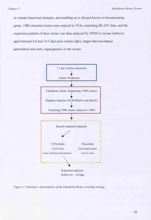
3.1.1 The endoderm library screen
Here I describe a screening strategy that uses the Beddington dissected mouse endoderm cDNA library to identity genes that are involved in early patterning events, and takes advantage of sequencing analysis and database searches. The endoderm library contains a number of tissues that have been implicated in establishing the body axes, namely the node (and its derivatives e.g. axial
mesendoderm), the definitive endoderm and the visceral endoderm (reviewed by Beddington and Robertson 1998, Martinez-Barbara and Beddington 2001). The strategy (see figure 1) involves using sequence data to pre-screen the endoderm library prior to expression analysis by WISH. A large-scale sequencing program, undertaken in collaboration with the Max Planck Institute for Molecular Genetics, produced 7000 sequences, which were then compared to sequence databases using the NCBI BlastN program, which compares nucleotide sequences to the database, and BlastX program, which translates the sequence through 6 frames and compares it to protein databases. The results from the database searches were then put through clustal analysis. Clustering grouped together any identical sequences and reduced the number of clones that were to be analysed further from 7000 to 1980. The 1980 independent sequences were then examined by scanning through the BLAST data for each clone, allowing selection of potentially interesting sequences, that may be novel
Chapter 3 Endoderm Library Screen
or contain functional domains, and enabling us to discard known or housekeeping genes. 1980 clustered clones were reduced to 74 by examining BLAST data, and the expression pattern of these clones was then analysed by WISH in mouse embryos aged between 6.0 and 10.5 days post coitum (dpc), stages that encompass
gastrulation and early organogenesis in the mouse.
7.5 dpc Em bryo dissection
I
Library Production
Endoderm Library Sequencing (7000 clones)
I
Database Searches (NCBI BlastX and BlastN)
I
Clustering (7000 clones reduced to 1980)
Results exam ined manually
Discarded: 74 Priorities:
N ovel C lones H ousekeep ing genes
Known G enes Clones with functional dom ains
Expression analysis
WISH 6 . 0 - lO.Sdpc
Chapter 3 Endoderm Library Screen
3.2 Results
3.2.1 Comparison of sequence from the endoderm library to sequence databases
In order to identify novel genes that are expressed specifically in tissues implicated in early axes formation, a screen was devised that used both sequence and expression analysis to analyse the Beddington dissected endoderm cDNA library. Initially, the gridded libraiy was sequenced, BLAST searched and clustered. The resulting 1980 clustered sequences were then analysed manually and clones were chosen according to their homology, or indeed lack of homology, to known sequences in the database. From the 1980 clustered sequences, 74 were prioritised for further study and fell into the catergories shown in figure 3.2. The majority of clones chosen represented novel genes that were not homologous to any sequences in the databases.
3.2.2 Whole mount RNA in situ hybridisation of prioritised clones
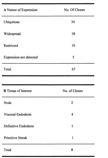
Whole mount RNA in situ hybridisation was performed on all 74 clones that were prioritised from the sequence analysis. Mouse embryos were dissected at 6.5, 7.5, 8.5 and 9.5dpc, stages which were chosen to encompass early pattern formation and the initial stages of organogenesis. Figure 3.3 A shows the nature of expression of the 74 mRNAs representing clones chosen from the sequence screen. Figure 3.3B shows the number of mRNAs that were restricted to tissues that are of particular interest to this study. The data shows that although around half of the mRNAs are ubiquitously expressed at the stages chosen, 12% (8/67) of the clones are restricted to the tissues
Chapter 3 Endoderm Library^ Screen
that are of particular interest here. Figure 3.4 shows the expression pattern of all of the restricted mRNAs in this study.
Catergory /
No. of Clones
Novel 33
Implicated in Human disorders 8
Transcriptional Regulators 5
Chromatin Structure 2
Inter or Intra Cell Signalling 6
Metabolism 5
Imprinted Genes 1
Splicing Regulators/RNA Processing 3
Cell Cycle Control 2
DNA Repair 2
Cytoskeleton 1
Clone Discarded (No growth/transcription) 5
Total 73
Chapter 3 Endoderm Library Screen
A Nature of Expression No. Of Clones
Ubiquitous 34
Widespread 18
Restricted 10
Expression not detected 5
Total 67
B Tissue of Interest No. of Clones
Node 2
Visceral Endoderm 4
Definitive Endoderm 1
Primitive Streak 1
Total 8
Figure 3.3 Table A shows the nature o f expression o f all o f the mRNAs investigated and Table B shows the number o f mRNAs whose expression was restricted to the tissues o f interest. Ubiquitous — expression detected in all tissues o f the embryo, Widespread — Expression detected in a number o f tissues o f the embryo, R estricted- Expression detected in only a small number o f specific tissues o f the embryo.
Chapter 3 Endoderm Library^ Screen
Figure 3.4. Whole mount in situ hybridisation analysis o f clones representing genes with restricted expression patterns isolated during the endoderm library screen (excluding C53, see following chapters). Early embryos (pre-turning) are all lateral \iews with anterior to the left. Later embryos are also lateral views with anterior to the top and dorsal to the right. Arrowheads point to areas o f restricted expression. Clones 23 and 3 are restricted to the ecoderm (extra-embryonic and embryonic respectively) at early stages. Clones 14 and 15 are restricted to the visceral
Expression Patterns of 9 Restricted mRNAs
e.Odpc 7.0dpc S.Odpc 9.0dpc
Clone 23
Ï
%
eoisc
Clone 3
Clone 14
*
#
$
Clone 15
#
»
Clone 17
Clone 29
Clone 42
Clone 57
Clone 33
h
&
Chapter 3 Endoderm Library’ Screen
3.3 Discussion
3.3.1 The endoderm library screen for novel genes implicated in early patterning.
Both embryonic and extra-embryonic endoderm are intimately involved in induction and patterning of the mouse embryonic axes. It has long been known that the node and its derivatives are capable of inducing anterior character (Beddington, 1984). Therefore, identification of novel genes expressed specifically in regions
encompassing the node would be particularly informative for the study of axes formation. Furthermore, the anterior VE is required for the proper specification of anterior structures (Beddington, 1998) and the posterior VE also takes part in inductive interactions with underlying tissues (Belaoussoff, 1998). However,
although a number of genes have been identified that are expressed specifically in the anterior VE, the identification of genes expressed specifically in the posterior VE has proved difficult. Therefore, genes whose expression is restricted asymmetrically in the VE would be of particular interest for the study of the formation of the A-P axis, and in particular, to understand the events that induce and pattern the posterior.
Chapter 3 Endoderm Library Screen
sequences can be generated and compared allowed us to take the library as a whole and enrich for novel or functional proteins essentially electronically, precluding the need for processes such as subtractive hybridisation.
Carrying out sequencing followed by database searches and clustering is novel in screening strategies of this type. Similar strategies to the one described here have used random expression analysis of libraries either in their entirety (Neidhardt et al,
2000; Gawantka et al, 1998) or after subtraction (Harrison et al, 1995; Neidhardt et al, 2000; Christiansen et al, 2001). The number of genes that were isolated with restricted expression patterns (the ultimate goal of the screens) was relatively high in our investigation (12%) compared to the other screens (Neidhardt - 8%, Christiansen - 8%, Gawantka - 25%[included ‘widespread’ genes]). The most direct comparison
can be made between the screen described here and that carried out by Nierdhart and colleagues. Nierdhart et al carried out random expression screening on a mouse 9.5dpc embryonic cDNA library in its original, subtracted and normalised forms, and obtained restricted expression in 5.7%, 7% and 17.8% of the clones investigated respectively. This indicates that subtraction and normalisation increases the chances of obtaining genes that display restricted expression, and the numbers compare favourably to our investigation. However, a large percentage of the genes in the Nierdhart screen (25%) were already characterised. In carrying out the sequencing analysis prior to screening, we were able to discard any clones that were already characterised, aswell as discarding housekeeping genes, a major advantage over other strategies.
Chapter 3 Endoderm Library Screen
3.3.2 Expression of ‘restricted’ mRNAs from this study.
From the 74 cDNA clones that were chosen for expression analysis, 10 represented mRNAs that were expressed specifically in the tissues of interest to this study, indicating the screening process was effective in fulfilling its aim. The expression patterns of 9 of the restricted mRNAs can be seen in figure 3.4. cDNA clones 23 and 3 represent mRNAs that are expressed specifically in the ectoderm, extra-embryonic and embryonic respectively. It is now clear that the ectoderm plays important roles in pattern formation, with the extra-embryonic ectoderm imparting proximal-posterior identity to the adjacent proximal epiblast prior to the initiation of gastrulation (Lu et al, 2001). cDNA clones 14, 15 and 17 represent mRNAs that are expressed
specifically in the VE, vyith 17 being expressed specifically in the AYE at early stages, a tissue integral in anterior pattern formation (Beddington and Robertson, 1999). cDNA clone 29 represents an mRNA that is specific to the primitive streak, and cDNA clones 42 and 57 represent mRNAs that are node specific, tissues fundemental for early axes formation (Beddington and Robertson, 1999). Finally cDNA clone 33 represents an mRNA that is expressed specifically in the definitive endoderm.
It was clear at the start of the investigation that the endoderm specific cDNA library was of good quality because mRNAs expressed in tissues implicated in axes
Chapter 3 Endoderm Library Screen
Stage of the screen (sequence and database analysis) indicating our strategy was effective in identifying genes of interest. The identification of the genes described here indicates that the strategy used is extremely effective in identifying novel genes expressed in the tissues of interest, and fulfilled many of the aims of this study. However, no novel genes were identified that were expressed asymmetrically in the VE, but mRNAs that did seem to be identified at high frequency were those
expressed in the node. The node was included in the tissue from which the endoderm library was constructed, and it seems the repertoire of genes it expresses is well represented within the endoderm library and may be a good resource with which to identify node specific mRNAs.
Once the screen was complete a number of clones were looked at in more detail, in order to narrow down the investigation. It was decided, from supplemental expression-analysis and additional sequencing data from a number of clones, that C53 would be the most interesting clone with which to continue the investigation. C53 is expressed in the VE, the node and hematopoietic tissues and its sequence is novel. Subsequent chapters deal with the analysis of C53.
Chapter 4
Chapter 4 cDNA, bioinformatics, expression
Chapter 4: € 53 cDNA cloning, bioinformatics and
expression analysis
4.1 Introduction
4.1.1 Full length cDNA cloning by rapid amplification of cDNA ends (RACE) PGR
Cloning of the full-length cDNA for C53 was carried out using 5’ rapid amplification of cDNA ends (RACE). The template for the RACE reaction is SMART (Switching Mechanism At 5’ end of RNA Transcript) race ready cDNA (Clontech) which has been prepared from adult mouse kidney and lung (tissues identified by northern blot to contain high levels of C53 mRNA expression, data not shown). The SMART technology is an efficient mechanism for generating full-length cDNAs in reverse transcription reactions (Chenchik et ai, 1998). When reverse transcriptase (RT) reaches the 5’ end of the mRNA, the enzymes terminal transferase activity adds a few deoxycytidine residues to the 3’ end of the first strand cDNA. The SMART oligo can then anneal to dC residues and serve as an extended template for RT, which the enzyme switches to. Since the dC tailing activity is most efficient if the enzyme has reached the end of the RNA template, the SMART sequence is only typically added to complete first strand cDNAs. Following reverse transcription, the first strand cDNA can be used directly in 5’ RACE reactions, preventing the need for time consuming second strand synthesis and amplification steps.
Chapter 4 cDNA, bioinformatics, expression
The C53 clone that was isolated from the Beddington Dissected Endoderm library was used to design gene specific primers just downstream of a convenient restriction enzyme site. The RACE reaction was carried out in two phases using nested primers and the resulting PGR product was cloned next to the library fragment.
4.1.2 The use of bioinformatics to assign function to unknown proteins
Chapter 4 cDNA, bioinformatics, expression
describe potential structural elements of the protein that may give valuable clues about its function.
One of the most informative tools on the web is SWISS-PROT, which can be found on the ExPASy Molecular Biology Server (http://www.expasv.ch/V SWISS-PROT is a protein sequence database that aims to provide a high level of annotations (such as the description of the function of a protein, its domains structure, post-translational modifications, variants etc), a minimal level of redundancy and high level of integration with other databases. TrEMBL is a computer-annotated supplement of SWISS-PROT that contains all of the translations of EMBL nucleotide sequence entries not yet integrated into SWISS-PROT. Its links with other database sources, such as Pfam (a protein family database from the Sanger Center
http://W W W .sanger.ac.uk/Software/Pfam/ (Bateman A et ai, 2000), means any
potential domains and protein family members can be easily identified. The most informative database entry for C53 (the C53 human ortholog, protein Accession No. NP_071437) indicated that it may be an integral membrane protein. This hint about potential protein structure can be investigated further using the programs SignalP, TMpred and TMHMM
SignalP (http://www.cbs.dtu.dk/servics/SignalP) is a world-wide-web based signal peptide prediction server that can predict the presence or absence of signal peptides in the first 50-70 amino acids of the protein in question. Signal peptides control the entry of virtually all proteins to the secretory pathway, both in eukaryotes and prokaryotes (Gierasch, 1989), and comprise the N terminal part of the amino acid
Chapter 4 cDNA, bioinformatics, expression
chain, which is cleaved off when the protein is translocated through the membrane. A strong interest in the automated identification of signal peptides (mainly due to the huge amount of unprocessed data available and the industrial need to find more effective vehicles for the production of proteins in recombinant systems) has led to a neural network approach to the identification of signal peptides and their cleavage sites. In essence, the SignalP server will return three scores between 0 and 1 for each position in the first 50-70 amino acids in the sequence. The C-score (raw cleavage site score) is the output from networks trained to recognise cleavage sites vs. other sequence positions, and are trained to be high at position +1 (immediately after the cleavage site) and low at all other positions. The S-score (signal peptide score) is the output from networks trained to recognise signal peptide vs. non signal peptide positions, and are trained to be high at all positions before the cleavage site and low