Dermoscopic Image Classification Using Image
Processing Technique For Melanoma Detection
Isha Patel
1, Sanskruti Patel
2, Atul Patel
3Faculty of Computer Science and Applications, Charotar University of Science and Technology, Changa,Gujarat,India.
1
Ph.D. Research Scholar, 2Assistant Professor, 3Dean & Professor
Email: [email protected], [email protected], [email protected]
Abstract- In recent days, melanoma cancer is seen as one of the most dangerous forms of the cancers found in people. Skin cancer is found in various types such as basal, squamous cell carcinoma among which melanoma skin cancer is the most unpredictable. The detection of the beginning stage of melanoma cancer can be helpful to cure it. Digital image processing can play important role in medical image diagnosis and also proved by many existing systems. In this paper, we presented a digital image processing method for the detection of melanoma Skin cancer and classify by machine learning techniques. The input to the method is the skin lesion image and then by applying image processing and classification techniques, it analyses it to conclude about the presence of skin cancer. The skin cancer image analysis techniques check for the various melanoma features like color, border, texture, and shape analysis for segmentation and feature extraction stage. The extracted features are used to classify the image as no cancer, beginning melanoma cancer, and highly melanoma cancer.
Index Terms-Melanoma Skin Cancer, Image Preprocessing, Segmentation, Feature Extraction, Classification.
1. INTRODUCTION
The malignant melanoma is caused due to ultraviolet radiation and damages the DNA indirectly. The reactive oxygen species and free radical are the two main causes of DNA damage. Melanoma is poisonous cancer and it can be cured at the beginning stages of cancer. The classification of skin cancer melanoma using images is a challenging task in the appearance of skin cancer [20]. Therefore, to overcome all these issues, digital image processing is applied for detecting melanoma skin cancer [2, and 4]. The techniques work on the image so there is no physical contact with skin, so this is non-invasive. The first stage is image preprocessing of the image of skin cancer which is followed by image segmentation after it is followed by feature extraction. The extracted features are used for classification the image as no cancer, beginning cancer and melanoma cancer [10, 17, and 21].
Paper is prepared as follows; section II presents related work on different classification techniques used for skin cancer detection. Section III includes the methodology and architecture flow diagram of Skin Cancer Disease. Section IV presents the image processing methods and classification techniques and its description. Section V provides the result analysis of melanoma cancer detection. Section VI contains a conclusion of the paper.
2. RELATED WORK
Suleiman Mustafa et. al. [3] proposed a computerized framework for recognizing melanoma skin cancer
from plain photos of influenced skin locales. Wilson F. Cueva et. al. [22] proposed a framework for the detection of melanoma by getting Asymmetry, Border,
while delivering the division result and the coarse characterization result.
Yading Yuan et. al. in [40], introduced a completely programmed strategy for skin lesion division by ideal use of a prepared 19-layer deep CNNs which doesn’t depends on earlier learning of the information. Here in [41] Supriya Joseph et. al., proposed a non-intrusive robotized skin lesion investigation framework for the beginning of melanoma utilizing digital image processing procedures and portable innovations. The test result is assessed on PH2 database. Yu-A Chung et. al. [42], proposed a deep Siamese CNN (SCNN) design that can be prepared with just parallel image match data to learn image portrayals with less supervision included. The aftereffects of the test demonstrate that creator's framework i.e. deep SCNN is practically identical to the cutting edge single administered CNN, and requires significantly less supervision for preparing. Haofu Liao [43], exhibited an examination on practicality of developing a general skin sickness finding framework utilizing CNN. Assessment aftereffects of framework demonstrate that, proposed system can accomplish as high as 73.1% Top-1 exactness and 91.0% Top-5 precision when testing on the Dermnet dataset. For the test on the OLE dataset, Top-1 and Top-5 correct nesses are 31.1% and 69.5%. N. C. F. Codella et. al. [44] The deep learning with set up machine learning approaches assessed utilizing the biggest freely accessible benchmark dataset of dermoscopic images. New cutting edge execution levels are illustrated, prompting a change in the territory under recipient working trademark bend of 7.5%, in normal exactness of 4%, and estimated 95%.
3. PROPOSED METHODOLOGY FOR
IDENTIFICATION AND DETECTION
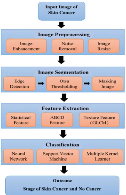
The methodology used for skin cancer melanoma detection using digital image processing is as available in following figure. The skin dermoscopic image is input first in raw form on which classification is applied to identify whether it is supposed to be a beginning cancer or melanoma cancer or no cancer. Then image preprocessing techniques are applied to increase the image quality. The edge detection techniques and Otsu thresholding method are used for segmentation of the input image. The image is then after made available to perform the feature extraction. The features considered for an experiment are color, size, statistical, abcd and texture. The statistical, abcd and texture features are the important features of skin cancer. After that extracted features are additionally
passed into an algorithm for classification which classifies beginning of cancer, skin cancer melanoma or normal by paralleling its feature value [2].
Fig. 1. Working of Proposed Methodology.
3.1. Image Pre-processing
The image given to the algorithm can be obtained by any camera such as a mobile or lighting condition. Therefore, it requires to be preprocessed. Here, the image preprocessing includes contrast adjustment and image denoising, image resizing [6, and 7]. The dermoscopic insert image is taken and the image is resized with 200X200 subsequently applied. In our work, the median filter is applied to eliminate the noise in the skin images [19].
3.2. Image Segmentation
proposed by Otsu applied [35]. Binary image for each plane is obtained and finally it produced for masking of cancer image [27]. We used the masking technique to increase the accuracy of the segmented image. The main extracting foreground form background is, cancer extracting from the skin [16, and 32].
3.3. Feature Extraction
Feature extraction is the way to characterize the arrangement of features which will be most effectively worked. The key features of skin cancer are statistical, texture and ABCD feature [5, and 6]. Hence, we extract the color, size, statistical, abcd and texture features from a segmented image of skin cancer [14, and 33]. Statistical features including mean, standard deviation, autocorrelation, median, and mode are extracted from the image [7]. Texture feature extracted as GLCM features from the curvelet domain and this feature includes contrast, correlation, energy, entropy, homogeneity [13]. The ABCD feature identifies Asymmetry (colour/structure), Border, Colour, and Diameter [11, and 28].
3.4. Classification
Classification is an essential stage for distinguishing skin cancer composes. Using image processing for skin cancer melanoma, implemented some available methods for image classification. There are two vital stages in the arrangement. They are preparing a training and testing stage. In the preparing stage, the pre-decided information and its related class names are utilized for classification [8]. In the classification step, it classifies images as beginning cancer or melanoma cancer or no cancer. In this work, many types of classification techniques are applied for detecting the skin cancer images and their performance are lastly compared [1, 31, and 34].
3.4.1. Using Neural Network
It is the supervised learning algorithm. In this work, the calculation is decayed in the accompanying four steps: Initialization of weights, after that feed-forward calculation, then forward propagation of errors, and at last biases and weights are updating [9]. In this work different features like color, size, texture and statistical feature are given to network. This algorithm gives 80.43% accuracy [24].
3.4.2. Using SVM
SVM is a kernel approach for constructing machine learning that minimizes the generalization errors. SVM constructed by a set of the hyperplane that
separate classes [15]. The SVM decision boundary is the line separating region containing them. Different features are applied to support vector machine [12, 29, and 30]. This algorithm gives 86.17% accuracy.
3.4.3. Using Multiple Kernel Learning
It is also a supervised learning method which refers to various ML techniques that uses multiple sets of kernels. MKL include (i) the capability to choice for parameters and an optimal kernel from multiple sets of kernels. (ii) Combining dataset from different bases that have different concepts of similarity. This algorithm gives 90.3% accuracy.
4. RESULT ANALYSIS AND DISCUSSION
4.1. Dataset
A dataset of Skin Cancer images is collected from the International Skin Cancer Images: Melanoma Project ISIC https://isic-archive.com. And another dataset of melanoma images is collected from the DermIS (http://www.dermis.net) and DermQuest (http://www.dermquest.com) database [18]. The dataset contains 1150 dermoscopic images of skin which is classified into three classes: beginning cancer or melanoma cancer or no cancer.
4.2. Result Analysis
The implementation work is carried out using image processing toolkit - MATLAB R2018a. Performance analysis helps to identify most efficient classification technique used for skin cancer image by analyzing parameters values. Performance analysis carried out based on application of three different classification methods, with consideration of accuracy, precision, f-measure, and recall [25] parameters. Table 1 shows the detail of cancer image test set experiment with its accuracy, precision, f-measure, and recall [23].
Fig. 2. Accuracy of correctly and incorrectly
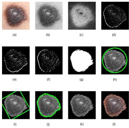
First, the image of skin cancer is input and then implemented preprocessing techniques. Then after, edge detection techniques and thresholding and masking method are applied to the image. It is presented in the following figures.
Fig. 3. No Cancer Image Segmentation (a) Original image (b) Remove noise (c) Grayscale image (d, e, f) Edge detection method (g) Otsu thresholding (h, i, j) Masking (k) Final grayscale image (l) Final color image
Fig. 4. Beginning Stage Cancer Image Segmentation (a) Original image (b) Remove noise (c) Grayscale image (d, e, f) Edge detection method (g) Otsu thresholding (h, i, j) Masking (k) Final grayscale image (l) Final color image
Fig. 5. Melanoma Cancer Image Segmentation (a) Original image (b) Remove noise (c) Grayscale image (d, e, f) Edge detection method (g) Otsu thresholding (h, i, j) Masking (k) Final grayscale image (l) Final color image
normal skin to melanoma cancer using the ABCD feature extraction.
Fig. 6. Melanoma Cancer Feature Extraction (a) Original image (b) Extract melanoma cancer (c) ABCD feature extraction
Third, the different classification techniques are implemented applied to the segmented image to classify the image. As shown in the flowing figure, the image of the skin is efficiently classifying for No cancer image, beginning cancer image and Melanoma cancer image using the classification method.
Fig. 7. Outcome of No Cancer Detection
Fig. 8. Outcome of Beginning Cancer Detection
Fig. 9. Outcome of Highly Melanoma Cancer Detection
5. CONCLUSION
This paper discusses an approach of identifying and classifying skin dermoscopic image using image
processing and classification techniques. An image is preprocessed, segmented and selected features are extracted. Different classification techniques are applied on these extracted features and their performance is measured by several parameters. Based on the experiment, it can be concluded that the MKL (multiple kernel learner) classification techniques are one of the efficient method used to diagnose the melanoma skin cancer more accurately. This work is more helpful for the research scholars where the medical field may not be available. Since the techniques are more robust and user-friendly for any conditions images, it can serve the purpose of automatic diagnostics of the Melanoma Skin Cancer. In future work, plan to explore different types of skin lesion images to better assess our lesion classification model. This will be done by considering other datasets or using images from the Internet. It would also be interesting to investigate other training algorithms for classification, and it may be useful to perform skin detection, especially when handling the varying skin
colors of people of different ethnicities. (A.1)
REFERENCES
[1] Navid Razmjooy, Fatima Rashid Sheykhahmad, Noradin Ghadimi, “A hybrid neural network world cup optimization algorithm for melanoma detection”, Navid Razmjooy et al., published by De Gruyter, September 26, 2017, pp. 9-16. [2] Shivangi Jaina, Vandana jagtap, Nitin Pise,
“Computer aided Melanoma skin cancer detection using Image Processing”, International Conference on Intelligent Computing, Communication & Convergence (ICCC-2015), Published by Elsevier, pp. 735 – 740.
[3] Suleiman Mustafa, Akio Kimura, "A SVM-based diagnosis of melanoma using only useful image features", 2018 International Workshop on Advanced Image Technology (IWAIT), IEEE 2018.
[4] S.S. Mane, S.V. Shinde, “Different Techniques for Skin Cancer Detection Using Dermoscopy Images”, International Journal of Computer Sciences and Engineering, Vol.5 (12), Dec 2017, pp. 159-163.
[5] Rosline Flex.G, “Skin Cancer Detection Using Air – PC with Inbuilt MATLAB”, International Journal of Innovative Research in Computer and Communication Engineering, Vol. 6, Issue 3, March 2018, pp. 2401-2405.
[6] M. Chaithanya Krishna, S. Ranganayakulu, Dr. P. Venkatesan,”Skin Cancer Detection and Feature Extraction through Clustering Technique”, International Journal of Innovative Research in Computer and Communication Engineering, Vol. 4, Issue 3, March 2016, pp. 3736-3742.
(IRJET), Volume: 04 Issue: 04, Apr -2017, pp. 2875-2881.
[8] Nabin K. Mishra and M. Emre Celebi, “An Overview of Melanoma Detection in Dermoscopy Images Using Image Processing and Machine Learning”, January 27, 2016, pp. 1-15.
[9] Ekta Singhal, Shamik Tiwari, “Skin Cancer Detection using Artificial Neural Network”, International Journal of Advanced Research in Computer Science, 6 (1), Jan–Feb, 2015, pp. 149-157.
[10]Ammara Masood and Adel Ali Al-Jumaily, “Computer Aided Diagnostic Support System for Skin Cancer: A Review of Techniques and Algorithms”, Hindawi Publishing Corporation, International Journal of Biomedical Imaging, Volume 2013, Article ID 323268, 22 pages. [11]Suneel Kumar, Ajit Singh, “Image Processing for
Recognition of Skin Diseases”, International Journal of Computer Applications (0975 – 8887) Volume 149 – No.3, September 2016, pp. 37-40. [12]Diwakar Gautam, Mushtaq Ahmed, “Melanoma
Detection and Classification Using SVM Based Decision Support System”, 2015 IEEE, pp. 1-6. [13]Li-sheng Wei, Quan Gan, and Tao Ji, “Skin
Disease Recognition Method Based on Image Color and Texture Features”, Hindawi, Computational and Mathematical Methods in Medicine, Volume 2018, Article ID 8145713, 10 pages.
[14]Chandrahasa M, Varun Vadigeri and Dixit Salecha,” Detection of Skin Cancer Using Image Processing Techniques”, International Journal of Modern Trends in Engineering and Research (IJMTER), Volume 03, Issue 05, May– 2016, pp. 111-114.
[15]Samy Bakheet, “An SVM Framework for Malignant Melanoma Detection Based on
Optimized HOG Features”,
www.mdpi.com/journal/computation, 2017, pp. 1-13.
[16]Esperanza Guerra-Rosas and Josué Álvarez-Borrego, “Methodology for diagnosing of skin cancer on images of dermatologic spots by spectral analysis”, BIOMEDICAL OPTICS EXPRESS 3876, Oct 2015, Vol. 6, No. 10. [17]Heydy Castillejos-Fernández, Omar
López-Ortega, Félix Castro-Espinoza and Volodymyr Ponomaryov, “An Intelligent System for the Diagnosis of Skin Cancer on Digital Images taken with Dermoscopy”, Vol. 14, No. 3, 2017, pp. 169-185.
[18]Skin Cancer Detection, VISION AND IMAGE PROCESSING LAB, https://uwaterloo.ca/vision- image-processing-lab/research-demos/skin-cancer-detection
[19]Sanjay Jaiswar, Mehran Kadri, Vaishali Gatty, “Skin Cancer Detection Using Digital Image Processing”, International Journal of Scientific Engineering and Research (IJSER), Volume 3 Issue 6, June 2015, pp. 138-140.
[20]Sheeju Diana B, Ramamurthy B, “Skin cancer detection and prediction using image processing techniques”, International Journal of Engineering & Technology, 7, 2018, pp. 204-209.
[21]Vedanti Chintawar and Jignyasa Sanghavi, “A Review on Computer-Aided Melanoma Skin Cancer Detection using Image Processing”, EasyChair Preprint, 584, October 24, 2018. [22]Wilson F. Cueva, F. Muñoz, G. Vásquez., G.
Delgado, "Detection of skin cancer melanoma‖ through Computer Vision", 2017 IEEE XXIV International Conference on Electronics, Electrical Engineering and Computing (INTERCON), IEEE 2017.
[23]VS. Sabeera, P. Vamsi Krishna, “Early Detection of Melanoma Skin Cancer Using Classifiers”, International Advanced Research Journal in Science, Engineering and Technology, Vol. 3, Issue 8, August 2016, pp. 191-196.
[24]Aswin. R.B, J. Abdul Jaleel, Sibi Salim, “Implementation of ANN Classifier using MATLAB for Skin Cancer Detection”, International Journal of Computer Science and Mobile Computing, ICMIC13, December 2013, pp. 87-94.
[25]Nidhal K. EL Abbadi and Zahraa Faisal, “Detection and Analysis of Skin Cancer from Skin Lesions”, International Journal of Applied Engineering Research, Volume 12, Number 19 (2017) pp. 9046-9052.
[26]Farzam Kharaji Nezhadian, Saeid Rashidi,"Melanoma skin cancer detection using color and new texture features",2017 Artificial Intelligence and Signal Processing (AISP), IEEE 2017.
[27]Neenu Paliwal, “Skin Cancer Segmentation, Detection and Classification Using Hybrid Image Processing Technique”, International Journal of Engineering and Applied Sciences (IJEAS) ISSN: 2394-3661, Volume 3, Issue 4, April 2016, pp. 71-73.
[28]Dr. S.Gopinathan, S. Nancy Arokia Rani, “The Melanoma Skin Cancer Detection and Feature Extraction through Image Processing Techniques”, International Journal of Emerging Trends & Technology in Computer Science (IJETTCS), Volume 5, Issue 4, July - August 2016, pp. 106-112.
[29]Poornima M S, Dr. Shailaja K, “Detection of Skin Cancer Using SVM”, International Research Journal of Engineering and Technology (IRJET), Volume 4, Issue 7, July - 2017, pp. 3021-3024. [30]Hutokshi Sui, Manisha Samala, Divya Gupta,
Neha Kudu, “Texture Feature Extraction for Classification of Melanoma”, International Research Journal of Engineering and Technology (IRJET), Volume: 05 Issue: 03, Mar-2018, pp. 1026-1029.
Journal of Engineering and Technology (IRJET), Volume: 05 Issue: 09, Sep 2018, pp. 613-617. [32]Meskini E., Helfroush M. S., Kazemi K.,
Sepaskhah M, “A New Algorithm for Skin Lesion Border Detection in Dermoscopy Images”, Journal Biomed Phys Eng 2018, pp. 117-126. [33]Catarina Barata, Margarida Ruela, Mariana
Francisco, Teresa Mendonc, Jorge S. Marques, “Two Systems for the Detection of Melanomas in Dermoscopy Images using Texture and Color Features”, IEEE, July 2013, pp. 1-13.
[34]P. Mohamed Sajid, Dr.A. Rajesh, “Performance Evaluation of classifiers for automatic Early Detection of skin cancer”, Journal of Adv Research in Dynamical & Control Systems, Vol. 10, 07-Special Issue, 2018, pp. 254-261.
[35]Reshu Bansal, Meenu Saini, “A Method for Automatic Skin Cancer Detection”, International Journal of Advanced Research in Computer Science and Software Engineering 5(9), September- 2015, pp. 529-533.
[36]Maglogiannis and C. N Doukas, "Overview of Advanced computer visions systems for skin lesions characterization", IEEE transactions of Information technology in Biomedicine, vol. 13,
no. 5, 2009.
https://doi.org/10.1109/TITB.2009.2017529. [37]Kulkarni and A. Panditrao, "Classification of
Lung cancer stages on CT scan images using image processing", IEEE International conference on Advanced communication control and computing techniques (ICACCCT), 2014. https://doi.org/10.1109/ICACCCT.2014.7019327. [38]Uzma Bano Ansari,Tanuja Sarode,"Skin Cancer
Detection Using Image Processing", International Research Journal of Engineering and Technology (IRJET), Volume: 04,Issue: 04, Apr-2017. [39]Yuexiang Li, Linlin Shen,"Skin Lesion Analysis
towards Melanoma Detection Using Deep Learning Network", p-1-16 Sensors 2018.
[40]Yading Yuan, Ming Chao, Yeh-Chi Lo, "Automatic Skin Lesion Segmentation Using Deep Fully Convolutional Networks with Jaccard Distance", IEEE Transactions on Medical Imaging, Volume: 36, Issue: 9, Sept. 2017, IEEE 2017.
[41]Supriya Joseph, Janu R Panicker,"Skin Lesion Analysis System for Melanoma Detection with an Effective Hair Segmentation Method", IEEE International Conference on Information Science (ICIS), IEEE Aug-2016.
[42]Yu-An Chung, Wei-Hung Weng, "Learning Deep Representations of Medical Images using Siamese CNNs with Application to Content-Based Image Retrieval",31st Conference on Neural Information Processing Systems (NIPS 2017).
[43]Haofu Liao,"A Deep Learning Approach to Universal Skin Disease Classification", Graduate Problem Seminar - Project Report, University of Rochester, 2015.
[44]N. C. F. Codella, Q.B. Nguyen, S. Pankanti, D. A. Gutman, B. Helba, A. C. Halpern, J. R.