Selective Inhibitory Effect of Adenosine A
1Receptor Agonists on
the Proliferation of Human Tumor Cell Lines
Hossein Hosseinzadeh
1, Mahmoud R. Jafari
1 & 2and Jamal Shamsara
*11
Pharmaceutical Research Center and 2Biotechnology Research Center, School of Pharmacy, Mashhad University of Medical Sciences, Mashhad, Iran
Received 3 September 2007; revised 19 April 2008; accepted 23 April 2008
ABSTRACT
Background: In this study, the effects of three structural analogues of adenosine upon proliferation of human tumor cells were investigated. Previous research showed a cytotoxic effect of adenosine via A3 receptor and A1 receptor and sometimes this effect was receptor independent. The researches showed a differential cytotoxic effect of adenosine and its A3 agonists on cancerous cells, while other studies demonstrated tumor promoting effect of adenosine and its A1 agonists. The purpose of the present study was the evaluation of the possible selective anti-tumor effect of A1 receptor agonists on cancerous cells. Methods: The substances of N6-cyclohexyl-adenosine (CHA, A1 agonist), R-isomer of N
6
-phenylisopropyladenosine (R-PIA, A1 agonist) and N5-ethylcarboxamido-adenosine (NECA, adenosine A1-A2 non-specific agonist) were tested for their anti-proliferative effect using 3-(4,5-dimethylthiazol-2-yl)-2,5-diphenyltetrazolium bromide (MTT) assay method. Hep G2, Hep2, CACO2, ACHN and L929 cell lines were used in this assay. Results: CHA inhibited cell proliferation in three cell lines (in concentration of 5-50 µM) and R-isomer of R-PIA in one cell line (in concentration of 10-50 µM). These effects were inhibited partially by addition of 1,3-Dipropyl-8-cyclopentylxanthine (A1 antagonist). The NECA analogue had no inhibitory effect on the cell proliferations. All of the substances had no cytotoxic effect on L929 cells (mouse connective tissue fibroblast cell line). Conclusion: CHA and R-PIA had inhibitory effect on the proliferation of human tumor cell lines partially via A1 receptor, while they didn't show such effect on fibroblast cells. These results suggest that A1 adenosine receptor agonists have a good potential of specific anti-tumor activity. Iran. Biomed. J. 12 (4): 203-208, 2008
Keywords: Adenosine, A1 receptor, Cytotoxicity, Anti-tumor effect, N6-cyclohexyl-adenosine (CHA), N6-phenylisopropyladenosine (R-PIA)
INTRODUCTION
denosine acts via A1, A2 (A2A and A2B) and A3 receptors which all of these subclasses are G-coupled receptors. The A2A and A2B receptors preferably interactwith members of the Gs family of G proteins and the A1 and A3 receptors with Gi proteins. However, other G protein interactionshave also been described. They are not sensitive to nucleotides such as ATP, ADP and AMP. When adenosine binds to A1 or A3 receptors, the intracellular level of cAMP is decreased, however A2 receptor acts through increasing of cAMP concentration in cell [1]. Effect of adenosine on proliferation of cells is very controversial.
Adenosine has potent cytoprotective functions that have been extensively studied in the context of two major organs, heart and brain. Studies showed a protective effect of adenosine and its A3 agonists on normal cells [2, 3] and other reports suggested that adenosine and its A1 agonists have also growth-promoting functions. Adenosine accumulates in solid tumors at high concentrations, and has been shown to stimulate tumor growth and angiogenesis and to inhibit cytokine synthesis suggesting the tumor promoting effect of adenosine [4-6]. However, few reports showed that adenosine induced cell death [1, 7, 8]. Activation of A1 receptor inhibited proliferation of LoVo colon carcinoma, MOLt-4 leukemia and three breast
A
cancer cell lines (T47D, HS578T and MCF-7) [7]. Mechanism of this effect was investigated and results demonstrated that receptor were responsible for this cytotoxic effect and rarely the effect was receptor independent [8, 11, 12] especially in solid tumors, A3 [9, 10] and in few cell lines (breast, colon and leukemia), A1 [6, 11]. Therefore, adenosine-induced cell death may occur via both receptor-dependent and receptor-inreceptor-dependent mechanisms. But adenosine has failed to exert anti-cancer or chemoprotective effects when given orally or intraperitoneally in mice [6]. These several studies suggested that adenosine acts as a potent regulator of normal and tumor cell growth.
Previous studies [2, 3, 6, 9] mainly focused on A3 receptor and also suggested that adenosine cytotoxic effects depends on the extracellular concentration and expression of different adenosine receptor subtypes and on the signal transduction mechanisms activated following the binding of specific agonists. For instance, A1 receptor activation protected kidney from the cisplatin-induced nephrotoxicity, while other study showed completely opposite results with the same cells [6]. Therefore, in this study we simultaneously evaluated the effect of adenosine A1 agonists on the proliferation of cancer cell lines, especially on cells which have medium level expression of A1 receptor, and also normal cells at the same condition.
MATERIALS AND METHODS
Materials. CHA (N6-cyclohexyl-adenosine),
R-PIA (R-isomer of N6-phenylisopropyladenosine), NECA (N5-ethylcarboxamido-adenosine), DPCPX (1,3-Dipropyl-8-cyclopentylxanthine), amphotericin B vials and 3-(4,5-dimethylthiazol-2-yl)-2,5-diphenyltetrazoliumbromide (MTT) were purchased from sigma-Aldrich company (Germany). EMEM (minimum essential medium with Earl's), trypsin powder and FCS were from Gibco (England), penicillin and streptomycin vials from Jaberebn Haiian (Iran), methotrexate ampule was from Ebewe (Austria). NaHCO3, NaCl, EDTA, KCl, DMSO and glycine were purchased from Merck (Germany).
Cell lines and culture conditions. The following
cell lines (from Pasteur Institute of Iran, Tehran) were used in this study: CACO2, human colon adenocarcinoma cell line; Hep G2, human Caucasian hepatocytes carcinoma cell line; ACHN, human renal adenocarcinoma cell line; Hep2, human
Caucasian larynx carcinoma cell line and L929, mouse connective tissue fibroblast cell line [13]. The cells were maintained in EMEM medium supplemented with 10% FCS, penicillin 100 u/mL, streptomycin 100 µ/mL and amphotericin B 100 microgram/mL, pH 7.4 at 37°C in a humidified atmosphere with 5% CO2 incubator.
Cell proliferation assay. Cell viability after
substance exposure was examined using the MTT assay. Metabolically, active mitochondrial dehydro-genases convert the tetrazolium salt MTT to insoluble purple formazan crystals, at a rate that is proportional to cell viability. The cultured cells were plated in 96-well microtitre plates at a concentration of 25 × 103 cells/ml for ACHN cells and 5 × 10³ cells/ml for other cells in a 200-µL volume. After overnight incubation, the medium was removed and the cells were incubated in the presence of 200 µL media supplemented with increasing concentrations of substances in 96-well plate for 72 h. At the end of incubation periods, 20 µL MTT solution (5 mg/ml in PBS) was added to each well for 4 h. Then, 200 µL of DMSO was added to each well to solubilize the formazan crystals. After the addition of 25 µL glycine buffer to each well, the OD was read at 570 nm using Dynatech MR 600 microplate reader (Dynatech Laboratories, Chantilly, VA, USA). Untreated negative controls were run together with the treated cells. Plates with reagent only were served as background controls. The results were expressed as OD after background subtraction [14].
Evaluation of A1 receptor antagonist on the
proliferation of tumor cell lines. To investigate the
mechanism responsible for A1-mediated inhibitory effect on cell proliferation, an A1 receptor antagonist was used. The cells were seeded into 96-well plates (5000 cells/well for ACHN cells and 1000 cells/well for other cells) and cultured for 24 h. Then, the medium was removed and replaced with fresh medium containing various concentrations of the compounds to be tested for another 72 h: agonists alone and agonists plus A1 antagonist (DPCPX). Cell viability was determined by MTT assay as explained before.
Statistics. One way ANOVA followed by Tukey
post test was used to determine significant differences between groups using Prism Software. Treatment-related differences were considered significant at P<0.05.
RESULTS
Effect of DMSO on cell viability. DMSO was
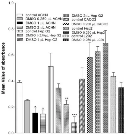
used as a co-solvent for adenosine agonists. We tested its toxicity effect on two cell lines. DMSO in concentration of 2 µL and 1 µL per well had clear cytotoxic effect on ACHN and Hep G2 cell lines but in 0.25 µL per well didn't show any significant difference with control in all cell lines used (Fig. 1). Therefore, we used DMSO in concentration of 0.25 µL per well as co-solvent in our experiment.
Fig. 1. Effect of DMSO on cell lines. Cells were cultured in 10% fetal calf serum medium and incubated without any treatment (control) and with different concentration of DMSO for 72 h then MTT added and absorbance read immediately after 4 h incubation in 570 nm. The significant difference between absorbance of wells containing DMSO and control are shown. N = 8, Mean ± SE, *P<0.05, **P<0.01 and ***P<0.001.
Effect of specific A1 agonists (with or without an
A1 antagonist) on cell viability. Results showed that
the addition of CHA was associated with decreased MTT turnover in three cells lines: Hep G2 (in concentration of 25-50 µM), CACO2 (in concentration of 5-50 µM) and ACHN (in concentration of 5-50 µM) (Fig. 2) following a 72-h exposure period. R-PIA was associated with decreased MTT turnover in one cell line: CACO2 (in concentration of 10-50 µM) (Fig. 3). DPCPX inhibited the cytotoxic effects of both CHA and R-PIA and there was no significant difference between absorbance of wells containing DPCPX plus A1 agonist and control well. Both of these agonists had no inhibitory effect on Hep2 and L929 cells (Fig. 4). R-PIA had no inhibitory effect on Hep G2, ACHN and Hep2 cells.
Fig. 2. Effect of CHA on ACHN (A) Hep G2 (B) and CACO2
(C) cell line. Cells were cultured in 10% fetal calf serum medium and incubated without any treatment (control), with 0.250 µL DMSO (control DMSO), with 0.250 µL DMSO and 0.1 µM DPCPX (control DMSO + DPCPX), with different concentration of CHA (A and B) and CHA or R-PIA (C) and finally with two concentration of CHA and 0.1 µM DPCPX for 72 h then MTT added and absorbance read immediately after 4 h incubation in 570 nm. The significant difference between absorbance of wells containing CHA and control DMSO are shown. N = 8, mean ± SE, *P<0.05, **P<0.01 and ***P<0.001.
0 . 0 0 0 . 0 5 0 . 1 0 0 . 1 5 0 . 2 0 0 . 2 5 0 . 3 0 0 . 3 5
* * * * * *
* * * * * *
M
e
an
V
a
lu
e
o
f ab
so
rb
an
ce
0 . 0 0 . 1 0 . 2 0 . 3 0 . 4 0 . 5 0 . 6 0 . 7
* *
M
ean
V
a
lu
e o
f ab
so
rb
an
ce
0 . 0 0 . 1 0 . 2 0 . 3 0 . 4 0 . 5 0 . 6 0 . 7
* *
* * * * * * * * *
M
e
an
V
a
lu
e o
f ab
so
rb
an
c
e
control control DMSO control DMSO+DPCPX CHA 5µM
CHA 10µM CHA 25µM CHA 50µM
CHA 25µM + DPCPX CHA 50µM + DPCPX
(A)
(B)
(C)
Effect of methotrexate on cell viability.
Methotrexate was added to the plate of L929 cells as positive control and showed a clear dose-dependent cytotoxic effect as expected (Fig. 4).
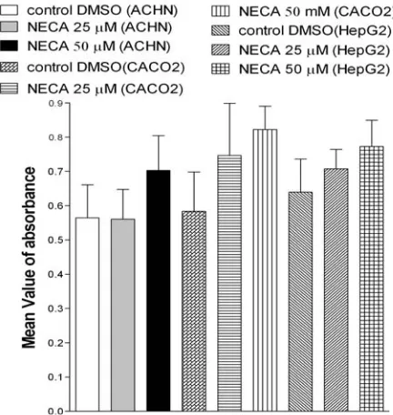
Effect of NECA on cell viability. To investigate
the effect of the non-specific adenosine agonist on tumor proliferation we used NECA. None of the cell lines demonstrated significant inhibition of MTT turnover, following a 72-h exposure to NECA, over the concentration range investigated (5-50 µM, P>0.05, Fig. 5). These results demonstrated that activation of A1 receptor has role in inhibition of proliferation of tumor cells but not in normal fibroblast cells.
Fig. 3. Effect of R-PIA on CACO2 cell line. Cells were cultured in 10% fetal calf serum medium and incubated without any treatment (control), with 0.250 µL DMSO (control DMSO), with 0.250 µL DMSO and 0.1 µM DPCPX (control DMSO + DPCPX), with different concentration of R-PIA and finally with two concentration of CHA or R-PIA and 0.1 µM DPCPX for 72 h then MTT added and absorbance read immediately after 4 h incubation in 570nm. The significant difference between absorbance of wells containing R-PIA and control DMSO are shown. N = 8, mean ± SE, **P<0.01 and ***P<0.001.
DISCUSSION
In this study, the effect of adenosine agonists on the proliferation of CACO2, human colon adeno-carcinoma cell line; Hep G2, human Caucasian hepatocytes carcinoma cell line; ACHN, human renal adenocarcinoma cell line and Hep2, human Caucasian larynx carcinoma cell line were evaluated using MTT method. A1 adenosine receptor has a medium expression level in kidney, colon and liver
Fig. 4. Effect of CHA, R-PIA and methotrexate on L929 cell line. Cells were cultured in 10% fetal calf serum medium and incubated without any treatment (control), with 0.250 µL DMSO (control DMSO), with different concentration of CHA, R-PIA or MTX and finally for 72 h then MTT added and absorbance read immediately after 4 h incubation in 570 nm. The significant differences between absorbance of wells containing CHA, R-PIA or MTX and control (for MTX) or control DMSO for (CHA or R-PIA) are shown. N n = 8, mean ± SE, *P<0.05 and ***P<0.001.
Fig. 5. Effect of NECA on ACHN, CACO2 and Hep G2 cell lines. Cells were cultured in 10% fetal calf serum medium and incubated with 0.250 µL DMSO (control DMSO), with wo concentration of NECA for 72 h then MTT added and absorbance read immediately after 4 h incubation in 570 nm. There is no significant difference between absorbance of wells containing NECA and control DMSO. n = 8, mean ± SE.
0 . 0 0 . 1 0 . 2 0 . 3 0 . 4 0 . 5 0 . 6 0 . 7
* * * * *
* *
M
e
an
V
a
lu
e
o
f a
b
so
rb
a
n
ce
control control DMSO control DMSO+DPCPX
R-PIA 5µM
R-PIA 10µM
R-PIA 25µM
R-PIA 50µM
R-PIA 25µM + DPCPX
R-PIA 50µM + DPCPX
[16] and also, a high expression in some cancerous tissues such as human colorectal adeno-carcinoma cell lines [7]. Therefore, these cell lines were used in our experiments to examine our hypothesis about cytotoxic effect of A1 agonists. Both CHA and R- PIA are potent A1 agonists [15] and they were evaluated for possible cytotoxic effects. Also, the same concentrations of NECA (non-specific A1 agonist) was used to compare its effects with cytotoxic effects of A1 specific agonists. Our study showed the inhibitory effect of CHA (A1 agonist [15]) on proliferation of CACO2, Hep G2, ACHN cell lines (in concentration of 5-50 µM) and R-PIA (another A1 agonist [15, 16]) on one CACO2 cell line (in concentration of 10-50 µM). CHA effects were in agreement with our expectation and demonstrated the ability of CHA in inhibition of proliferation in cell lines which have higher level expression of A1 receptor than Hep2 cell line [16].
Also, a previous report showed that activation of A1 receptor inhibits proliferation of LoVo colon carcinoma cell line [7]. These cytotoxic effects of CHA and R-PIA were inhibited by addition of DPCPX (a specific A1 antagonist) to culture media. Furthermore, NECA (non-specific adenosine agonist [15, 16]) showed no inhibitory effect on any cell lines up to 50 µM concentration. Also, one study showed that in some solid tumors, the cytotoxicity order of some adenosine agonists is CPA = R-PIA>NECA [6] that this had correlation with order of affinity to A1 receptor. Therefore, NECA, a non-specific agonist, needs higher concentration than A1 specific agonists to inhibit cell proliferation. One recent study showed that adenosine can induce apoptosis in Hep G2 cells via activating caspase 3 mainly by accumulation in cells rather than activating A1 receptor [12]. Difference between these results and ours might be related to using high concentrations of adenosine (3 mM) in this study and physicochemical difference between adenosine and its agonists which we used.
Difference between effects of CHA and R-PIA on cells (in despite of their similarity in affinity for A1 receptor [15]) might be related to difference of these two substances in activation of second messengers. Activation of A1 receptor results in decreasing concentration of cAMP or increasing concentration of phospholipase C [6] and A1 receptor agonists showed agonist-specific G protein activation, Gi or Gq [17]. R-PIA can protect neuron against death by increasing concentration of phospholipase C possibly via A1 receptor [18] which was possibly coupled to Gq protein, while one study showed
adenosine induces RCR-1 astrocytoma cell death partially via an A1 adenosine receptor, Gi protein signaling pathway [19]. These two experiments showed that Gi activation has a main role in adenosine-inducing cell death while R-PIA acts at least partly through Gq signaling pathway and accordance to our experiment, it has a less potential to induce cell death.
We also investigated the effect of CHA and R-PIA on fibroblast cells of mice as normal cells. CHA and R-PIA had no inhibitory effect on proliferation of fibroblast cells of mice up to 50 µM. Low concentration of adenosine leaded to proliferation in some normal cells such as fibroblast, murine bone marrow (which is mediated through A3 and A1 receptors), muscle cells and IM-9 lymphocytes [10]. A1 receptor activation protected human proximal tubular cells from the direct cytotoxic effect of H2O2 [6].
In attention to partially neutralization of inhibitory effect of CHA and R-PIA (two A1 agonists) by addition of DPCPX (A1 antagonist), effect of NECA (non-specific agonist) which didn't show cytotoxic effect and results of other studies about cytotoxic effect of adenosine and A1 receptor, we concluded that R-PIA and specially CHA have inhibitory effect on human tumor cell lines via A1 receptor. CHA and R-PIA didn't have inhibitory effect on proliferation of fibroblast cells (in same concentration). Because of this potential of selective toxicity of CHA and R-PIA on cancerous cells, we select them as a good choice for further in vivo studies, especially on tumors which over expresses A1 receptor such as colorectal adeno-carcinoma.
REFERENCES
1. Linden, J. (1991) Structure and function of A1 adenosine receptors. FASEB J. 5: 2668-2675. 2. Fishman, P., Bar-Yehuda, S., Madi, L. and Cohn, I.
(2002) A3 adenosine receptor as a target for cancer therapy. Anticancer Drugs 13: 437-443.
3. Ohana, G., Bar-Yehuda, S., Barer, F. and Fishman, P. (2001) Differential effect of adenosine on tumor and normal cell growth: focus on the A3 adenosine receptor. J. Cell. Physiol. 186: 19-23.
4. Fotheringham, J.A., Mayne, M.B., Grant, J.A. and Geiger, J.D. (2004) Activation of adenosine receptors inhibit tumor necrosis factor- release by decreasing TNF- mRNA stability and p38 activity. Eur. J. Pharmacol. 497: 87-95.
5. Spychala, J. and Shugar, D. (2000) Tumor promoting function of adenosine. Pharmacol. Ther. 87: 161-173.
6. Merigli, S., Mirandola, P., Varani, K., Gessi, S., Leunge, E., Giovanni, P., Aghazadeh, M.T. and Borea, P.A. (2003) A glance at adenosine receptors: target for antitumor therapy. Pharmacol. Ther. 100: 31-48.
7. Catherine P. and Barry E. (2000) Adenosine mediated killing of cultured epithelial cancer cells. Cancer Res. 60: 1887-1894.
8. Saitoh, M., Nagai, K., Nakagawa, K., Yamamura, T., Yamamoto, S. and Nishizaki, T. (2004) Adenosine induces apoptosis in the human gastric cancer cells via an intrinsic pathway relevant to activation of AMP-activated protein kinase. Biochem. Pharmacol. 67: 2005-2011.
9. Fishman, P., Bar-Yehuda, S., Barer, F., Madi, L., Multani, A.S. and Pathak, S. (2001) The A3 adenosine receptor as a new target for cancer therapy and chemoprotection. Exp. Cell Res. 269: 230-236. 10. Colquhoun, A. and Newsholme, E.A. (1997)
Inhibition of human tumor cell proliferation by analogues of adenosine. Cell Biochem. Funct. 15: 135-139.
11. Wakade, A.R., Przywyne, D.A. and Wakade, T.D. (2001) Intracellular nonreceptor-mediated signaling by adenosine. Mol. Neurobiol. 23: 137-154.
12. Wu, L., Li, G.P., Feng, J.L. and Pu, Z.j. (2006) Molecular mechanism of adenosine-induced apoptosis in human Hep G2 cells. Acta Pharmacol. Sin. 27: 477-484.
13. ECACC.gov.uk [homepage on the internet]. England: European Collection of Cell Culture. Available from http://www.ECACC.gov.uk/
14. Pulmb, J.A., Milory, R. and Kaye, J.B. (1989) Effect of the pH dependence of 3-(4,5-dimethylthiazol-2-yl)–2,5–diphenyl–tetrazolium bromide formazan absorbtion on chemosensitivity determined by a novel tetrazolium-based assay. Cancer Res. 49: 4435-4440.
15. Hosseinzadeh, H. and Stone, T.W. (1996) Adenosine in the central nervous system. Med. J. Islam. Rep.
Iran 9: 362-365.
16. Fredholm, B.B., Ijzerman, A.P., Jacobson, K.A., Klotz, K.N. and Linden, A. (2001) Nomenclature and classification of adenosine receptors. Pharmacol. Rev. 53: 527-546.
17. Cordeaux, Y., Ijzerman, A.P. and Hill S.J. (2004) Coupling of adenosine receptor to different heteromeric G proteins: evidence for agonist-specific G protein activation. Br. J. Pharmacol. 143: 705-714. 18. Rogel. A. (2005) Phospholipase C is involved in the
adenosine-activated signal transduction pathway conferring protection against iodoacetic acid-induced injury in primary rat neuronal cultures. Neurosci. Lett. 373: 218-221.
19. Sai, K., Yang, D., Yamamoto, H., Fujikawa, H., Yamamoto, S., Nagata, T., Saito, M., Yamamura, T. and Nishizaki, T. (2006) A1 adenosine receptor signal and AMPK involving caspase-9/-3 activation are responsible for adenosine-induced RCR-1 astrocytoma cell death. Neurotoxicology 27: 458-467.