Ultrastructural Study of Rotavirus Replication and Localization
of the Intermediate Capsid Protein VP6
Shahin Ahmadian
Electron Microscope Laboratory, Institute of Biochemistry and Biophysics, University of Tehran, Tehran, Iran
ABSTRACT
Rotavirus, a triple-layered non-enveloped member of the Reoviridae family, obtained a transient membrane envelope when newly synthesized subviral particles bud into the endoplasmic reticulum (ER). As rotavirus particles mature, they lose their transient membrane and obtain outer layer. It is mostly believed that only double layered particles bud into the ER. The present study describes that the single layered particles can also bud into the ER and become the immediate precursors of the mature virions. Virus replication was studied within a line of African green monkey kidney (BSC-1) cells infected with simian rotavirus SA11. The virus intermediate capsid protein (VP6) was localized within the infected cells using protein A-gold. Monospecific antibody to VP6 was the primary antibody. The electron micrographs of budding sites of ER showed two different sizes of subviral particles. The gold particles were seen on the double layered particles and very little in the cytoplasm and some on ER or very close to it. These results indicate that the single layered particles are also capable of being the precursor of the triple layered and obtain the VP6 while budding into the ER. Iran. Biomed. J. 7 (1): 7-11, 2003
Keywords: Rotavirus, Morphogenesis, Immunoelectron microscopy
INTRODUCTION
otavirus is a major cause of infantile gastroenteritis in humans and other mammalian species. The simian strain, SA11, grows readily in tissue culture, and its virion is composed of 11 segments of double-stranded RNA surrounded by a triple layered protein capsid [1, 2]. During viral maturation, double layered particles (previously known as single–shelled particles) assembled from VPI, VP2, VP3 and VP6 in the cytoplasm, interact with NS28, a virus–
encoded transmembrane protein that reside in the endoplasmic reticulum (ER) and functions as a receptor for VP6 [3, 4]. As the double layered particle buds through the ER membrane, it transiently acquires an ER membrane derived envelope. In the ER, the particle loses this membrane and acquires the outer capsid consisting of VP7 and VP4 [5, 6]. VP2 proteins form the core of the virus, which is surrounded by VP6, form characteristic double layered particles. Calcium is very important for rotavirus replication. The physical integrity of rotavirus particles requires calcium [7, 8]. It has been suggested that in viral replication, the double layered particles act as the
precursor of mature virions and bud into the ER [9, 10].
In the present study, we used African green monkey kidney cell line (BSC-I) to study virus replication. Since viral non-structural glycoprotein NSP4 (NS28) functions as an intracellular receptor in the ER membrane and binds newly made subviral particles via VP6 [11, 4], we used immunocy-tochemical technique to localize VP6 in per-meabilized infected cells. The morphological observations and the results from the immuno-cytochemical experiments presented in this study suggest that viral core particles can serve as immediate precursor of the mature virions.
MATERIALS AND METHODS
Cells and purification of virus. The African
green monkey kidney cell line (BSC-l) was grown as a monolayer in DMEM supplemented with 100 U of penicillin and 100g of streptomycin per ml, and 8% fetal bovine serum as described [12]. Stockes of simian rotavirus SA11 were propagated in BSC-l cells and purified from the infected cells by harvesting the cells in serum free medium after
R
8 infection, frozen and thawed three times and sonicated briefly. The supernatant of a low speed spin of the lysate was pooled and pelleted through a 40% sucrose cushion by centrifugation in centricon TST 28.38 rotor for 2 h at 42,000 g. The pellet was then banded by gradient centrifugation in CsCl in a centricon TST 55.5 rotor for 3 h at 85,000g. The visible band was collected and dialyzed against 10 mM Tris-buffered saline [13,14].
SDS-PAGE. Viral proteins were analyzed by 10%
SDS-PAGE under reducing conditions according to the method described by Laemmli [15].
Antisera. The monospecific antiserum against
VP6 was prepared by injecting the New Zealand white rabbits with purified protein eluted from polyacrylamide gels [16]. Briefly, the purified protein was mixed with complete adjuvant and injected the rabbits subcutaneously. Three injections were given at weekly intervals and a booster was given three weeks later. Animals were bled 10 days following the last injection. Antibody titration was determined by ELISA and its specificity was confirmed by Western-blot analysis [17].
Electron microscopy. The cell infected with rotavirus were taken at 4 h intervals (4, 8, 12, 16, 20 and 24 h) post-infection, fixed in 2.5% glutaraldehyde in 0.1M phosphate buffer, post-fixed in 1% osmium tetroxide and finally embedded in epoxy resin. Thin sections were stained with uranyl acetate followed by lead citrate and examined using a hitachi-HU-12A electron microscope at 75 KV.
Pre-embedding immunogold labeling. Infected
cells were collected 4 to 20 h post-infection. The cells were permeabilized either by freezing and thawing or by treatment with 0.1% Triton X -100 [18]. After incubation with 1% gelatin in PBS for 10 min, the cells were exposed to specific antibody diluted 1/20 in PBS containing 1% BSA. Pre-immune serum was used similarly as control. After incubation at 37oC for l h, cells were washed three times with PBS-BSA and exposed to protein A that was coupled to 10 nm gold particles and diluted 1/20 in PBS containing 1% BSA. After l h at 37oC the cells were washed three times in PBS to remove excess gold conjugates, fixed in 2% glutaraldehyde in PBS for l h and post-fixed in 1% osmium tetroxid. The fixed cells were rinsed several times in distilled water, incubated in 2% aqueous uranyl acetate for l h, dehydrated in a ascending series of ethanol and embedded in Araldite. Ultrathin
sections were prepared and stained with lead citrate [19].
RESULTS AND DISCUSSION
Morphology of rotavirus infected cells. The
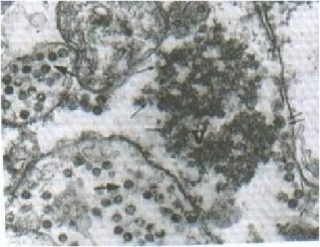
BSC-1 cell line was chosen for a detailed study of SA11 morphogenesis, as SA11 grows to a higher titer in BSC-l cells than in a variety of other cell culture lines. Samples of SA11-infected BSC-1 cells were fixed for electron microscopy at 4, 8, 12, 16, 20, 24 h following the end of the 60 min absorption period. The first detectable change that could be observed in the infected cells was the appearance of cytoplasmic dense granular material termed viroplasms that could be seen 4 h post-infection (p.i.). As the post-infection progressed, the size and number of viroplasmic inclusions increased. At 8 h p.i., single and double layered particles emerged from the viroplasms. By 12 h p.i., these particles were seen adjacent to the ER in process of budding through the ER membrane and having envelop. These particles apparently lost their membranes and matured as triple-layered virions inside the cisternae of the ER (Figs. 1 & 2). The same process was also seen at 16, 20 and 24 h p.i. Lysed cells were seen first at 12 h p.i., and increasingly in 16, 20 and 24 h. Virus particles were never seen budding into the golgi apparatus or into the plasma membrane.
Fig. 1. Electron micrograph of thin sections of infected cells at 12 h p.i. Viroplasm (V), viral cores (Single arrows) and double layered particle at the stage of budding through the ER (double arrows) and the mature particles in the lumen of the ER (large arrows) can be seen. (magnification,55,000).
Fig. 2. Electron micrograph of infected cells at 16 h p.i. Viral core particles (Single layered) at various stages of budding (arrows) (magnification,55,000).
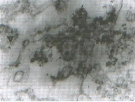
Fig. 3. Immunogold labeling of SA11-infected cells with antibody to VP6. Unlabeled viroplasm (v) with labeled subviral particles. Mature particles within the ER (arrows head) are not labeled. Arrows indicate areas where some gold grains are on the ER membrane. (magnification,60,000).
Ultrastructural localization of VP6. SA11 infected cell cultures were processed for ultrastructural localization of virus antigen VP6 between 16 to 20 h p.i., a time when virus–induced cytological alterations were widespread. Permeab-ilization of the infected cells with freezing and thawing procedure has been successfully used to localize rotavirus VP7 antigen [10], therefore, we used this procedure through out the experiments. Morphological preservation was satisfactory and the intracellular membranes remained intact. A monospecific antibody to VP6 was used to localize this protein within SA11 infected cells. The results
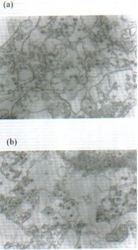
showed that subviral particles at the periphery of the viroplasm were labeled with gold, but the viroplasms themselves did not appear to be labeled (Fig. 3). Neither the enveloped virus particles nor the mature particles in permeabilized cell reacted with the antibody to VP6. Some gold particles located on and adjacent to the membranous structures of the ER (Figs. 3 and 4) indicating the presence of VP6 in these areas. However, the amount of gold was weak. The infected cells similarly treated with pre-immune serum or pretreated antibody with excess antigen and gold conjugate and found no labeling, (Figs. 5 a and b) indicating the specificity of the assay.
Fig. 4. Immunogold labeling of SA11-infected cells with antibody to VP6. Gold grains on the exoplasmic side of the ER membrane can be seen. (magnification,50,000).
In conclusion, the electron microscopic examination of samples taken at 4 h intervals confirmed that SA11 follows the pattern considered typical for rotavirus morphogenesis. All of the five particle types described by Chasey [20] in the cells infected with calf and pig rotavirus were seen in our experiments. The earliest particles formed following the 4 and 8 h post-infection were small particles (core like particles) among the viroplasm. At the periphery of the viroplasm, double layered particles were observed (Fig. 1). It has been reported that only the double layered particles bud into the ER [9, 10], but in our experiments electron micrographs of budding sites on the ER indicated both viral single and double layered particles are the precursor of mature virions. Since viral cores, unlike double layered particles, lack VP6, one would expect this protein to be present at the budding sites. However, Previous work using immunogold has failed to localize VP6 on the ER [18]. In our experiment, in permeabilized cell culture using monospecific
10 antibodies to VP6, gold conjugates were located mostly on double layered virus particles. We also observed some gold particles in adjacent or on the ER membrane (Fig. 4). These results indicate that single layered (core like layer) particles like double layered particles are capable of budding into the ER and simultaneously obtain the VP6.
Fig. 5. Immunogold controls. (a), SA11–infected cells with pre-immune rabbit serum (55,000); (b), SA11–infected cells with Ab to VP6 after adsorption in the presence of excess Ag (40000).
ACKNOWLEDGMENTS
This work was supported by the Research Council of the University of Tehran. The author thanks Dr. M.S. Shahrabadi for his comments and helpful discussion. The technical assistance of Mrs. M. Jamshidi is gratefully acknowledged.
REFERENCES
1. Cukor, G. and Blacklow, N. (1984) Human viral gastroenteritis. Microbiol. Rev. 48: 157-179. 2. Kapikian, A. and Chanock, R.M. (1985) Rotaviruses
In: Virology. (Fields, B.N. ed.), Raven Press, New York. pp. 863-905.
3. Au, K.S., Chan, W.K., Burns, J.W. and Estes, M.K. (1989) Receptor activity of rotavirus nonstructural glycoprotein NS28. J. Virol. 63: 4553-4562. 4. Tian, P., Ball, J.M., Zeng, Q.Y. and Estes, M.K.
(1996) The rotavirus nonstructural glycoprotein NSP4 possesses membrane destabilization activity. J. Virol. 70 (10): 6973-6981.
5. Altenburg, B.C., Graham, D.Y. and Estes, M.K. (1980) Ultrastructural study of rotavirus replication in cultured cells. J. Gen. Virol. 46:75-85.
6. Estes, M.K., Palmer, E.L. and Obijesky, J.F. (1983) Rotaviruses: a review curr. Top. Microbiol. Immunol. 105: 123-184.
7. Shahrabadi, M.S. and Lee, W.K. (1986) Bovine rotavirus maturation is a calcium dependent process. Virology 152: 298-307.
8. Ahmadian, S. and Shahrabadi, M.S. (1999) Mophological study of the role of calcium in the assembly of the rotavirus outer capsid protein VP7. Biotech. Histochem. 74: 266-273.
9. Poruchynsky, M.S. and Atkinson, P.H. (1991) Rotavirus protein rearrangements in purified membrane–enveloped intermediate particles. J. Virol. 65: 4720-4727.
10. Estes, M.K. (1996) Rotavirus and their Replication. In: Fields Virology. (Fields, B.N. ed.), Lippincott– Raven, Philadelphia. pp. 1640-1646.
11. Au, K.S., Mattion, N.M. and Estes, M.K. (1993) A subviral particle binding domain on the rotavirus nonstructural glycoprotein NS28. Virology 194: 665-673.
12. Kabcenell, A.K., Poruchynsky, M.S., Bellamy, A.R., Greenberg, H.B. and Atkinson, P.H. (1988) Two forms of rotavirus VP7 are involved in assembly of SA11 rotavirus in endo plasmic reticulum. J. Virol. 62: 2929-2941.
13. Sabara, M., Babiuk, L.A., Gilchrist, J. and Misra, V. (1982) Effect of tunicamycin on rotavirus assembly and infectivity. J. Virol. 43: 1082-1090.
14. Tian, P., Estes, M.K., Hu, Y., Ball, J.M., Zeng, Q.Y. and Schilling, W.P. (1995) The rotavirus nonstructural glycoprotein NSP4 mobilizes Ca2+ from the endoplasmic reticulum. J. Virol. 69 (9): 5763-5772.
15. Laemmli, U.K. (1970) Cleavage of structural proetins during the assembly of the head of bacteriophage T4. Nature 227:680-685.
16. Estes, M.K., Graham, D.Y. and Mason, B.B. (1981) Proteolytic enhancement of rotavirus infectivity: Molecular mechanisms. J. Virol. 39: 879-888. 17. Burns, J.W., Chen, D., Estes, M.K. and Ramig,
R.F. (1989) Biochemical and immunological
characterization of a simian rotavirus SA11 variant with an altered genome 4. Virology 169: 427-435. 18. Petrie, B.L., Greenberg, H.B., Graham, D.Y. and
Estes, M.K. (1984) Ultrastructural localization of rotavirus antigens using colloidal gold. Virus Res. 1: 133-152.
19. Hayat, M.A. (1989) Colloidal gold for microbiological immunocytochemistry. In: Colloidal Gold: Principles, Methods and Applications. Vol. L, Academic Press, New York, pp. 417-423.
20. Chasey, D. (1977) Different particle types in tissue culture and intestinal epithelium infected with rotavirus. J. Gen. Virol. 37: 443-451.