Vol. 1, No. 3 (2013): 80-88 Research Article
Open Access
I
ISSSSNN::22332200--22224466
Bioactive molecules from bacteria strains: case of
bacteriocins producing bacteria isolated from foods
Essodolom TAALE*, Aly SAVADOGO, Cheickna ZONGO, Andre J ILBOUDO and Alfred S
TRAORE
Centre de Recherche en Sciences Biologiques, Alimentaires et Nutritionnelles (CRSBAN), University of Ouagadougou, 03BP7131 OUAGADOUGOU03, Ouagadougou, Burkina Faso
* Corresponding author: Essodolom TAALE, email: [email protected]; Tel: (+226) 78764090, Fax: (+226) 50337373
ABSTRACT
21 bacteria strains were isolated from traditional fermented food (Soumbala, Bikalga, indigenous yoghurt and fermented milk) using microbiology standard methods. Morphological and biochemistry analyses were done. Nine strains were selected as presumed belonging to lactic acid bacteria (LAB). These strains were characterized at species level by amplifying 16S-23S intergenic spacer region of different LAB using Polymerase Chain Reaction (PCR) with specific primers. Strains S3, S4, Y6 and Lf1 belonged to the genus Lactobacillus; and strains S2, S5, Y1, Y3 and Y5 to genus Lactococcus. The nine strains were tested for their antimicrobial activity. These activities were detected in cell-free culture supernatant fluids against Micrococcus luteus as indicator strain. The agar well diffusion methods was used for this purpose. All the nine strains were able to inhibit the growth of Micrococcus luteus. The strain S3 produced antimicrobial compounds that showed the highest inhibition zone (20.83 mm) and strain Y6 showed the lowest inhibition zone (08.25mm). The bacteriocin-like gene was amplified using specific primers. The antimicrobial compounds produced by the strains except strain Y1 can be assimilated to bacteriocins according to the tests performed. The kinetics studies were performed for optimum pH and determination of growth temperature of each strain. The result showed that strain Y5 can grew at pH 5.5 and 37°C and the maximum specific growth rate are respectively 0.175 h-1 and
0.150 h-1.
Keywords:
Bacteriocins, PCR, Specific primers, Kinetics, Inhibition zoneINTRODUCTION
Soumbala, Bikalga, fermented milk, yoghurt and attieke are fermented foods which are widely consumed in Burkina Faso. Soumbala and Bikalga are condiments produced by traditional uncontrolled fermentation of Parkia biglobosa and Hibiscus sabdariffa seeds respectively. They are excellent sources of proteins with essential amino acids and also contain lipids, carbohydrates, essential fatty acids and vitamins [1-2]. Soumbala and Bikalga are used as a low-cost meat substitute by many families. These condiments improve nutritional values of foods as well as sensory properties as taste enhancer [3]. Lactic acid bacteria (LAB) are found naturally in a variety of environmental habitats, including dairy, meat, fish, vegetable, cereal and plant
environments, where fermentation can occur. LAB is typically involved in a large number of spontaneous food fermentations [4]. Historically, the traditional roles for many LAB have been as starter cultures to drive food and dairy fermentations, leading to their widespread human consumption and generally recognized as safe (GRAS) [5].
The LAB can produce antimicrobial substances able to inhibit the growth of pathogenic and spoilage microorganisms in foods. The primary antimicrobial effect exhibited by LAB is the production of lactic acid and reduction of pH [6]. In addition, LAB can produce various antimicrobial compounds, which can be
classified as low-molecular-carbon mass compounds such hydrogen peroxide (H2O2), carbon dioxide (CO2), diacetyl(2,3-butanedione), uncharacterized compounds and high-molecular-mass compounds like bacteriocins [7-9]. Bacteriocins are ribosomal synthesized antimicrobial peptides produced by one bacterium that are active against other bacteria, either in the same species (narrow spectrum), or across genera (broad spectrum) and, as with host defense peptides, cell signaling mechanism can also be involved [10].
The location of bacteriocins genes fear to be chromosomal [11-12] or plasmid [13-14]. Genes involved in the production of bacteriocins are generally organized as operon. Structure genes and immunity genes are generally co-transcribed except when they are disjoint as in the carnobacteriocin A system [15]. Regardless of their membership class, all bacteriocins synthesized by LAB are secreted by ABC transporters [16-17]. All bacteriocins are produced by ribosome in the cytoplasm of the producing cell. They are always synthesized as inactive precursor called "prebactériocin". This peptide is mature during or immediately after its secretion into the external environment and is recognized by the carrier. The peptide "signal" or "leader" provides for secretion of the bacteriocin in the environment and protects the bacteria against the action of its own bactériocin [16]. The production of bacteriocins is often regulated by a quorum sensing system, a mechanism allowing certain genes to be expressed in terms of the density of the bacterial population in the medium.
The objective of this study was to characterize by molecular techniques, bacteria strains from fermented foods producing bioactive molecules as bacteriocins isolated from some fermented foods widely consumed in Burkina Faso.
MATERIAL AND METHODS
Biological material21 strains were isolated from different fermented foods at Microbiology Laboratory of CRSBAN. Strains S1, S2, S3 and S4 were isolated from Soumbala. Strains B1, B2, B3, B4 and B5 were isolated from Bikalga. Strains Y1, Y2, Y3, Y4, Y5, Y6 were isolated from traditional yoghurt. Strains Lf1, Lf2, Lf3, Lf4, Lf5 were isolated from local fermented milk. The strains were stored at +4°C in brain heart infusion broth supplemented with 15% of glycerol. This study was conducted from June 2011 to January 2012 at Microbiology Laboratory and Laboratory of Molecular Biology of CRSBAN.
Isolation of lactic acid bacteria
21 strains were inoculated by streaking tight on PCA medium and incubated at 37°C for 24 hours. Each isolated colony was reinoculated in MRS broth [18],
incubated at 37°C under CO2 atmosphere and checked for purity by streaking on MRS agar [18] and incubated under CO2 atmosphere at 37°C for 24 to 72 hours. Before experimental use, all LAB strains were
recovered in MRS broth and were incubated at 37°C for 16 to 24 hours.
Screening for antagonistic activity and determination of antimicrobial spectra
The agar well diffusion assay (AWDA) [9, 19] was used to determine antibacterial activities against Micrococcus luteus. PCA (Plate Count Agar, Liofilchem, Italy) plates were overlaid with 5 ml Brain-heart Infusion broth (BioMarieux sa, France) inoculated with an overnight culture of the indicator microorganism, Micrococcus luteus (106 UFC/ml). Wells (6 mm in diameter) were cut in the plates. LAB strains were grown in MRS broth at 37°C for 16 hours. Overnight bacterial culture was harvested by centrifugation (6000rpm, 15 min at room temperature) to obtain a cell-free supernatant (CFS). Samples were adjusted to pH 6.5 with 10N of NaOH to rule out acid inhibition.
Afterwards, 5μl of CFS were placed in each well. The
plates were then incubated aerobically for 24 h at 37°C and were subsequently examined for zones of inhibition. Inhibition was recorded as negative if no zone was observed around the agar well. Each antagonistic activity was related to the area of the inhibition zone displayed.
Biochemical and physiological characterization of selected isolates
Gram staining, oxidase activity [20], catalase activity, production of acetoin (VP) [21] and sporulation were biochemical tests performed. Cell morphology was determined with cells grown in MRS broth at 37°C for 16 hours by using optical microscopy with objectives X40 and X100 to see cell form, cellular arrangement and their motility. CO2 and H2 production from glucose were performed using Kliger Iron Agar (Liofilchem, Italy). Carbohydrate fermentation patterns were determined by the API 20E (API, BioMerieux, France) as described by the manufacturer and the identification was done by a computerized database programme provided by the same manufacturer.
Genetic identification of bacteriocin-producing LAB strain
Extraction of bacterial total DNA
The microbial isolate from which the CFS maintained its antibacterial activity throughout the characterization of antibacterial compounds was selected for genetic identification. Total cellular DNA was isolated according to QIAamp® DNA Mini and Blood Mini Handbook (Third Edition, 2010; QIAgen) as described in page 56.
PCR amplifications
DNA amplification was conducted in a DNA thermal cycler TC-412 (Serial No.: 137370-2, United Kingdom) in order to determine the species affiliation of the tested bacterial isolates (Table 1). All specific-primers are provided by Eurofins MWG Operon (Germany). 50
μl of volume was used: 30 μl of Taq PCR Master Mix Kit (QIAgen, France), 4 μl of each specific-primer, 8 μl of
The amplification profile was 94°C for 5 min, 94°C for 45 s, 53°C-60°C for 45 s, 72°C for 1 min and 72°C for 10
min, 30 cycles.
Table 1. Primers and PCR products for the nine studied lactic acid bacteria strains
ND: Not detected bp: pairs of bases
Electrophoresis
The presence of PCR products was determined by gel electrophoresis in 0.7% agarose gel containing ethidium bromide. Electrophoresis in Tris-borate-EDTA was performed at 150 volts for 30 min, and photographed under ultraviolet light illumination.
Kinetics study of selected isolates
Modeling the reactions
The kinetic model describes two reactions: effect of pH and temperature on the growth of selected isolates. Growth experiments were conducted in MRS broth using Microtest plates (Heinz Herenz Medizinalbedarf GmbH, Germany).
Growth rate model
The specific growth rate was given by the equation:
(DO’/DO) = µmax .t + K
Where:
OD = value of the optical density at the start of the exponential phase of growth
OD’= the value of the optical density at time t during the exponential growth phase.
µmax = maximum specific growth rate, t = time, K =
constant.
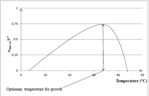
Fig.1 describes the curve of the pH-effect on specific growth rate. The growth pH range between 5-7. Fig.2 describes the curve of temperature-effect on growth rate. The optimal temperature growth of bacteria range between 30°C-40°C.
Figure 1. Description curve of pH-effect on specific growth rate for bacteria.
Figure 2. Description curve of the temperature-effect on specific growth rate
Influence of pH and temperature
The medium was inoculated with 1% v/v of inoculum. Test was made in triplicate. Growth was monitored for 24 hours by measuring each hour optical density (OD)
at 650 nm using a typical reader μquant (Bio-Tek
Species/Strain Primer Sequence Target PCR products Ref
Lactobacillus sp./
S3, S4, Y6, Lf1
5’- GGA ATC TTC CAC AAT GGA CG - 3’ 5’- CGC TTT ACG CCC AAT AAA TCC GG - 3’
16-23S rRNA gene spacer
450 bp [22]
Lactococcus sp./
S2, S5, Y1, Y3, Y5
5’- CTT TGA GTG ATG CAA TTG CAT C - 3’ 5’- CAC CGC TAC ACA TGG AG - 3’
16-23S rRNA gene spacer
<1000 bp [23]
Pediococcus sp./
S2, S5, Y1, Y3, Y5
5’- GTA AAG TGG CGT GTG TAC CTC AAG- 3’ 5’- CAC CGC TAC ACA TGG AG- 3’
16-23S rRNA gene spacer
ND [23]
Bacteriocins / S3, S4, Y6, Lf1 S2, S5,
Y1, Y3, Y5
5’- AAG AGT TTG ATC CTG GCT CAG - 3’ 5’- CTA CGG CTA CCT TGT TAC GA - 3’
instrument, Serial No.: 157904, USA) coupled to the computer managed by the KC junior (v1.31.5) software. The influence of pH on growth was studied at pH of 5; 5.5; 6.0; 6.5; 7.0 and 7.5 ± 0.2. The pH was controlled by the addition of 10N NaOH or 6N HCl. The influence of temperature was studied at temperature of 25; 30; 37; 40 and 44°C at the optimum pH of growth of each selected isolates.
RESULTS AND DISCUSSION
Biochemical, morphological and physiological characteristics of isolated strains
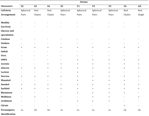
The results for the study of biochemical, morphological and physiological characteristics of the nine selected strains as belonging to lactic acid bacteria are shown in Table 2. The cells form are spherical or rod, with pairs and chains arrangement. One train Lf1 is single rod. All the strains were Gram positive, oxidase and catalase negative. No strain produced gas from glucose. Four strains were rods and five were spherical (Table 2). All strains were non motile and did not form spores (Table 2). Indeed, the lactic acid bacteria in their majority do not form spores but still manage to live in extreme conditions (Depletion of nutrient in the medium, high or very low variation of the salt concentration etc.) developing effective systems of protection allowing them to avoid death after stopping their growth [25-26].
Sept strains produce acetoin (Table 2). Acetoin is one of intermediate compounds (volatile acetoin, acetaldehyde, diacetyl and lactone). These intermediates are responsible of flavor and aroma found in fermented foods especially dairy, as appreciated by consumers. Only strains S2 and S3 do not produce this metabolite. However, this study shows that lactic acid bacteria can produce acetoin not only fermenting lactose but also other sugars: this is the case of S4 and S5, strains isolated from soumbala, a non-dairy product.
The existence of a betagalactosidase in bacteria confers the ability to ferment lactose effectively regardless of the bacterial permease. Ortho Nitro Phenyl Galactopyranoside (ONPG) reagent was able to detect this enzyme in strains Y1, Y3, Y5, Y6 and Lf1 (Table 2). These strains were isolated respectively from yoghurt (Y1, Y3, Y5 and Y6) and fermented milk (Lf1) two media rich in lactose. Betagalactosidase is absent in the four strains (S2, S3, S4 and S5) isolated from soumbala, a condiment produced by spontaneous alkaline fermentation of nere seeds. These results show that lactic acid bacteria use nutrients in their environment by conforming their respiratory chain. All nine strains ferment glucose and sucrose. But only strains Y1 and Y5 use lactose.
Antibacterial activities of selected lactic acid bacteria
The agar well diffusion assay (AWDA) [9, 19] was used to detect strains with antibacterial activities against Micrococcus luteus. The supernatant of nine strains of
lactic acid bacteria gave zones inhibition onto the indicator pathogenic strain (Micrococcus luteus) tested (Table 3).
comprise a heterogeneous group of physicochemical diverse ribosomally-synthesized peptides or proteins
showing a narrow or broad antimicrobial activity spectrum against Gram-positive bacteria.
Table 2. Biochemical, morphology and physiological characters of selected strains
+ = positive reaction; - = negative reaction; S = strains from Soumbala (S2, S3, S4, S5); Y = strains from yoghurt (Y1, Y3, Y5, Y6) ;
Lf = strains from fermented milk (Lf1); Lb. = Lactobacillus; Lc = Lactococcus
Table 3. Inhibition of Micrococcus luteus by bacteriocin produced by nine selected strains identified as lactic acid bacteria.
Strains pH of the
supernatant pH adjusted
ΔpH Diameter of
inhibition (mm)
S2 6.12 6.5 0.38 16.17 ± 2.31
S3 6.06 6.5 0.44 20.83 ± 2.84
S4 6.02 6.5 0.48 13.00 ± 0.00
S5 4.83 6.5 1.67 20.33 ± 1.53
Y1 4.40 6.5 2.10 10.00 ± 1.80
Y3 6.03 6.5 0.47 15.00 ± 1.41
Y5 4.34 6.5 2.16 13.09 ± 0.83
Y6 5.77 6.5 0.73 08.25 ± 0.35
Lf1 4.32 6.5 2.18 12.50 ± 0.00
pH: potential of hydrogen mm: millimeter ± : more or less
Figure 3. Inhibition of Micrococcus luteus by lactic acid bacteria strain S5 culture supernatant by the agar well diffusion assay. T: Negative control
Strains
Characters S2 S3 S4 S5 Y1 Y3 Y5 Y6 Lf1
Cell form Spherical Rod Rod Spherical Spherical Spherical Spherical Rod Rod
Arrangement Pairs Chains Chains Pairs Pairs Pairs Pairs Chains Single
Motility - - - -
Gas from
Glucose and
sporulation
- - - -
Catalase - - - -
Oxidase - - - -
Gram + + + + + + + + +
Indole - - - -
Urea - - - -
ONPG - - - - + + + + +
Acetoin - - + + + + + + +
Glucose + + + + + + + + +
Lactose - - - - + - + - -
Sucrose + + + + + + + + +
Mannitol - - - - + + + + -
Inositol + + + + + + + + +
Sorbitol + + + + + + + + +
Rhamnose - - - - + + + + -
Melibiose - + - - - + + - +
Arabinose - - - - + + + - -
Citrate - - - + -
Presumptive
identification
Identification of selected lactic acid bacteria to genus level and screening of bacteriocins genes by PCR
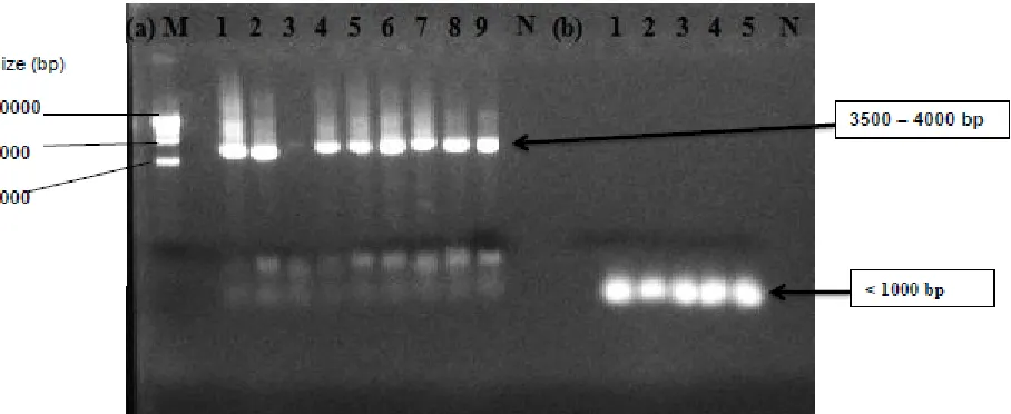
The strains S3, S4, Y6 and Lf1 were amplified with the Lactobacillus species specific-primers, producing a product of 450bp, indicating that they were members of Lactobacillus species (Fig.4). The same primers pairs were used by [22] but got product of 200bp.
Figure 4. Species-specific PCR with primers pairs Lb1 and Lb2. Lane M: 100-bp DNA molecular mass marker, Lane 1: strain Lf1, Lane 2: strain Y6, Lane 3: strain S4, Lane 4: strain S3, Lane N: negative control.
Figure 5. Species-specific PCR amplification with primers pairs Ped1 and Ped2. Lane N: negative control, Lane1: Strain S2, Lane2: strain S5, Lane3: strain Y1, Lane4: strain Y3, Lane 5: strain Y5.
The product of less 1000 bp were found when the strains S2, S5, Y1, Y3 and Y5 were amplified by Lactococcus species specific-primers, showing that these strains with spherical form belong to Lactococcus species [Fig.6-(b)]. But no result was found with the specific-primer designed for Pediococcus species (Fig.5).
The strains S2, S3, S4, S5, Y1, Y3, Y5, Y6 and Lf1were amplified with the bacteriocins primers, producing a product 3500 – 4000bp. Only strain Y1 gave a negative result [Fig.6-(a)]. Those results indicate that the eight strains can produce antimicrobial compounds which can be assimilated to bacteriocins because they have bacteriocins genes.
Kinetics study of selected lactic acid bacteria: Determination of pH and temperature of growth
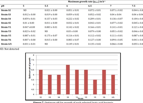
For all strains the specific growth rate at pH value of 5 was low. Thus, the optimum pH for growth of the nine selected lactic acid bacteria range between 5.5 to 7: pH 5.5 for strain Y5, pH 6 for strains S3, S4 and Lf1, pH 6.5 for strains S2, Y1, Y3 and Y6 and pH 7 for strain S5 (Table 4, Fig.7). The maximum specific growth rate varies from 0.007 h-1 (lowest value observed for strain S2 at pH 6.5) to 0.222 h-1 (highest value observed for strain S4 at pH 6). The pH value of 7 found for Lactococcus strain S5 are compatible because that strain were isolated from soumbala, a condiment obtained by uncontrolled alkaline fermentation of nere seeds.
Table 4. Maximum growth rate of selected lactic acid bacteria at uncontrolled pH
Maximum growth rate (µmax) in h-1
pH 5 5.5 6 6.5 7 7.5
Strain S2 ND 0.021 ± 0.00 0.051 ± 0.01 0.072 ± 0.01 0.071 ± 0.02 0.044 ± 0.00
Strain S3 0.012 ± 0.00 0.019 ± 0.00 0.059 ± 0.02 0.052 ± 0.02 0.04 ± 0.04 0.04 ± 0.04
Strain S4 0.059 ± 0.01 0.157 ± 0.03 0.222 ± 0.02 0.209 ± 0.01 0.136 ± 0.07 0.104 ± 0.01
Strain S5 0.01 ± 0.00 0.011 ± 0.00 0.032 ± 0.01 0.042 ± 0.01 0.077 ± 0.02 0.045 ± 0.00
Strain Y1 0.047 ±0.00 0.083 ± 0.01 0.142 ± 0.02 0.166 ± 0.01 0.141 ± 0.01 0.121 ± 0.01
Strain Y3 0.023 ± 0.02 ND 0.03 ± 0.00 0.079 ± 0.00 0.051 ± 0.02 0.046 ± 0.01
Strain Y5 0.087 ± 0.01 0.175 ± 0.07 0.116 ± 0.01 0.112 ± 0.02 0.111 ± 0.01 0.087 ± 0.03
Strain Y6 0.007 ± 0.00 0.015 ± 0.00 0.083 ± 0.07 0.127 ± 0.00 0.094 ± 0.05 0.041 ± 0.01
Strain Lf1 0.031 ± 0.01 ND 0.149 ± 0.01 0.135 ± 0.02 0.066 ± 0.00 0.055 ± 0.00
ND: Not detected
Figure 7. Optimum pH for growth of each selected lactic acid bacteria
The specific growth rate for each selected lactic acid bacteria at temperature value of 25°C and 44°C was low. The higher specific growth rate (0.259 h-1) was observed at temperature values of 40°C (Table 5) for strain S3 (Lactobacillus sp.). Temperature 30°C, 37°C and 40°C are adequate temperatures (Table 5, Fig.8) of growth for the nine selected lactic acid bacteria strains subjected to this study. Indeed, among Lactobacillus species, strains S3, S4 and Y6 have an optimum growth temperature of 40°C, whereas it is 30°C for strain Lf1. The strain Lf1 is mesophilic Lactobacillus while S3, S4
and Y6 are thermophilic Lactobacillus. S2, S5 and Y5, three (03) Lactococcus species strains grew better at temperature of 37°C. 30°C of temperature is suitable for other Lactococcus species strains (Table 5, Fig.8). The maximum growth rate varies between 0.068 h-1 (lowest value observed for Y3 strain at 30°C) to 0.259 h-1 (highest value observed for strain S3 at 40°C). According to the literature, Lactococcus grow better at temperatures between 30 and 37°C.
Table 5. Maximum growth rate of selected lactic acid bacteria at controlled pH and uncontrolled temperature.
Maximum growth rate (µmax in h-1)
Temperature 25°C 30°C 37°C 40°C 44°C
Strain S2 (pH6.5) 0.069 ± 0.04 0.093 ± 0.02 0.096 ± 0.05 0.068 ± 0.04 0.031 ± 0.04
Strain S3 (pH6) 0.042 ± 0.02 0.081 ± 0.01 0.151 ± 0.01 0.259 ± 0.02 0.047 ± 0.00
Strain S4 (pH6) 0.025 ± 0.02 0.029 ± 0.00 0.032 ± 0.00 0.097 ± 0.05 0.025 ± 0.00
Strain S5 (pH7) 0.002 ± 0.00 0.145 ± 0.04 0.153 ± 0.03 0.099 ± 0.02 0.044 ± 0.01
Strain Y1 (pH6.5) 0.079 ± 0.01 0.114 ± 0.08 0.078 ± 0.06 0.037 ± 0.00 0.026 ± 0.00
Strain Y3 (pH6.5) 0.057 ± 0.03 0.068 ± 0.01 0.026 ± 0.00 0.024 ± 0.02 0.024 ± 0.00
Strain Y5 (pH5.5) ND 0.092 ± 0.01 0.150 ±0.01 0.077 ± 0.01 0.052 ± 0.00
Strain Y6 (pH6.5) ND 0.010 ± 0.00 0.013 ± 0.00 0.123 ± 0.01 0.083 ± 0.00
Strain Lf1 (pH7) 0.080 ± 0.02 0.162 ± 0.02 0.057 ± 0.06 0.021 ± 0.01 ND
Figure 8. Optimum temperature for growth of each selected lactic acid bacteria
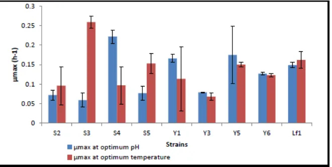
Figure 9. Maximum specific growth rate of each selected lactic acid bacteria at controlled pH and temperature
The strain S3 showed a higher maximum specific growth rate with a value of 0.259 h-1 at 40°C and strain Y3 showed the lowest maximum specific growth rate as 0.068 h-1 at 30°C (Fig.8). The strain S4 showed a higher maximum specific growth rate (0.259 h-1) at pH=6 and the lowest (0.068 h-1) at pH=6 observed for strain S3 (Fig.9).
CONCLUSION
Lactic acid bacteria play an important role in food fermentation, as the products obtained with their aid are characterized by hygienic safety, storage stability and attractive sensory properties. These properties are due to the compounds they synthetize, especially bacteriocins. Nine lactic acid bacteria were isolated and characterized by standard biochemical, microbiology and PCR methods from Soumbala, Yoghurt and Fermented milk. All the nine strains can produce active antimicrobial compounds against Micrococcus luteus. The diameter of inhibition and results gave by PCR after amplifiying bacteriocins genes show that those compounds can be assimilated to bacteriocins. The study of effect of pH and temperature on growth of the selected lactic acid bacteria strain shows that our Lactobacillus species can be divided into two groups: mesophilic Lactobacillus (Strain Lf1) and thermophilic Lactobacillus (strains S3, S4, Y6). Only Strain Y5, isolated from Yoghurt has an optimal pH on growth at value of 5.5.
In view of the results obtained in this study, it would be interesting to perform a complete genome sequencing of the strain Y5.
ACKNOWLEDGEMENTS
We thank PACER-UEMOA for funding this work.
REFERENCES
1. Odunfa SA (1985). Biochemical changes in fermenting African locust bean (Parkia biglobosa) during “iru” fermentation. J Food Technol. 20: 295 – 303
2. Ouoba LII, Rechinger KB, Barkholt V et al. (2003). Degradation of proteins during the fermentation of African locust bean (Parkia biglobosa) by strains of Bacillus subtilis and Bacillus pumilus for production of soumbala. J Appl Microbiol. 94 (3): 396– 402
3. Savadogo Aly and Alfred S Traore (2011). La flore microbienne et les propriétés fonctionnelles des yaourts et laits fermentés. Int J Biol Chem Sci. 5(5) : 2057-2075 4. Stiles ME and Holzapfel WH (1997). Lactic acid bacteria of
foods and their current taxonomy. Int J Food Microbiol. 36(1): 1 – 29
5. Klaenhammer TR, Barragou R, Buck LB et al. (2005). Genomic features of lactic acid bacteria effecting bioprocessing and health. FEMS Microbiol Rev. 29: 393 – 409
6. Daeschel MA (1989). Antimicrobial substances from lactic acid bacteria for use as food preservatives. Food Technol. 43(1): 164-167
7. Klaenhammer TR (1988). Bacteriocins of lactic acid bacteria. Biochimie. 70: 337 – 349
© 2013; AIZEON Publishers; All Rights Reserved
This is an Open Access article distributed under the terms of the Creative Commons Attribution License which permits unrestricted use, distribution, and reproduction in any medium, provided the original work is properly cited.
9. Khay E, Idaomar M, Castro LMP et al. (2011). Antimicrobial activities of the bacteriocin-like substances produced by lactic acid bacteria isolated from Moroccan dromedary milk. Afr J Biotechnol. 10(51): 10447 – 10455
10. Cotter PD, Hill C and Ross RP (2005). Bacteriocins: developing innate immunity for food. Nat Rev Microbiol. 3: 777–788
11. Cintas LM, Casaus P, Havarstein LS et al. (1997). Biochemical and genetic characterization of enterocin P, a novel sec-dependent bacteriocin from Enterococcus faecium P13 with a braod antimicrobial spectrum. Appl Environ Microbiol. 63(11): 4321 – 4330
12. Cocolin L and Rantsiou K (2007). Sequencing and expression analysis of sakacin genes in Lactobacillus curvatus strains. Appl Genet Mol Biotechnol. 76: 1403 – 1411
13. Marrugg JD, Gonzales CF, Kunka BS et al. (1992). Cloning, expression, and nucleotide sequence of genes involved in production of pediocin PA-1 and bacteriocin from
Pediococcus acidilactici PAC1.0. Appl Environ Microbiol. 58(8): 2360 -2367
14. Héchard Y, Derijard B, Letellier F et al. (1992). Characterization and purification of mesentericin Y105, an anti-Listeria bacteriocin from Leuconostoc mesenteroides. J Gen Microbiol. 138(12) :2725 – 273
15. Franz CMP, Belkum JV, Worobo RW et al. (2000). Characterization of the genetic locus responsible for production and immunity of carnobacteriocin A: the immunity gene confers cross-protection to enterocin B. Microbiology. 146: 621 – 631
16. Nes IF, Diep DB, Havarstein LS et al. (1996). Biosynthesis of bacteriocin in lactic acid bacteria. Antonie Van Leeuwenhoek. 70(2-4): 113 – 128
17. McAuliffe O, Ross RP and Hill C (2001). Lantibiotics: structure, biosynthesis and mode of action. FEMS Microbiol Rev. 25(3): 285 – 308
18. Man JD, Rogosa MA and Sharpe ME (1960). A medium for the cultivation of lactobacilli. J Appl Bacteriol. 23: 130 – 135 19. Castro MP, Palavecino NZ, Herman C et al. (2011). Lactic acid
bacteria isolated from artisanal dry sausages: Characterization of antibacterial compounds and study of the factors affecting bacteriocin production. Meat Sci. 87 : 321– 329
20. Kovacs N (1956). Identification of Pseudomonas pyocyana by oxydase reaction. Nature (Lond). 178:703
21. Clark W and Lubs H (1915). The identification of bacteria of the colony aerogenes family by use of indicators. J Inf Dis. 17:160-173
22. Bakar AF, Nordin N, Yoke TS et al. (2010). Detection and quantification of probiotic bacteria using optimized DNA extraction, traditional and real-time PCR methods in complex microbial communities. Afr J Biotechnol. 9(10): 1481-1492
23. Savadogo A, Ouattara CAT, Savadogo PW et al. (2004). Identification of exopolysaccharides-producing lactic acid bacteria from Burkina Faso fermented milk samples. Afr J Biotechnol. 3(3): 189 – 194
24. Diop MD, Dubois-Dauphin R, Dortu C et al. (2008). In vitro detection and characterization of bacteriocin-like inhibitory activity of lactic acid bacteria (LAB) isolated from Senegalese local food products. Afr J Microbiol Res. 2: 206 – 216 25. Glaasker E, Tjan FS, Ter Steeg PF et al. (1998). Physiological
response of Lactobacillus plantarum to salt and nonelectrolyte stress. J Bacteriol. 180(17): 4718 – 4723 26. Guchte M, Serror P, Chervaux C et al. (2002). Stress responses
in lactic acid bacteria. Antonie Van Leeuwonhoek. 82: 187 – 216.
27. Lade H, Chitanand M, Gyananath G et al. (2009). Studies On Some Properties Of Bacteriocins Produced By Lactobacillus
Species Isolated From Agro-Based Waste. The Internet Journal of Microbiology. Volume 2: Number 1
28. Jiang J, Shi B, Zhu D et al. (2012). Characterization of a novel bacteriocin produced by Lactobacillus sakei LSJ618 isolated from traditional Chinese fermented radish. Food Control. 23: 338 – 344
29. Badr S, Karem H, Hussein H et al. (2005). Characterization of nisin produced by Lactococcus lactis. Int J Agric Biol. 7(3): 499 – 503
30. Tuncer Y and Ozden B (2010). Partial biochemical characterization of nisin-like bacteriocin produced by
Lactococcus lactis subsp. lactis YBD11 isolated from Boza, a traditional fermented Turkish beverage. Rom Biotechnol Lett. 15(1): 4940 – 4948
31. Klaenhammer TR (1993).Genetics of bacteriocins produced by lactic acid bacteria. FEMS Microbiol Rev. 12(1-3): 39 – 85 32. Bhunia AK, Johnson MC, Ray B et al. (1991). Mode of action of
pediocin AcH from Pediococcus acidilactici H on sensitive bacterial strains. J Appl Bacteriol. 70: 25 – 33
33. Martinez-Cuesta MC, Palaez C, Juarez M et al. (1997). Autolysis of Lactococcus lactis ssp. lactis and Lactobacillus casei ssp. casei. Cell lysis induced by a crude bacteriocin. Int J Food Microbiol. 38(2 – 3):125 – 131
34. Dortu C and Thonart P (2009). Les bactériocines des bactéries lactiques : Caractéristiques et intérêts pour la bioconservation des produits alimentaires. Biotechnol Agron Soc Environ. 13(1):143 – 154