_____________________________________________________________________________________________________ 14(3): 1-8, 2019; Article no.JAMB.44635
ISSN: 2456-7116
Electricity Production Potential of Decayed
Tectona
grandis
Using Microbial Fuel Cell
D. V. Adegunloye
1and I. M. Ojo
1*1
Department of Microbiology, Federal University of Technology, Akure, P.M.B. 704, Nigeria.
Authors’ contributions
This work was carried out in collaboration between both authors. Author IMO designed the study, performed the statistical analysis, wrote the protocol and wrote the first draft of the manuscript. Authors DVA and IMO managed the analyses of the study. Author DVA managed the literature searches. All authors read and approved the final manuscript.
Article Information
DOI: 10.9734/JAMB/2019/44635 Editor(s): (1)Dr. Simone Aquino, Professor, Universidade Nove de Julho, São Paulo, Brazil.
Reviewers: (1)Glauber Cruz, Federal University of Maranhão, Brazil. (2)P. Saravana Kumari, Rathnavel Subramaniam College of Arts and Science, India. Complete Peer review History:http://www.sdiarticle3.com/review-history/44635
Received 02 August 2018 Accepted 21 October 2018 Published 11 February 2019
ABSTRACT
The potential of decayed Tectona grandis wood to generate current and voltage due to the inherent microorganism present in it was determined in this study. The decayed wood was collected from the Federal University of Technology Akure forest plantation. Microorganisms were isolated from the decayed Tectonagrandis wood and the organisms were identified using both cultural and molecular methods. The microbial fuel experimental set up was carried out for 14 days. The microbial fuel cell was made up of two chambers which are the anodic i.e where bacteria oxidise the organic matter present in the wood and cathodic chamber, this contained the substrate (decayed wood) and water respectively. Current and voltage generated by the decayed wood was measured using a multimeter. Results revealed that the microorganisms isolated include Bacillus licheniformis, Micrococcus luteus, Bacillus sp, Acinetobacter iwoffii Bacillus cereus, Pseudomonas putida, Bacillus thuringiensis, Penicillum notantum, Rhizopus stolonifer, Aspergillus penicilloides, Rhizopus oryzae
and Aspergillus flavus. It also showed that there was a continuous increase in the current generated which was within the range from (0.032 ± 0.00 to 0.441 ± 0.02) mA. The highest voltage was generated on day 12 with the value (0.369 ± 0.02) mV. It was shown that there was a progressive increase in the voltage generated from day 1 to day 12 with the range of values from 0.023 ± 0.01 to
Adegunloye and Ojo; JAMB, 14(3): 1-8, 2019; Article no.JAMB.44635
0.369 ± 0.02) mV. Findings from this study affirmed that decayed Tectona grandis wood has the ability to generate current and voltage using microbial fuel cell due to the microorganisms present in them which initiate oxidation reaction.
Keywords: Tectona grandis; microbial fuel cell; electricity; decayed; potential.
1. INTRODUCTION
Microbial fuel cell technology is a new type of renewable and sustainable method for the production of electric energy from the microbial breakdown of organic matter [1]. It has also been considered a promising technology for power generation [2,3]. A Microbial Fuel Cell (MFC) is a device that converts chemical energy from bio-convertible organic substrate, directly into electrical energy through the metabolic activity of microorganisms [4]. Fuel cells are able to generate electricity from many different chemicals by oxidation of the chemicals at the anode and reduction at the cathode [5]. Tectona grandis Linn. commonly known as teak tree is known in the world for its dimensional stability, extreme durability and hardness in timber production [6]. Following the current global energy crises in relation to increasing demand for fossil fuels (particularly oil, coal and gas), as well as inadequate electricity supply various human services in a country like Nigeria, evaluating for newer sources of meeting required demand cannot be over emphasised as future economic growth crucially depends on this. However, this study attempts to isolate the organisms present in decayed wood and determine the feasibility of using
Tectona grandis wood to produce current and voltage generation in a microbial fuel cell.
2.METHODOLOGY
2.1 Collection and Preparation of Samples
Decayed Tectona grandis wood was collected from the forest plantation located in the Federal University of Technology, Akure, (FUTA) at Obanla. The decayed wood was collected into a sterile polythene bags and were transferred to the Microbiology Postgraduate Laboratory,
Obanla, Federal University of Technology, Akure for microbiological analyses. The decayed
wood sample was then crushed into small pieces.
Plate 1. FUTA forest plantation
2.2 Isolation of Microorganism
Five fold serial dilution was carried out on 1gram of decayed Tectona grandis. One millilitre of each diluent was pipeted into Petri dishes and pour plated with molten nutrient agar and potato dextrose agar media. Nutrient agar plates were incubated at 37ºC for 24 hours for bacteria and 28ºC from 3 to 5 days for fungi on potato dextrose agar plates respectively in duplicate before examination for microbial growth. The bacterial isolates were purified by streaking on fresh sterile nutrient agar before sub culturing. Fungal isolates were also sub cultured to obtain pure isolates. The pure isolates were stored temporarily on agar slants and kept at 4ºC for further use [7]. Colony counting was carried out on plates (in duplicates) by using colony counter (TT-02 Techmel USA). Colony counting was expressed as colony forming unit (CFU) x 105 and spore forming unit (sfu) x 104 per gramme of decayed wood for bacteria and fungi respectively [7].
2.3 Identification of Microorganism
further identified using molecular methods ascertain their identities.
2.4Molecular Identification of Bacteria Isolated
Molecular identification of the bacteria isolates were determined using sequencing method as
described by Ologun et al.
deoxyribonucleic acid (DNA) of each isolates was extracted in accordance with the procedure of Zymo bacterial DNA Mini-prep kit. The extracted genomic DNA was stored at 4º use of polymerase chain reaction (PCR) was employed in the amplification of the extracted DNA portion encoding 16SrRNA using universal bacterial primers.
Plate 2. A constructed microbial fuel set up
2.5 Microbial Fuel Construction
The microbial fuel cell (MFC) was constructed according to Yoganathan and Ganesh Adegunloye and Olotu [10]. The
constructed using two screw capped plastic bottles which is made of two chambers; the anode (anaerobic) which contains the decayed wood and the cathode (aerobic) which contains water. In the case of the control, the decayed wood which was contained in the anode was sterilized which killed all microorganism present in it. Both anode and cathode chambers were connected with 1.2 cm in diameter and 6 cm long tube which was filled up with a salt bridge made of sodium chloride and agar in the ratio of 1:2 Agar salt bridge acted as a barrier between the anode and cathode chambers. The Purpose of an agar salt bridge is to provide an internal electrical connection between the ch
while minimizing the transfer of ions from the electrical environment. The carbon rods of 1.5 cm diameter and 13.5 cm long served as anode
Adegunloye and Ojo; JAMB, 14(3): 1-8, 2019; Article no.
ing molecular methods
Molecular Identification of Bacteria
Molecular identification of the bacteria isolates were determined using sequencing method as
Ologun et al. [9]. The
(DNA) of each isolates was extracted in accordance with the procedure prep kit. The extracted genomic DNA was stored at 4ºC. The use of polymerase chain reaction (PCR) was employed in the amplification of the extracted n encoding 16SrRNA using universal
Plate 2. A constructed microbial fuel set up
2.5 Microbial Fuel Construction
The microbial fuel cell (MFC) was constructed Yoganathan and Ganesh [1], . The MFC was constructed using two screw capped plastic bottles which is made of two chambers; the anode (anaerobic) which contains the decayed wood and the cathode (aerobic) which contains water. In the case of the control, the decayed in the anode was ed which killed all microorganism present in it. Both anode and cathode chambers were connected with 1.2 cm in diameter and 6 cm long tube which was filled up with a salt bridge made of sodium chloride and agar in the ratio of 1:2. Agar salt bridge acted as a barrier between the anode and cathode chambers. The Purpose of an agar salt bridge is to provide an internal electrical connection between the chambers, ing the transfer of ions from the e carbon rods of 1.5 cm diameter and 13.5 cm long served as anode
and cathode. Before the MFC operation the electrodes were soaked in 1 mol/L HCl solution
for a day to remove possible metal
contamination, and after the MFC operation, the electrodes were washed with 1 mol of sod dioxide solution to sterilize the attached cells. The electrodes were externally connected with copper wire and all exposed metal surface was sealed with non-conductive epoxy. A digital multimeter (DT9205A) was connected to the copper wires and it was used to read the current and voltage produced.
3. RESULTS
3.1 Total Microbial Load of Decayed Wood Samples
Microbial load of the decayed wood indicated that there was a significant difference (p
in total viable Bacterial and fungal counts of the decayed wood, the bacterial load of
grandis was 4.5 x10⁵ Cfu/g, while the fungal load was 4.2 x 10⁴ Sfu/g.
3.2 Morphological, Biochemical Characteristics and Identification of Bacterial Isolates from Decayed Wood
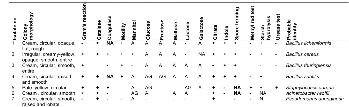
The biochemical tests carried out on the bacterial isolates (Table 1) are; Gram stain, Catalase, Coagulase, Motility, Citrate, Indole, Spore forming, Starch Hydrolysis and Urease. All the isolates showed different biochemical reactions and were morphologically characterized. The isolates were identified as Bacillus thuringiensis, Acinetobacter iwoffii, Stapphylococcus aureus, Bacillus licheniformis Bacillus sp Pseudomonas putida, Bacillus cereus, Rhizopus stolonifer,
Penicilliun notantum, Aspergillus flavus
Aspergillus penicilloides, Rhizopus oryzae.
3.3 Molecular Identification of Bacteria Isolate from Decayed Tectona grandis
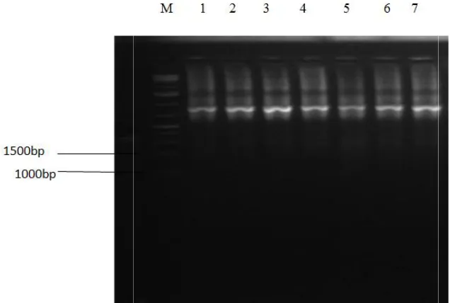
Molecular identification of the bacterial isolates are shown in Table 3. The lengths of amplified products were 1412, 1000, 1499, 1425, 1512, 1419, 1525 base pair for Pseudomonas putida,
Bacillus sp, Bacillus cereus, Bacillus
thuringiensis Acinetobacter iw
licheniformis, Micrococcus luteus
(Plate 1). The sequence obtained was analysed with BLAST in National Centre for Biotechnology Information (NCBI) database. Based on the 16SrRNA sequences, the bacteria
; Article no.JAMB.44635
and cathode. Before the MFC operation the electrodes were soaked in 1 mol/L HCl solution
for a day to remove possible metal
contamination, and after the MFC operation, the shed with 1 mol of sodium e the attached cells. The electrodes were externally connected with copper wire and all exposed metal surface was conductive epoxy. A digital multimeter (DT9205A) was connected to the pper wires and it was used to read the current
Total Microbial Load of Decayed
Microbial load of the decayed wood indicated that there was a significant difference (p ≤ 0.05) in total viable Bacterial and fungal counts of the decayed wood, the bacterial load of Tectona
while the fungal load
Morphological, Biochemical
Characteristics and Identification of Bacterial Isolates from Decayed Wood
The biochemical tests carried out on the bacterial isolates (Table 1) are; Gram stain, Catalase, Coagulase, Motility, Citrate, Indole, Spore forming, Starch Hydrolysis and Urease. All the isolates showed different biochemical reactions ally characterized. The
Bacillus thuringiensis, Acinetobacter iwoffii, Stapphylococcus aureus, Bacillus licheniformis Bacillus sp Pseudomonas putida, Bacillus cereus, Rhizopus stolonifer,
Penicilliun notantum, Aspergillus flavus.
Aspergillus penicilloides, Rhizopus oryzae.
Molecular Identification of Bacteria
Tectona grandis
Molecular identification of the bacterial isolates are shown in Table 3. The lengths of amplified products were 1412, 1000, 1499, 1425, 1512,
Pseudomonas putida,
Bacillus sp, Bacillus cereus, Bacillus
thuringiensis Acinetobacter iwoffi, Bacillus
Bacillus subtilis, Acinetobacter iwoffi, Bacillus
cereus, Staphylococcus aureus, Bacillus
thuringiensis, Pseudomonas aeuriginosa
confirmed to be Bacillus licheniformis Bacillus sp
strainVP9 Acinetobacter iwoffi, strain HAMBI 97
Bacillus cereus strain 20UPMNR, Micrococcus
luteus strain NCTC 2665 Bacillus thuringiensis
strain SP-17_SP-15 and Pseudomonas putida
strain TCA4. The phylogenetic of the organisms isolated is shown in plate.
Plate 3. PCR amplification of genomic DNA targeted to amplify the 16SrRNA gene of 7 bacterial isolate on 1.0% agarose gel electrophoresis
Plate 4. Phylogenetic tree of bacteria isolate from
Adegunloye and Ojo; JAMB, 14(3): 1-8, 2019; Article no.
subtilis, Acinetobacter iwoffi, Bacillus
cereus, Staphylococcus aureus, Bacillus
thuringiensis, Pseudomonas aeuriginosa, were
Bacillus licheniformis Bacillus sp
strain HAMBI 97 , Micrococcus Bacillus thuringiensis and Pseudomonas putida
strain TCA4. The phylogenetic of the organisms
3.4 Voltage and Current Generated from the Decayed Tectona grandis
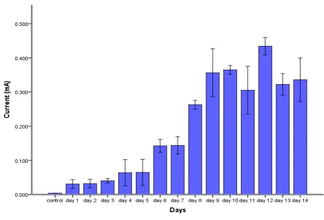
The voltage generated from the decayed wood during the period of 14 days is represented in Fig. 1. The voltages generated were within the range of (0.023± 0.01 to 0.369±0.02) mV. Fig. 2 shows the current generated from the decayed wood. The current produced was within the range of (0.032±0.00 to 0.441±0.02) mA.
Plate 3. PCR amplification of genomic DNA targeted to amplify the 16SrRNA gene of 7 bacterial isolate on 1.0% agarose gel electrophoresis
Key: M = Molecular marker
Plate 4. Phylogenetic tree of bacteria isolate from Tectona grandis wood
; Article no.JAMB.44635
Voltage and Current Generated from
Tectona grandis Wood
voltage generated from the decayed wood during the period of 14 days is represented in Fig. 1. The voltages generated were within the range of (0.023± 0.01 to 0.369±0.02) mV. Fig. 2 shows the current generated from the decayed d was within the range of (0.032±0.00 to 0.441±0.02) mA.
Plate 3. PCR amplification of genomic DNA targeted to amplify the 16SrRNA gene of 7 bacterial
Adegunloye and Ojo; JAMB, 14(3): 1-8, 2019; Article no.JAMB.44635
5
Table 1. Morphological, biochemical characteristics and identification of isolates from decayed Tectona grandis wood
Isolate
no
Col
ony
m
orpho
lo
gy
Gram’s
re
a
ction
Catala
s
e
Coa
gul
as
e
Motil
it
y
Ma
nni
tol
Glu
cose
Fruct
ose
Ma
ltos
e
Lacto
se
Galactose Citr
ate
In
dole
Spore
for
m
in
g
Me
thyl
red t
es
t
Sta
rch
hydrolys
is
Urease t
e
st
Pr
obabl
e
id
entity
1 Cream, circular, opaque, flat, rough
- + NA + A A A A - A + + + - + - Bacillus licheniformis
2 Irregular, creamy-yellow, opaque, smooth, entire
+ + + + + A A A - NA + + + - + - Bacillus cereus
3 Cream, circular, smooth, entire
+ - + - A A A A A - + + - - Bacillus thuringiensis
4 Cream, circular, raised and smooth
+ + NA + A AG AG A A A + + + - + Bacillus subtilis
5 Pale yellow, circular + + + A AG AG A + - NA + + + Staphylococcs aureus
6 Cream , circular, smooth + + - AG A A A + - NA - NA Acinetobacter iwoffii
7 Cream, circular, smooth, raised and lobate
- + - - A - - - + - - N Pseudomonas aueriginosa
Keyword: (+) = positive, (AG) = Acid and Gas, (-) = negative, (A) = Acid, (NA) = not applicable
Table 2. Fungal isolates obtained from the decayed wood
Cultural and microscopy description Isolates
Hyphae broad, not or scarcely septate; rhizoids and stolons present; sporangiophores brown, solitary or in tufts on the stolons, diverging from the point at which the rhizoids form; sporangia rather round; apophysis absent or scarcely apparent;
sporangiophores ovoid.
Rhizopus stolonifer
Yellowish green to dark green hyphae. Conidiophores arising from the mycelium singly or less often in synnemata, branched near the apex, penicillate, ending in a group of phialides
Penicillium notatum
Stipes are smooth, brown and pigmented, vesicles are globose, phialide biseriate, conidia are globose, conidial head are dark green and radiate.
Adegunloye and Ojo; JAMB, 14(3): 1-8, 2019; Article no.JAMB.44635
Table 3. Molecular identification of isolated bacteria from decayed Tectona grandis
Cultural and biochemical identification
Gene sequence identification
Max identity Accession number
Bacillus sp Bacillus licheniformis 100 MH605438.1
Bacillus subtilis Bacillus spp 100 JX025734.1
Acinetobacter 1woffii Acinetobacter iwoffi 98 LT899953.1
Bacillus cereus Bacillus cereus 100 KJ729602.1
Staphylococcus aureus Micrococcus luteus 98 NR_075062.2
Bacillus thuringiensis Bacillus thuringensis 100 JQ289048.1
Pseudomonas aueriginosa Pseudomonas putida 100 JQ782505.1
Fig. 1. Voltage produced from decayed Tectona grandis wood within 14 days
Adegunloye and Ojo; JAMB, 14(3): 1-8, 2019; Article no.JAMB.44635
4. DISCUSSION
In this study, the potentials of decayed Tectona
grandis Linn. to produce current and voltage was
evaluated. The microbial load obtained in this study has shown that decayed wood harbour bacteria and fungi. However, there were differences in total viable count bacterial and fungal counts of Tectona Grandis. It was observed that bacteria counts of 4.5 x10⁵ Cfu/g. were higher than fungal counts 4.2 x 10⁴ Sfu/g. High microbial counts in the decayed woods could be attributed to high moisture content and nutrients such as minerals present in the soil where the woods are fallen. These findings are in agreement with the reports of Janet and Kelechi [11].
Morphological, biochemical and cultural
characteristics of bacterial and fungal isolate revealed the microorganisms that were isolated to include; Bacillus thuringiensis, Acinetobacter
iwoffii, Micrococcus luteus, Bacillus sp
Pseudomonas putida, Bacillus cereus, Bacillus licheniformis while the fungi isolates includes;
Rhizopus stolonifer, Penicilliun notantum and
Aspergillus penicilloides. These
micro-organsisms were probably found on these decayed wood due to high moisture content, the nutrients they derive and their attachacment with the soil since soil harbors many organism. However, the presence of Pseudomonas putida
and Acinetobacter iwoffii is highly uncommon and could have been as a result of contamination or environmental factors such as anthropogenic activities as reported by Chenhui et al. [12] who
confirmed that the microbial community
compositions of fallen logs are affected by both
physicochemical wood properties and
environmental factors. In addition most of the isolated bacteria (Bacillus licheniformis, Bacillus
sp, Acinetobacter iwoffi, Bacillus cereus,
Pseudomonas putida, Micrococcus luteus and
Bacillus thuringiensis) from the decayed wood owned their origin from air and soils this is in agreement with the findings of Singh et al. [13].
Polymerase Chain Reaction revealed that the molecular weight of the genomic DNA of sequenced bacteria in this study is 1500bp. According to the 16S rDNA analyses, selected bacteria showed more than 80% similarity in National Centre for Biotechnology Information (NCBI) database. Results, the isolates confirmed were Bacillus licheniformis, Bacillus sp,
Acinetobacter iwoffi, Bacillus cereus,
Pseudomonas putida, Micrococcus luteus and
Bacillus thuringiensis. The result also revealed a difference in cultural identification of Micrococcus luteus, Bacillus subtilis and Bacillus cereus. This was also reported by Akinyemi and Oyelakin [14], who reported differences in conventional method and molecular method of bacteria identification. However, the results of this study demonstrate clearly the interest and feasibility to introduce the 16S rDNA gene sequencing method in the identification of bacteria, combination of conventional techniques and molecular approach will improve bacteriological investigation and authentication, allowing specific and efficient identification of microorganisms as against cultural method that is probable.
There was a progressive increase in the current generated within the period of 14 days. It was observed that as the current generated increased, there was a decrease at some point in the current generated. This could be as a result of low proton transfer between the anode and cathode when the decayed woods were immersed in water and was kept in the same position throughout the experiment which limited power generation. This is in agreement with the findings of Liu et al. [15]. The highest current generated from the decayed Tectona grandis
was recorded on the day (12) twelve (Figure 2) after which it started decreasing gradually. This is similar with the finding of Chonde et al. [16], who used waste water to generate current and had the highest current generated on day 8 after which there was a decrease in current generated.
Voltage generated from the decayed wood was recorded daily for the entire time period of 14days. The results showed a general increase across the number of days. The maximum voltage generated within 14days was on day (11) eleven, after this was noticed (Figure 1) a definitive increase which there was a definitive decrease, was noticed. The result obtained is comparable with that of Parkash [5] who reported a similar result, for example initially the voltage was raised rapidly but after voltage started falling down.
5. CONCLUSION
This study evaluated the potential of generating alternative electrical energy from decayed
Adegunloye and Ojo; JAMB, 14(3): 1-8, 2019; Article no.JAMB.44635
matter to produce electrical energy using this technology. Also current and voltage production was comparable to those reported for other substrates. Hence, electricity generation using such waste wood by means of MFC technology proffers a promising alternative for electricity generation. We further recommend its trial at large scale as a means to harness an alternative and additional sources of electricity.
COMPETING INTERESTS
Authors have declared that no competing interests exist.
REFERENCES
1. Yoganathan K, Ganesh P. Electrogenicity assessment of Bacillus subtilis and
Bacillus megaterium using microbial fuel cell technology. International Journal of Applied Research. 2015;1(13):435-438.
2. Lee SC, Shih LH. Renewable energy
policy evaluation using real option model-The case of Taiwan. Energy Econ. 2010; 32:567-578.
3. Refaat A. Correlation between the
chemical structure of biodiesel and its physical properties. Int. J. Environ. Sci. Tech. 2009;6(4):677-694.
4. Sharma K, Bulchandani B. Comparative
study of various substrates and
microorganisms in a laboratory designed microbial fuel cell. Int. J Res Chem Environ. 2012;2(3):168-174.
5. Parkash A. Microbial fuel cells: A source of bioenergy. J Microb Biochem Technol. 2016;8:247-255.
6. Sherifat A, Akinsola A, Guido F. Chemical constituents, toxicity and antimicrobial activities of the essential oil from the leaves of Tectona grandis. Elixir Bio Technology. 2013;61:6795-16798.
7. Fawole M, Oso B. Laboratory manual of microbiology. Spectrum Books Limited. 2012;2:112-121.
8. Samson R, Varga J. Aspergillus systematic
in the genomic era. CBS fungal
Biodiversity centre Utrecht. 2007;206.
9. Ologun O, Boboye B, Owoyemi O.
Molecular identification and antibiotic sensitivity pattern of bacteria associated with decompose domestic food waste from Akure Metropolis. Microbiology Research Journal International. 2018;24(3):1-11.
10. Adegunloye D, Olotu T. Generating
electricity using microbial fuel cell powered by benthic mud collected from two locations in Akure, Nigeria. European Scientific Journal. 2017;13(18).
[ISSN: 1857 – 7881]
11. Janet OW, Kelechi H. Microorganisms associated with dumpsites in Port Harcourt Metropolis, Nigeria. Journal of Ecology and the Natural Environment. 2015;8(2):9-12.
12. Chenhui C, Fuzhong W, Wanqin Y,
Zhenfeng X, Rui C, Wei H, Bo T, Meta F. The microbial community in decaying fallen logs varies with critical period in an alpine forest. Environ Sci Technol. 2017;6:3. 13. Singh P, Kumar V, Agrawal S. Evaluation
of phytaseproducing bacteria for their plant growth promoting activities. Int J Microbiol. 2014;1:7.
14. Akinyemi A, Oyelakin O. Molecular
characterization of bacteria isolates from farm-raised cat fish Clarias garipinus. British Microbiology Research Journal. 2014;4(12):1345-1352.
15. Liu H, Cheng S, Logan B. Power
generation in fed-batch microbial fuel cells as a function of ionic strength, temperature and reactor configuration. Environ Sci Technol. 2005;39:5488-5493.
16. Chonde S, Mishra A, Raut P. Bioelectricity production from wastewater using Micro-bial Fuel Cell (MFC); 2013.
_________________________________________________________________________________
© 2019 Adegunloye and Ojo; This is an Open Access article distributed under the terms of the Creative Commons Attribution License (http://creativecommons.org/licenses/by/4.0), which permits unrestricted use, distribution, and reproduction in any medium, provided the original work is properly cited.
Peer-review history: