B I O C H E M I S T R Y
Control of protein function through oxidation
and reduction of persulfidated states
É. Dóka1*, T. Ida2*, M. Dagnell3*, Y. Abiko4*, N. C. Luong4,5*, N. Balog1, T. Takata2, B. Espinosa3,
A. Nishimura2, Q. Cheng3, Y. Funato6, H. Miki6, J. M. Fukuto7, J. R. Prigge8, E. E. Schmidt8†,
E. S. J. Arnér3†, Y. Kumagai4†, T. Akaike2†, P. Nagy1†‡
Irreversible oxidation of Cys residues to sulfinic/sulfonic forms typically impairs protein function. We found that persulfidation (CysSSH) protects Cys from irreversible oxidative loss of function by the formation of CysSSO1-3H derivatives that can subsequently be reduced back to native thiols. Reductive reactivation of oxidized persulfides by the thioredoxin system was demonstrated in albumin, Prx2, and PTP1B. In cells, this mechanism protects and regulates key proteins of signaling pathways, including Prx2, PTEN, PTP1B, HSP90, and KEAP1. Using quantitative mass spectrometry, we show that (i) CysSSH and CysSSO3H species are abundant in mouse liver and enzymatically regulated by the glutathione and thioredoxin systems and (ii) deletion of the thioredoxin-related protein TRP14 in mice altered CysSSH levels on a subset of proteins, predicting a role for TRP14 in persulfide signaling. Furthermore, selenium supplementation, polysulfide treatment, or knockdown of TRP14 mediated cellular responses to EGF, suggesting a role for TrxR1/TRP14-regulated oxidative persulfidation in growth factor responsiveness.
INTRODUCTION
The formation of hydropersulfide (─SSH) and hydropolysulfide (─SSnH)
moieties on protein cysteine (Cys) residues has gained attention as a primary element in hydrogen sulfide (H2S) biology (1–4).
Accumu-lating evidence indicates that per- or polysulfide species can be abundant protein modifications that may possibly act as regulators of enzymatic functions and signaling processes (4–7). In addition, on the basis of their chemical properties, it has been hypothesized that these modifications could serve as a means to protect critical Cys residues against irreversible oxidative inactivation under ele-vated oxidative stress (3, 8, 9). In equivalent conditions, persulfide groups are better nucleophiles than thiols because of the presence of a lone electron pair on the vicinal sulfur atom in the persulfide, resulting in the commonly known alpha effect, which increases reactivity toward oxidants and electrophiles. The stepwise oxidation of a persulfide group leads to the consecutive formation of perthio-sulfenic acid, perthiosulfinic acid, and perthiosulfonic acid (─SSOH, ─SSO2H, and ─SSO3H, respectively; hereafter ─SSO1-3H); the
equivalently oxidized forms of a thiol moiety are sulfenic, sulfinic, and sulfonic acids (─SOH, ─SO2H, and ─SO3H) (3). These two sets of
modifications differ in the presence of an intrinsic disulfide bond in the persulfide derivatives, which renders them reducible by enzymatic or small inorganic disulfide reductants.
Early experimental support of the concept of regulated protein protection by persulfidation by Greiner et al. (8) showed that, upon H2O2 oxidation, the PTEN active-site mutant C71A could only be
reactivated by dithiothreitol (DTT) if it had been preconditioned with NaHS before H2O2 exposure, causing persulfidation of C124
before oxidation. Furthermore, the prevalence of in vivo CysSSOH residues was recently reported, along with the important finding that these species react with dimedone (10). Dimedone labeling was hitherto considered specific for sulfenic acids and has been widely used for their detection (11). The relative contribution of CysSOH versus CysSSOH to dimedone labeling outcomes thus needs to be revisited.
In a cellular context, the important biological oxidant H2O2
reacts primarily with peroxiredoxins (Prxs) (12). On the basis of their high abundance and fast as well as specific reactivity with H2O2, Prx1 and Prx2 likely capture most cytosolic H2O2 (13). Prxs
therefore serve as the cellular frontline antioxidant defense and also act as central hubs in catalytically transmitting oxidative redox signals to other proteins (14). Prx1 and Prx2 are so-called 2-Cys Prxs, having a highly reactive peroxidative (Cp) and a resolving (Cr)
Cys residue. The extraordinary reactivity of Cp toward H2O2 stems
from the unique juxtapositioning of proximal electron-donating and electron-withdrawing functional groups (15). Upon oxidation, the generated Cp sulfenic acid derivative reacts with the Cr on
another Prx subunit within the multimeric enzyme to form an intermolecular disulfide link. The disulfide is recycled to the active reduced form by the thioredoxin (Trx) system. The reaction of Cp
-SOH with Cr is, however, relatively slow; therefore, in the presence
of excess peroxide, Cp can be further oxidized to the corresponding
Cp-SO2H form. This process serves as a redox switch of the
peroxi-dase activity, which can only be reactivated back to the active thiol form in a very slow ATP (adenosine 5′-triphosphate)– and cytosolic Trx1-consuming process catalyzed by sulfiredoxin (16).
Protein tyrosine phosphatase 1B (PTP1B) provides a classic example for redox regulation of a signaling protein (17, 18). As a member of the class I PTP superfamily of phosphatases, PTP1B features an HCX5R active-site motif with a highly nucleophilic Cys residue at
1Department of Molecular Immunology and Toxicology, National Institute of
Oncology, 1122 Budapest, Hungary. 2Department of Environmental Medicine and
Molecular Toxicology, Tohoku University Graduate School of Medicine, 980-8575 Sendai, Japan. 3Department of Medical Biochemistry and Biophysics, Division of
Biochemistry, Karolinska Institutet, SE-171 77 Stockholm, Sweden. 4Environmental
Biology Section, Faculty of Medicine, University of Tsukuba, 305-8575 Tsukuba, Japan. 5Faculty of Pharmacy, Hue University of Medicine and Pharmacy, Hue
University, 06 Ngo Quyen, Hue, Vietnam. 6Department of Cellular Regulation,
Research Institute for Microbial Diseases, Osaka University, Suita, Osaka 565-0871, Japan.
7Department of Chemistry, Sonoma State University, Rohnert Park, Sonoma,
CA 94928, USA. 8Department of Microbiology and Immunology, Montana State
University, Bozeman, MT 59717, USA.
*These authors contributed equally to this work. †These authors should be considered joint senior authors. ‡Corresponding author. Email: [email protected]
Copyright © 2020 The Authors, some rights reserved; exclusive licensee American Association for the Advancement of Science. No claim to original U.S. Government Works. Distributed under a Creative Commons Attribution NonCommercial License 4.0 (CC BY-NC).
on September 17, 2020
http://advances.sciencemag.org/
position 215. Because of its low pKa (where Ka is the acid dissociation
constant) (~5.4), Cys215 is susceptible to oxidation by H
2O2, leading
to enzymatic inactivation (17, 19, 20). Both the Trx and the gluta-thione (GSH) systems have been implicated in the reactivation of PTP1B (17, 18, 21–23). A unique redox property of PTP1B is the reported formation of a sulfenyl amide intermediate via the reaction of the primary sulfenic acid with an adjacent backbone nitrogen. The cyclic sulfenyl amide is readily converted back to the active thiol form by small thiols (DTT and GSH), thus providing a potential transient protection route for the enzyme from overoxidation (19). In addition, the active-site Cys can acquire a variety of modifica-tions inside the cell, such as S-nitrosylation, S-glutathionylation, or S-persulfidation, which similarly inhibit the activity of PTP1B yet allow reactivation by the disulfide reductase systems (23–25). Of particular relevance to this study, Krishnan and coworkers (23) reported that persulfidation of Cys215 is a prevalent modification of
PTP1B in response to endoplasmic reticulum stress, which reversibly inhibits the enzyme. Here, we hypothesized that reduction of the persulfidated Cys215and its oxidized states in PTP1B can provide a reversible regulatory mechanism of control also under oxidative conditions that would otherwise irreversibly destroy PTP1B function. It would also be possible that persulfidation states could thereby affect cellular responses to endogenous oxidative bursts such as those oc-curring during growth factor stimulation.
In the study presented here, we show that persulfidation of protein thiols is reversible by the action of disulfide reductase machineries and demonstrate using genetically modified mouse liver samples that this mechanism regulates the persulfidation status of low–molecular weight (LMW) Cys species and protein Cys residues. We show that persulfidation plays a universal role in protecting protein Cys under oxidative stress in cells. We developed a new mass spectrometry
(MS) method, which allowed quantitative detection of CysSO3H
and CysSSO3H modifications on protein Cys. Using this method,
we demonstrated that protein CysSSO3H derivatives are abundant
modifications in vivo and their levels are mediated by the Trx system in mouse liver samples. Our data also suggest that oxidation and re-duction of persulfidated functional Cys thiols are likely to play a role in redox regulation of cellular signaling via reversible persulfidation and persulfide oxidation of Prx Cp residues and the active-site Cys
of PTP1B. In line with this proposal, we present evidence that, in a cytosolic thioredoxin reductase 1 (TrxR1)– and thioredoxin-related protein of 14 kDa (TRP14)– dependent manner, persulfidation is involved in modulation of epidermal growth factor (EGF)–induced signaling processes in cells.
RESULTS
Persulfidation status is orchestrated by the disulfide reductase machineries in vivo
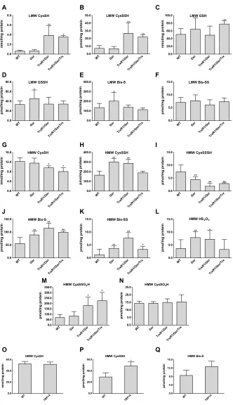
MS-based metabolomics experiments were carried out using mouse liver tissue samples to assess the relative abundance of Cys persulfide modifications and other sulfur species. The mice used in these ex-periments had livers that were genetically engineered so that all hepatocytes were either wild type (WT), homozygous glutathione disulfide reductase (Gsr)–null, double-homozygous TrxR1/Gsr-null, or triple-homozygous TrxR1/Gsr/Trx1–null (26, 27). Consistent with previous data indicating a regulatory role of the Trx and the GSH systems in protein [high–molecular weight (HMW)] persulfide homeostasis (28), we here used a more quantitative approach that
can also assess persulfidation of metabolite (LMW) fractions (29). This showed that persulfidation levels of HMW and LMW mole-cules in the liver samples were critically and in a species- dependent manner responsive to disruptions of either the Trx or the GSH systems (Fig. 1). In the TrxR1-null livers, Cys persulfide (CysSSH) was significantly elevated (Fig. 1B), whereas in the Gsr-null livers, the concentrations of GSH-persulfide (GSSH) and sulfide anion (Bis-S) were significantly higher compared to the corresponding WT levels (Fig. 1, D and E). In the HMW pool, about two times larger protein CysSSH concentrations were measured in Gsr-null and TrxR1/Gsr-null compared to WT livers (Fig. 1H). Moreover, the levels of sulfide and inorganic disulfide (Bis-SS) were at least doubled in these samples, up to approximately fourfold higher levels of HMW Bis-SS in TrxR1/Gsr-null livers (Fig. 1, J and K). The levels of the sulfide oxidation product thiosulfate (HS2O3) also increased in
Gsr- and TrxR1/Gsr-null livers as compared to WT livers (Fig. 1L). These latter LMW sulfur species that appeared in the HMW pool were most likely the result of Cys-polysulfide chain cleavage by the alkylating agent or other artifactual oxidation/hydrolysis liberating these species from proteins, as we demonstrated previously (30). The HMW Cys trisulfide (CysSSSH) pool was significantly smaller in all the mutant samples (Fig. 1I). This may possibly relate to elevated oxidative stress as a result of the disrupted reductase activities (27), because longer polysulfide chains are more prone to oxidation (3, 5). This explanation is also supported by the TrxR1/Gsr/Trx1-null samples, wherein the oxidative burden is highest (27), showing significantly lower Cys levels than in the WT samples (Fig. 1G) and lower CysSSH than in Gsr- or TrxR1/Gsr-null livers (Fig. 1H). These findings provided a foundation for our hypothesis that persulfidation of Cys residues under oxidative stress may play a role in protecting protein thiols from irreversible damage.
To investigate the in vivo relevance of this model, we developed a new quantitative MS method to measure CysSO3H and CysSSO3H
concentrations in liver proteins. The method uses internal standards
that were quantified using authentic CysSO3H and CysSSO3H
compounds (see Materials and Methods and fig. S1). Measurements with this method demonstrated that protein CysSSO3H (Fig. 1M)
was comparable to Cys persulfidation levels (Fig. 1H) in the WT and different mutant mouse liver samples, which were 5 to 20 times
higher compared to the corresponding CysSO3H concentrations
(Fig. 1N). In addition, the elevated levels of CysSSO3H in TrxR1/
Gsr and TrxR1/Gsr/Trx1 but not in Gsr compared to WT livers (Fig. 1M) indicate that the TrxR1-dependent Trx1 system plays a key role in regulating oxidized protein persulfide levels in vivo.
The role of TRP14 in regulation of protein persulfidation in vivo
TRP14 is a unique member of the Trx family because most protein disulfides are not substrates for this protein, in contrast to Trx1, while TRP14 likely plays roles in recovering thiols from protein CysSNO and Cys persulfide derivatives (28, 31). These observations led us to hypothesize that TRP14 may be specific to only a subset of potentially redox-regulated signaling proteins.
To assess whether TRP14 functions in protein persulfide reduction in vivo, we generated a conditional-null allele of the mouse Txndc17 gene encoding TRP14 (fig. S2). Preliminary phenotypic assessment of both full-body and liver-specific homozygous disruption of TRP14 revealed that, unlike the constitutive Trx1-null state, which is embry-onic lethal (27, 32), both liver-specific and constitutively TRP14-null
on September 17, 2020
http://advances.sciencemag.org/
LMW CysSH WT Gsr TrxR1/Gsr TrxR1/Gsr/Trx 0.0 2.0 4.0 6.0 8.0 ** * nm ol /m g pr otein LMW CysSSH WT Gsr TrxR1/Gsr TrxR1/Gsr/Trx 0.0 10.0 20.0 30.0 40.0 50.0 ** ** pm ol /m g pr otein LMW GSH WT Gsr TrxR1/Gsr TrxR1/Gsr/Trx 0.0 20.0 40.0 60.0 80.0 100.0 ** nm ol /m g pr otein LMW GSSH WT Gsr TrxR1/Gsr TrxR1/Gsr/Trx 0.0 20.0 40.0 60.0 80.0 * pm ol /m g pr otein LMW Bis-S WT Gsr TrxR1/Gsr TrxR1/Gsr/Trx 0.0 100.0 200.0 300.0 400.0 * pm ol /m g pr otein LMW Bis-SS WT Gsr TrxR1/Gsr TrxR1/Gsr/Trx 0.0 5.0 10.0 15.0 pm ol /m g pr otein HMW CysSH WT Gsr TrxR1/Gsr TrxR1/Gsr/Trx 0.0 4.0 8.0 12.0 16.0 * * nm ol /m g pr otein HMW CysSSH WT Gsr TrxR1/Gsr TrxR1/Gsr/Trx 0.0 100.0 200.0 300.0 400.0 ** ** pm ol /m g pr otein HMW CysSSSH WT Gsr TrxR1/Gsr TrxR1/Gsr/Trx 0.0 5.0 10.0 15.0 ** ** ** pm ol /m g pr otein HMW Bis-S WT Gsr TrxR1/Gsr TrxR1/Gsr/Trx 0.0 50.0 100.0 150.0 ** ** ** pm ol /m g pr otein HMW Bis-SS WT Gsr TrxR1/Gsr TrxR1/Gsr/Trx 0.0 5.0 10.0 15.0 ** ** * pm ol /m g pr otein
HMW HS2O3
WT Gsr TrxR1/Gsr TrxR1/Gsr/Trx 0.0 5.0 10.0 15.0 ** * pm ol /m g pr otein
HMW CysSSO3H
WT Gsr TrxR1/Gsr TrxR1/Gsr/Trx 0.0 50.0 100.0 150.0 200.0 250.0 300.0 350.0 * * pmol /m g pr otei n
HMW CysSO3H
WT Gsr TrxR1/Gsr TrxR1/Gsr/Trx 0.0 5.0 10.0 15.0 20.0 25.0 pmol /m g pr otei n HMW CysSH WT TRP14 0.0 20.0 40.0 60.0 nm ol /m g pr ot ei n HMW CysSSH WT TRP1 4 0.0 20.0 40.0 60.0 * pm ol /mg prot ei n HMW Bis-S WT TRP1 4 0.0 4.0 8.0 12.0 16.0 pmol/mg pr ot ei n C B A F E D I H G L K J N M Q P O
Fig. 1. Quantitative assessment for the roles of Trx and GSH systems in control of oxidized sulfur species in vivo. Concentrations of LMW (A to F) and protein-
derived (HMW) (G to Q) thiol or persulfide and other oxidized sulfur species were determined using previously described high-sensitivity liquid chromatography–MS/MS protocols with appropriate modifications, as well as a newly developed LC-MS/MS method to detect protein CysSO3H and CysSSO3H (see Materials and Methods). Hepatocytes
in the mouse liver tissue samples were WT, homozygous Gsr-null (Gsr), double-homozygous TrxR1/Gsr-null (TrxR1/Gsr), or triple-homozygous TrxR1/Gsr/Trx1-null (TrxR1/ Gsr/Trx1). Livers of full-body knockout TRP14-null mice were used in (O) to (Q). The indicated LMW (A to F) and protein-derived (G to Q) sulfur species were extracted from deep-frozen tissue samples as described in Materials and Methods. Analyte levels were normalized to total protein concentrations. Data values and errors are means ± SD of measurements from n = 9 (WT), 8 (Gsr), 7 (TrxR1/Gsr), 5 (TrxR1/Gsr/Trx1), and 3 (TRP14) animals, all gender- and age-matched controls (young female adults) with similar feeding and lightning conditions. Significant differences in values compared to WT are indicated (*P < 0.05 and **P < 0.01).
on September 17, 2020
http://advances.sciencemag.org/
mice developed normally and showed no overt defects. Using livers from adult TRP14-null mice, we carried out quantitative liquid chromatography (LC)–MS measurements to assess the impacts of TRP14 on global protein persulfide levels. Moderate but significant elevation of the HMW protein-bound Cys persulfide level was ob-served in the TRP14- null livers compared to age- and sex-matched WT controls (Fig. 1P). These data suggest that TRP14 participates in reduction of specific persulfidated proteins. No significant dif-ferences were found either in LMW Cys persulfide levels or in other sulfide species that we measured (fig. S3), which supports the hy-pothesis that TRP14 is likely responsible for reducing only a subset of persulfidated proteins in vivo.
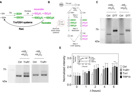
The protective effect of persulfidation illustrated on human serum albumin model protein
The chemical rationale of the hypothesized protective function of persulfidation is depicted in Fig. 2A, illustrating how previous persulfidation provides a possibility of functional reversibility in oxidation of Cys residues together with cellular reductase systems. We used human serum albumin (HSA) as a model protein to investigate the potential Cys-protecting role of per/polysulfidation under oxidizing conditions. The single surface–exposed and non–disulfide- linked Cys residue of HSA, Cys34, is readily polysulfidated upon inorganic polysulfide treatment (28). Here, polysulfide-treated HSA was subsequently further oxidized with a high concentration of H2O2
to generate the corresponding ─SxSOH, ─SxSO2H, and ─SxSO3H forms
(x ≥ 1, referring to a mixture of different chain lengths). A nonpoly-sulfidated control was included to yield the expected ─SOH, ─SO2H,
and ─SO3H oxidation products. The protein samples with these
oxidized modifications on Cys34 were subsequently treated with
DTT, and the reductive liberation of free HSA thiol form was finally monitored by labeling with the thiol-reactive fluorescent agent 5-iodoacetamido fluorescein (5-IAF), followed by nonreducing denaturing electrophoretic separation [SDS–polyacrylamide gel electrophoresis (SDS-PAGE)] and fluorimetric detection (Fig. 2B). DTT reduction led to an increased level of fluorescent labeling when the protein had been persulfidated in step 1 before H2O2 oxidation
(Fig. 2C). This observation corroborates the hypothesis that, because of the presence of an intrinsic disulfide bond within the oxidative modification, precursor polysulfidation of Cys in proteins may yield reducible oxidation products, in contrast to the same oxidative challenge on the corresponding native Cys thiol.
Enzymatic reduction of oxidized HSA polysulfide species We previously reported that TrxR1 has polysulfide reductase capacity, further potentiated by the TrxR1 substrate Trx1 or TRP14 (28). On the basis of this, we here tested whether these members of the Trx system can also reduce oxidized HSA polysulfide species. Using the same workflow as shown in Fig. 2B, DTT was substituted in step 3 by NADPH (reduced form of nicotinamide adenine dinucleotide phosphate) and TrxR1. The higher recovered thiol content in the polysulfide-treated samples compared to the untreated controls further illustrated the protective role of polysulfidation and, at the
same time, demonstrated that TrxR1 can reduce HSA-SxSO1-3H
species (Fig. 2D). Control experiments revealed that the recovery of the thiol groups from oxidized persulfide species by TrxR1 was approximately 60% under these conditions (fig. S4B). Next, we investigated whether the addition of Trx1 or TRP14 could further accelerate the reduction, which was not the case under these conditions,
and all samples with TrxR1 were recovered at similar rates (Fig. 2E). Using mutant TrxR1 enzymes in which its active-site selenocysteine (Sec) residue was replaced with Ser or Cys, we demonstrated that the direct HSA-SxSO1-3H reductase activity of TrxR1 is Sec dependent
(fig. S4A).
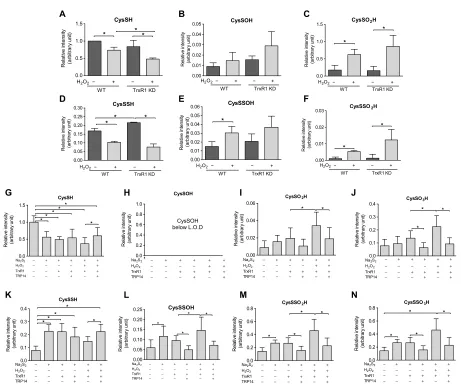
Persulfidation of the peroxidative cysteine protects Prx2 against overoxidation
Prxs are the first line of defense against H2O2 and other peroxides in
cells. MS revealed that approximately 15% of Prx2 Cp was detected in
its persulfidated form in normally cultured human embryonic kidney (HEK) 293 cells under the applied conditions (Fig. 3, A and D). The persulfidated fraction of Cp decreased upon treating cells with
H2O2 (Fig. 3D) with a concomitant increase in the corresponding
oxidized perthiosulfenic acid derivative (Cp-SSOH; Fig. 3E) and the
perthiosulfinic form (Cp-SSO2H; Fig. 3F). Following knockdown
(KD) of TrxR1, both the persulfidated (Fig. 3D) and perthiosulfinic acid (Fig. 3F) derivatives of Prx2 Cp were elevated compared to normal
controls, suggesting a role for the Trx system in recovering the active Cp thiol from the persulfidated Prx Cp. Oxidation of the native
thiol form of Cp followed a similar pattern (Fig. 3, B and C).
The reversal of the Prx2 oxidized persulfide species with or without exogenously added H2O2 display different, but not significantly
different, steady-state levels between WT and TrxR1 KD cells (see Fig. 3, E and F). In addition, the role of TrxR1 in this system is rather complicated because, as detailed in Introduction, it is also an electron donor for Prxs via Trx1. Therefore, to investigate the Prx persulfide and oxidized persulfide reducing capacity of the Trx system, we performed MS-based experiments on isolated Prx2. As shown in Fig. 3 (G to N), when Prx2 was treated with Na2S2 followed by H2O2,
TrxR1 alone (in the presence of NADPH) cannot reduce CysSSO2-3H
modifications, but in the presence of TRP14, significantly less of these derivatives were detected. Despite this, it is well known that the Trx system cannot reduce CysSO2-3H species; significantly higher
levels of CysSO2H and CysSO3H were detected after consecutive
Na2S2 and H2O2 treatment followed by treatment with TrxR1 alone
compared to when TRP14 was also added. Therefore, we checked the possibility whether the reason of the increased CysSO3H levels
could be due to decomposition or reorganization of a fraction of the elevated levels of CysSSO3H species in these samples [this type of
chemistry has been reported previously (33)]. Using the authentic
CysSSO3H compound, we observed the formation of CysSO3H
upon incubation in phosphate buffer or upon the addition of formic acid under oxidative conditions, which may serve as an explanation for the increased levels of sulfinic and sulfonic acid forms in samples that contain larger amounts of CysSSO2-3H derivatives.
Moreover, we observed a drop in the levels of the peroxidative Cys thiol upon consecutive treatment of Prx2 with Na2S2, H2O2,
and NADPH/TrxR1/TRP14. This might be due to more Prx2 dimer formation upon polysulfide treatment (see fig. S5I), which cannot be reduced by TrxR1/TRP14 (34). The fact that the perthio- modifications were observed in samples, which had no polysulfide added to them, may be the result of noncomplete reduction of endogenous peroxidative Cys persulfides by the low amount (1 mM) of DTT during the protein purification phase.
On technical grounds, note that it is challenging to adequately measure the redox states of Prx Cp. Normally, large concentrations
of fast-reacting alkylating agents must be used to outcompete cell lysis–induced artifactual oxidation of this extremely reactive thiol
on September 17, 2020
http://advances.sciencemag.org/
(35). However, because under these conditions protein Cys per- and polysulfides are destroyed via alkylating agent–induced polysulfur chain cleavage (30, 36), we here used mild alkylating conditions to capture the perthio- derivatives. Even milder electrophiles such as iodoacetamide have the potential to disrupt polysulfide chains on longer time scales (30). Figure S5 shows a case when tryptic digest was carried out in the absence of iodoacetamide (for 10 hours), in which case a high amount of ─SSO3H form was detected upon H2O2
treatment of HEK293 cells, which was significantly elevated in TrxR1 KD compared to WT cells (also in the absence of H2O2 treatment).
In addition, the formation of CysSO3H was significantly impaired
in TrxR1 KD compared to WT cells, further corroborating the role of TrxR1 in the persulfide-mediated protection of the Prx2 Cp thiol.
However, it has to be noted that, using these mild alkylating conditions, the relative amounts of oxidation observed on the Cys residues of Prx compared to the reduced thiol form are likely to be overestimated. Nevertheless, our data indicate that persulfidation of the peroxidative Cys residue of Prx2 is a potential protecting mechanism against overoxidation and provide strong support that TRP14 coupled to TrxR1, which cannot reduce Prx2 dimers, is likely to be involved in the recovery of the active peroxidative thiol from its persulfide and oxidized persulfide derivatives. This model could potentially give
another layer of regulation for Prx functions not only in cellular antioxidant protection but also in mediating redox signaling (see Discussion), which is worthy and require future comprehensive investigations to make firm mechanistic conclusions. These are outside the scope of the present study.
Persulfidation is a universal protective mechanism of protein Cys residues in oxidative stress
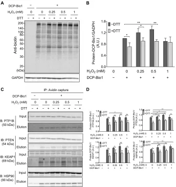
The reversible formation and physiological relevance of perthiosulfenic acid (─SSOH) species was recently reported by Heppner et al. (10). Historically, dimedone has been used to discriminate sulfenic (─SOH) derivatives from thiol (─SH) groups during oxidation mediated by reactive oxygen species (ROS) (11). However, we recently demonstrated that dimedone also reacts with perthiosulfenic acid (─SSOH) groups (10). In the study presented here, we subjected A431 cells to oxidative stress in the form of H2O2 treatment (Fig. 4), and oxidized protein
modifications were captured by labeling with DCP-Bio1, a biotin- tagged dimedone-based probe. We found that about 30% of the DCP-Bio1–labeled untreated A431 cellular proteome was reducible by DTT, representing the fraction of the dimedone-labeled proteome that contained a disulfide bond and therefore was the portion corresponding to perthiosulfenic acid rather than sulfenic acid
A B C
D E
Fig. 2. Reversible oxidation of HSA upon previous persulfidation. (A) Hypothesized role of the Trx or GSH system in reduction of excessively oxidized persulfide
species, which become functionally reversible oxidation states (green), while nonpersulfidated Cys become irreversibly oxidized (magenta). (B) Experimental workflow for generation and detection of HSA-SSO1-3H species (for details, see Materials and Methods) (C and D) Fluorescent gels (n = 3 and 5, respectively) resulting from an
experiment presented in (B) using DTT or TrxR1 as reducing agent. Excessively oxidized polysulfide species are more prone to DTT and NADPH (reduced form of nicotinamide adenine dinucleotide phosphate)/TrxR1 reduction compared to their thiol counterparts. (E) Addition of Trx1 or TRP14 does not increase the TrxR1 catalyzed reduction rate of HSA-SSO1-3H species. A representative time-resolved gel with WT TrxR1 is shown in fig. S4A (upper gel). Fluorescent signal intensities were determined densitometrically
at each time point and normalized to the respective intensities at t = 0. Data points and error bars represent means ± SD of n = 5 experiments (*P < 0.05, **P < 0.01, and ***P < 0.001). The inset shows the derived kinetic curves, and the dashed lines represent a linear fit of the control (Ctrl) dataset and a single exponential least-square fit of all data points from the enzymatically reduced samples.
on September 17, 2020
http://advances.sciencemag.org/
moieties (Fig. 4, A and B). As we reported earlier, protein labeling could result from either persulfide oxidation or polysulfide hydrolysis (30). To resolve the relative contributions of the oxidative effects, the samples were titrated with H2O2. The increase in dimedone-
labeled proteins with increased H2O2 (up to 0.5 mM H2O2) indicated
that a substantial proportion of the observed DTT-reducible perthio-sulfenic acid pool arose from oxidative processes. Thus, the proposed mechanism in Fig. 2A might universally protect protein Cys residues during oxidative stress. The observed drop in Cys-dimedonylation at the highest H2O2 concentration (1 mM) is indicative of further
oxidation to give, among others, perthiosulfinic or perthiosulfonic
acid derivatives, as proposed in our model (Fig. 2A), which would not be labeled with dimedone. To identify specific protein targets under these experimental conditions, we affinity-captured the biotinylated fraction (representing dimedone-labeled protein Cys derivatives) and performed Western blot analyses against proteins well known to be subjected to redox regulation or have significant roles in antioxidant protection (Fig. 4, C and D). The protective effect of persulfidation against irreversible oxidation of Cys residues was apparent for all investigated proteins with redox-active thiols, namely, PTP1B, PTEN, KEAP1, and HSP90. The fact that PTP1B was affected by perthiosulfenylation was particularly interesting because previous
A B C
D E F
H
G I J
L
K M N
Fig. 3. Persulfidation of the peroxidative cysteine of Prx2 is modulated by the Trx system. (A to F) WT and TrxR1 KD HEK293 cells were exposed to 600 M H2O2
in Hanks’ balanced salt solution buffer at 37°C for 5 min and lysed in radioimmunoprecipitation assay (RIPA) buffer without SDS containing 5 mM iodoacetamide. The lysates were immunoprecipitated with anti-PRDX2 antibody. The eluted protein was subjected to tryptic digest in the presence of 5 mM iodoacetamide. (G to N) Recombinant human Prx2 (hPrx2) (12 M) was incubated with or without 100 M Na2S2 for 10 min, followed by 1 mM H2O2 for 10 min at 25°C. Samples were
desalted and treated with or without TrxR1 (250 nM), TRP14 (4 M), and NADPH (1 mM) for 1 hour at 37°C. The samples were alkylated with 1 mM iodoacetamide for 30 min, followed by tryptic digestion (37°C, 10 hours). Following LC–quadrupole time-of-flight (Q-TOF)–MS analyses, Mascot search refined the active-site Cys51-containing
tryptic peptide (see at the top). Cys modifications were identified as shown in the panel headlines. The obtained intensities for modified peptides were normalized to the 93-EGGLGPLNIPLLADVTR-109 peptide fragment of Prx2 from the corresponding tryptic digest. Intensity values are plotted as a relative value compared to the CysSH peptide form from the untreated WT cells (A to F) or recombinant Prx2 (G to N). Data points and error bars represent means ± SD of n = 3 (A to F) and n = 9 experiments (G to N). *P < 0.05.
on September 17, 2020
http://advances.sciencemag.org/
findings suggested that Prx2 cannot transfer oxidative equivalents to PTP1B (22). Although redox regulation of PTP1B has major effects on growth factor signaling, the actual mechanisms of its oxidative inhibition are still unknown. Therefore, we further investigated how persulfidation and the Trx system can modulate PTP1B activities.
Protective effect of persulfidation against irreversible oxidative inhibition of PTP1B
First, we tested whether polysulfide pretreatment could alleviate H2O2-induced irreversible inhibition of pure PTP1B in vitro.
Consistent with a previous report (23), incubation of PTP1B with 50 M Na2S2 for 10 min decreased its catalytic activity by about 70%
(Fig. 5A). Subsequent treatment of this incubation mixture with 100 M H2O2 resulted in further inhibition of PTP1B activity (Fig. 5A).
DTT could partially restore PTP1B activity that was diminished by H2O2 alone (to about 25% of the initial activity after 30-min incubation)
but more robustly recovered H2O2-dependent activity loss when the
enzyme had been preconditioned with Na2S2 (Fig. 5A). However,
when Na2S2 addition to the reaction mixture was subsequent to H2O2
treatment, it had no significant effect on the recovery of enzymatic activity by DTT. These data indicate that preemptive persulfidation can preserve reduction-mediated recovery of oxidation- induced PTP1B activity loss.
Reduction of oxidized persulfidated PTP1B by the Trx system
Previous findings show that the Trx system has prominent roles in reactivation of oxidized PTP1B (21, 22). On the basis of our results
A B
C D
−
KEAP1 KEAP1 (A.U.) HSP90 (A.U.)
HSP90
Fig. 4. H2O2-dependent formation of cellular protein SxSOH in A431 cells. (A and B) DTT-reversible formation of oxidative modifications susceptive to dimedone labeling from whole-cell lysates. (A) A431 cells were exposed to increasing amount of H2O2 and disrupted by lysis buffer containing DCP-Bio1, a specific sulfenic acid
probe containing a biotin tag. Control samples were included without the labeling agent. Cell lysates were then incubated with or without DTT, subjected to nonreducing SDS-PAGE, and immunoblotted (IB) against biotin. (B) Band intensities of the Western blot results (obtained by densitometric analyses) were plotted as a ratio of the untreated sample (─H2O2 and ─DTT). A.U., arbitrary units. (C and D) DCP-Bio1–labeled proteins were affinity-captured on avidin beads from whole-cell lysates generated
as in (A), and specific proteins were identified from the eluates by Western blotting. (D) Band intensities of the membranes shown in (C). Western blots shown in (A) and (C) are representative of n = 3 independent experiments. The band intensities were assessed by densitometry using ImageJ software and normalized to (B) glyceraldehyde- 3-phosphate dehydrogenase (GAPDH) or (D) the respective protein band intensities from the input gels [whole-cell lysate before immunoprecipitation (IP)]. Each value is the mean ± SD of n = 3 independent experiments. *P < 0.05 and **P < 0.01.
on September 17, 2020
http://advances.sciencemag.org/
with HSA, we hypothesized that the Trx system would more efficiently restore oxidation-diminished PTP1B activity when the oxidative insult is preceded by persulfidation of the active-site Cys215. We found that Trx1, TRP14, and the thioredoxin-related protein of 32 kDa (TRP32) each potentiated reactivation of oxidized PTP1B-persulfide
species as compared to the corresponding oxidized thiol form that had not been pretreated with polysulfide (Fig. 5, A and B). In contrast to its activity on HSA, TrxR1 alone was inefficient at reactivating PTP1B after consecutive treatment with Na2S2 and H2O2, but in the
presence of Trx1, TRP14, or TRP32, it became highly efficient. In A
C
G
K
O
D
H
L
P
E
I
M
Q
F
J
N
R B
Fig. 5. Reversible inhibition of PTP1B and concomitant formation of active-site CysSSO1-3H derivatives upon inorganic polysulfide and H2O2 treatment. Recombinant
human PTP1B [(A and B), 13.5 M; (C to R), 20 M] was incubated with or without 50 M Na2S2 for 10 min followed by 100 M H2O2 for 10 min at 25°C. Samples were then
treated with or without Trx1 (15 M), TRP14 (15 M), or TRP32 (15 M) with TrxR1 (250 nM) in NADPH [(A and B), 500 M; (C to R), 1 mM] or 20 mM DTT for 30 min (A and C to J) or 60 min (B and K to R) at 37°C. (A and B) Phosphatase activity of PTP1B was measured in spectrophotometric assays using para-nitrophenyl phosphate (pNPP) as substrate. The control activity (PTP1B stock; last bar to the right) represents the untreated sample. (C to R) Protein samples were alkylated with 1 mM iodoacetamide for 30 min, digested with trypsin (37°C, 10 hours), and analyzed by LC-Q-TOF-MS. Peptides with modified active-site Cys derivatives were determined using Mascot; their levels were normalized to the intensities of the corresponding 157-QLELENLTTQETR-169 tryptic peptides and shown as relative values to the Cys thiol form in nontreat-ed PTP1B set as 1.0. Data values are means ± SD of n = 3 (A to J) or n = 6 (K to R) experiments; *P < 0.05; **P < 0.01, and ***P < 0.001. L.O.D., limit of detection.
on September 17, 2020
http://advances.sciencemag.org/
the presence of NADPH, TrxR1 seemingly catalyzed the inactivation of PTP1B compared to the control sample. This latter observation may be due to the previously reported NADPH oxidase activity of TrxR1, which is apparent when there is no reductase substrate around (37). Trx1 and TRP14 had the largest PTP1B reactivating capacity under the applied experimental conditions.
Formation of active-site Cys215 persulfide derivatives
in PTP1B
To gain further insights into how polysulfide treatment aids reduction- mediated recovery of oxidatively inhibited PTP1B activity, we used ultra-performance liquid chromatography-tandem mass spectrom-etry (UPLC-MS/MS) to investigate the posttranslational modifications of the active-site Cys215 residue upon polysulfide and/or H
2O2
treat-ments. As shown in fig. S6 (A and B), incubation with Na2S2
lowered the amount of the CysSH form and induced concomitant formation of the persulfidated species. This modification was fully reversible by DTT. Corroborating our model on Fig. 2A, consecu-tive Na2S2 and H2O2 treatment converted the PTP1B Cys215-SH thiols
to oxidized persulfide species, some of which were susceptible to dimedone labeling (fig. S6, A to D). These dimedone-labeled deriv-atives were entirely reducible by DTT (fig. S6D). DTT reduction should result in thiodimedone (dimedone-SH) release. To optimize detection of thiodimedone upon release from derivatized persulfide species, we used a well-characterized thiol-activated Sepharose 4B bead system, with which these oxidative modifications were previously demon-strated (9). The activated resin was persulfidated and then treated with H2O2 and dimedone to obtain R-SS-dimedone arms on the Sepharose
beads. Next, DTT was added to the incubation mixture to cleave the generated disulfide bonds. The retention time, mass, and fragmenta-tion pattern of the released thiodimedone product were finally charac-terized by UPLC-MS analyses (fig. S6, E to I). Using similar LC-MS/MS conditions, the dimedone-SH product could be detected using PTP1B samples pretreated with polysulfide and oxidized with H2O2 followed
by reduction with DTT (fig. S6, E and J to L), thus corroborating the formation of PTP1B-SxSOH species during Na2S2 and H2O2 treatment.
To further characterize the generated oxidative posttranslational modifications on Cys215 and their relevance in the Trx system–
mediated recovery of PTP1B activity, we used high-resolution LC-MS with quantitative quadrupole time-of-flight (LC-MS-Q-TOF) detection under similar conditions that were applied for the kinetic assays. On the basis of measured exact molecular masses, retention times, and fragmentation patterns of the Cys215-containing tryptic peptides, we validated the formation of Cys persulfide and oxidized perthio- derivatives at the active site of PTP1B and found additional evidence that these modifications are reduced by TrxR1 coupled to Trx1, TRP14, or TRP32 (Fig. 5, I, J, Q, and R). In line with the enzyme activity data (Fig. 5, A and B) at the 60-min time point after the addition of the Trx system, reduction of the oxidized persulfidated forms (Fig. 5, Q and R) was found to be more prominent compared to the 30-min time point (Fig. 5, I and J), which indicates that (similar to the HSA system) the enzymatic recovery of the PTP1B active-site Cys215 thiol
from its CysSO2-3H forms is relatively slow. The slight but significantly
lower sulfinic or sulfonic acid forms in some of the samples that were treated with different compositions of the Trx system may be due to similar effects that are discussed at the Prx2 section. The elevated levels of CysSO3H in TRP32-treated samples at the 60-min time
point were unexpected, yet it occurred in all replicates and is worthy of further investigations.
The formation and reduction of Cys215 oxidized persulfide products corroborate that the recovery of PTP1B activity shown in Fig. 5 (A and B), which was measured under similar experimental conditions, is likely to be largely attributed to the model proposed in Fig. 2A, wherein the restoration of enzymatic activity is due to cleavage of polysulfide-induced disulfide bonds in ─SSH, ─SSOH, and ─SSO2,3H
functional groups on Cys215. The fact that other LMW thiol–containing reducing agents such as Cys 2-mercaptoethanol (2-ME), and GSH functioned in recovery of PTP1B activity upon Na2S2 and H2O2
treatments further supports this proposal (fig. S7). In the case of these monothiols, some mixed disulfides can be generated; therefore, these small monothiols are slightly weaker at restoring activity to PTP1B as compared to DTT, for which thiols are the sole protein Cys–containing product (38).
Effect of TRP14-mediated persulfidation on EGF-induced protein phosphorylation
EGF stimulation of A431 cells is known to lead to PTP1B oxidation and inactivation, which enables EGF-triggered intracellular phos-phorylation cascades (17). Using the A431 cell model, we found that pretreatment of A431 cells with polysulfide before EGF stimulation markedly increased both total EGF-dependent protein phosphorylation and specific EGF receptor (EGFR) phosphorylation (Fig. 6, A to C). As reported before and shown above, oxidized PTP1B can be reactivated
by TrxR1 together with Trx1 or with TRP14 (21). Work by the
Tonks laboratory identified a greater potency of the Trx system to recover enzymatic activity of persulfidated as compared to H2O2-
inactivated recombinant PTP1B (23). In addition, our studies
demonstrate that the TrxR1/TRP14 system potently reduces specific persulfidated proteins in cells (28) and in mouse liver (see above) and can reverse the activity of PTP1B by reducing its persulfidated and oxidized persulfide derivatives (see above). Moreover, the increases seen in phosphorylation upon polysulfide and EGF treatment suggest that EGFR and the involved protein kinases are more resistant to inhibi-tion by persulfidainhibi-tion than PTP1B and potentially other phosphatases. Considering the importance of TRP14 and its dependence on the selenoprotein TrxR1, we hypothesized that either KD of TRP14 or lack of selenium supplementation (required to yield adequate activity of TrxR1) would potentiate the effect of polysulfide treatment on protein phosphorylation upon EGF stimulation. In addition, in HEK293 cells, we found that polysulfide pretreatment potentiated EGF-dependent phosphorylation, and in these cells, we could knock down TRP14, which also potentiated the effects of EGF stimulation (Fig. 6D). Moreover, fully supplementing the cells with selenium inhibited early responses to EGF in A431 cells, and more polysulfide treatment was needed to yield protein phosphorylation compared to cells that had not been saturated with selenium (fig. S8).
DISCUSSION
Here, we have shown that persulfidation can protect Cys residues from irreversible overoxidation and that the GSH and Trx system, particularly TrxR1 together with TRP14, can reduce oxidized persulfidated proteins. We furthermore found that persulfidation can modulate protein phosphorylation cascades in response to growth factor treatment, and we propose that persulfidation and redox regulation of PTPs such as PTP1B underpin these observations.
Our study should be considered in light of redox regulatory pathways in general, but perhaps especially observations related to
on September 17, 2020
http://advances.sciencemag.org/
EGF 0 IB: Total pTyr
0
2 4 6 2 4 6 min
IB: EGFR IB: p1148 IB: EGFR EGF 0 IB: p992 0
2 4 6 2 4 6 min
IB: EGFR
EGF 0 2 4 6 0 2 4 6 min
EGF −
TRP14 KD
− − + + + − − − + + +100 WT
500 100 500 100 500 100 500 IB: Total pTyr
WT WT 10
0 WT 50
0 WT EG
F
WT EGF 1 00
WT EGF 50014KD
14KD 100 14KD 500 14KD EGF
14KD EGF 10 0
14KD EGF 50
0 0 20 40 60 80 Total pTyr/P on ceau * * * Contr ol EGF 2
′ EGF 4
′ EGF 6
′ 0 5 10 15 p992 /E GF R * Contr ol
EGF 2 ′
EGF 4 ′
EGF 6
′
′
Contr ol Na
2
S3
EGF 2 ′ Na2
S3
EGF 4 ′ Na2
S3
EGF 6 ′ Na2
S3 0 2 4 6 8 10 To ta l pT yr /E GF R * Contr ol
EGF 2 ′
EGF 4 ′
EGF 6 ′ 0 2 4 6 8 10 p1148 /E GF R *
Na2S3
Na2S3
Na2S3
Na2S3
Contr ol Na
2
S3
EGF 2 ′ Na2
S3
EGF 4 ′ Na2
S3
EGF 6 ′ Na2
S3
Contr ol Na
2
S3
EGF 2 ′ Na2
S3
EGF 4 ′ Na2
S3
EGF 6 ′ Na2
S3 A
B
C
D
Fig. 6. Effect of persulfidation on EGF-induced phosphorylation cascades. (A) Overnight starved A431 cells were pretreated with 500 M sodium polysulfide (Na2S3)
for 10 min and subsequently stimulated with EGF ligand (100 ng/ml) for the indicated times. (B and C) A431 cells were stimulated as in (A) and immunoblotted against EGFR phosphorylation sites, p1148 and p992. (D) Overnight starved WT and TRP14-deficient HEK293 (TRP14 KD) cells were pretreated with the indicated amounts of sodium polysulfide (Na2S3) for 10 min and subsequently stimulated with EGF ligand for 2 min. Total EGFR phosphorylation and EGFR expression were determined
by immunoblotting using antibodies against phosphotyrosine (4G10) and EGFR, respectively (left). Equal protein loadings were confirmed by Ponceau S staining. Phosphorylation intensities were measured by densitometric analyses (right side of the panels) and normalized to respective EGFR expression levels. Western blots are representative of n = 3 (B to D) or n = 4 experiments (A). Data points are means ± SD of the indicated number of repeats; *P < 0.05. Significance refers to the untreated control samples (Control), unless otherwise specified.
on September 17, 2020
http://advances.sciencemag.org/
H2S signaling. Soon after it was first proposed that H2S could play
important mediatory functions in biology, a number of investigations reported that it participates in different pathophysiological conditions by alleviating oxidative stress–induced damage. This included protection against ROS-induced damage in brain, gastric mucosa and hepatic ischemia-reperfusion injury, as well as vascular endothelium in hypoglycemia [see (4) and references therein]. Initially, those effects were proposed to be due to direct scavenging of ROS. However, we and others argued that, because of the low physiological concentrations of free sulfide, even the fastest ROS scavenging reactions (e.g., the reduction of HOCl by sulfide, which is almost diffusion-controlled) would not be kinetically competitive in the presence of millimolar concentrations of GSH and protein thiols (39). We proposed that a potential mechanism to explain the protecting effects of sulfide, and of polysulfide species, could be associated with their reactions with metal centers of metalloproteins (4), as demonstrated with myeloperoxidase (40) or hemoglobin (41). Sulfide efficiently reduces the highly oxidizing and reactive ferryl heme intermediates of these enzymes, with concomitant production of polysulfide species (42) that are the most effective persulfidating agents of protein Cys side chains. Alternative redox-mediated events could also potentially induce protein per/polysulfidation under oxidative stress, as we reviewed previously (4). Thus, we proposed that per/polysulfidation contributes to protection against oxidative stress. In the present study, we have systematically addressed the physiological relevance of persulfidation- mediated protection of protein Cys residues.
Cys persulfidation and oxidation of persulfides
We previously introduced the ProPerDP method as a convenient tool for protein persulfide detection (28). In the present study, state-of-the-art LC-MS–based methods allowed a more quantitative determination of Cys persulfide and trisulfide species, which confirmed the elevated levels of both protein-derived and LMW CysSSH in liver tissues of mice with disrupted functions of the NADPH-dependent reductases glutathione reductase (Gsr) and/or TrxR1. As we measured lower steady-state concentrations of the more oxidant-sensitive CysSSSH species in livers in which the NADPH reductase machineries were disrupted, this may possibly suggest that these species play roles in scavenging a proportion of the elevated ROS in these livers. To corroborate this hypothesis, we developed a quantitative MS method to measure protein CysSO3H and CysSSO3H
levels in tissue samples. Our method revealed that CysSSO3H is an
abundant protein modification in mouse liver tissue. The fact that, compared to WT livers, CysSSO3H levels were significantly elevated
in TrxR1/Gsr and TrxR1/Gsr/Trx1-null livers, in which cells are exposed to excessive oxidative stress, supports our model that per-sulfidation protects cellular protein Cys residues during oxidative stress in vivo. The fact that, compared to WT, CysSSO3H levels were
not elevated in Gsr livers, in which only the GSH-based reduction was impaired, suggests that oxidized persulfide species are primarily reduced by the Trx system.
A large body of literature reports the intracellular formation of protein sulfenic acids under mild oxidative stress (11). Most of these studies are based on the reactivity of dimedone derivatives with CysSOH. However, Heppner et al. (10) recently showed that these conditions also generate abundant Cys-SSOH modifications, which are labeled by dimedone and thereby result in disulfide-linked Cys-SS-dimedone products. Moreover, we hypothesized that the presence of dimedone in a biological system is capable of shifting
the hydrolysis equilibria of protein persulfides toward the formation of ─SSOH species, meaning that a fraction of the putative sulfenylated proteome originates from the protein persulfide pool (30). Here, we have further corroborated that considerable amounts of reducible dimedone-reactive protein modifications are generated by extracellular H2O2, which is attributable to the process of perthiosulfenylation.
Note that CysSSO2H and CysSSO3H are unreactive toward dimedone,
so the method does not account for those species. As we identified perthiosulfenylated species of protein targets connected to antioxidant protection (KEAP1), chaperone activity with various posttranslational modification (HSP90), and redox-regulated signaling proteins (PTP1B and PTEN), it seems clear that this reducible protein modification can have major biological roles especially in light that it is likely to be enzymatically regulated and controlled.
Roles of the Trx1 system in regulating
persulfidation-mediated protein thiol protection
Components of the cytosolic Trx system (TrxR1 alone or coupled with Trx1 or TRP14) were previously shown to exhibit protein per-sulfide reductase activity (28). Here, we found that TrxR1 can directly reduce oxidized persulfide species into thiols. The classical TrxR1 substrates Trx1 and TRP14, generally involved in protein disulfide or persulfide reduction reactions (28, 31, 43), had no additional effects on the reduction rates of oxidized HSA polysulfide species. Although its biological relevance may be disputed considering that HSA is found in serum and TrxR1 is cytosolic, in case this activity is not unique to HSA, then it could potentially represent a previously unrecognized property of TrxR1.
The Trx system was previously found to be more effective in reactivating both H2O2-treated and persulfidated PTP1B compared
to the GSH system (21–25). In mouse embryonic fibroblasts, we found that the alternative TrxR1 substrate TRP14 also has the capability to reactivate PTP1B (21, 31). Under similar conditions, the oxidized form of SHP2, a nonreceptor type PTP, remained unaffected by both Trx1 and TRP14. In addition, recently, we reported that TrxR1 alone protects PTP1B from excessive oxidation, most likely by directly reducing the sulfenic acid intermediate (22). These studies collectively suggest that the members of the Trx family contribute to the regulation of phosphorylation signaling in a substrate-specific targeted manner. Here, we found that TrxR1 has no significant reducing activity toward oxidized persulfide derivatives of Cys215 in enzymatic assays of PTP1B. We also found that, unlike the case for oxidized persulfidated HSA, TRP14 or Trx1 coupled to TrxR1 increased the reducing efficiency. Using the livers from TRP14-null mice generated for this study, we could assess quantitative information of the persulfide reductase activity of TRP14, noting only slightly elevated protein CysSSH levels in TRP14-null livers compared to WT, while no difference was detected in the LMW persulfide or related reactive sulfur species pools. These observations further corroborate the delicate substrate specificity of different components of the disulfide reductase systems. We hypothesize that the GSH system is primarily responsible for protein persulfide homeostasis, while the Trx system, perhaps especially TRP14, can target a specific subset of proteins. Here, note that TRP14 catalyzed the recovery of persulfidated Prx2 and PTP1B (Figs. 3, L to N, and 5, I, J, Q, and R) but not similarly modified HSA (Fig. 2E). A comprehensive proteomics approach will be necessary in the future to identify protein persulfide modifications specifically reduced by TRP14 or other members of the Trx or GSH systems.
on September 17, 2020
http://advances.sciencemag.org/
Cellular implementation of the persulfide-based thiol protection mechanism
As a potential antioxidant defense mechanism, the relative importance of persulfidation was investigated on Prxs, the primary cellular peroxide-detoxification enzyme family. Note that cytosolic Prx2 reduces H2O2 with a rate constant of ~107 M−1 s−1 and can transmit
redox signals to redox-regulated proteins, thus interlinking oxidative stress protection and signaling. Note, in this context, that a considerable fraction of the peroxidative Cys of Prx2 is persulfidated in HEK293 cells at steady-state levels and that its persulfidation is further
in-creased upon TrxR1 KD. Our experiments also showed that H2O2
treatment generated overoxidized Cys51-SSO
2H and Cys51-SSO3H
species in normal cells. In vitro experiments on purified Prx2 con-firmed that persulfidation of the peroxidative Cys and its oxidized persulfide forms are reduced back to the active thiol by TRP14 coupled to TrxR1 but not with TrxR1 alone. These findings suggest that persulfidation of Prx2 may protect the enzyme from permanent loss of activity due to overoxidation of Cys51 and that TRP14 is
potentially involved in Prx2 functions by modulating its persulfi-dated states.
The current study shows that protein persulfidation normally exists in vivo and can, together with the cellular reducing systems, protect the proteins from irreversible overoxidation. However, the question arises: How is this protective mechanism executed in cells? Once an oxidative event has initiated, it is likely too late to begin persulfidating critical proteins, because the most critical thiols (e.g., in the active sites of Prxs, PTPs, and others) will also be the most reactive with H2O2 and will therefore become irreversibly oxidized
before they could be protectively persulfidated. It is, in that context, important that even in unstressed states, we found that roughly 30% of protein thiols are persulfidated in vivo. It is thus possible that, rather than being a reactive defense, protein persulfidation is a pre-emptive mechanism that continuously invests ~30% of key proteins in inactive persulfidated state at the active site such that, following an oxidative event, these proteins can be rapidly reactivated to aid recovery. Depending on disulfide reductase–mediated on and off rates, this persulfidated pool could be achieved either by having a discrete subset of proteins on reserve or by having each protein spend 30% of its time in the persulfidated form and 70% of its time in the active thiol form. In this regard, it is interesting that we recently found that Cys persulfides are also cotranslationally inserted into proteins (29). This could ensure that even during events that would immediately overoxidize reactive Cys residues in newly synthesized proteins, these proteins have the ability to be activated at a later time by the cellular disulfide reductase systems.
Possible roles of thiol persulfidation beyond thiol protection Protein thiol persulfidation, in particular of a catalytic active-site Cys, introduces a bulky extra sulfur atom into this site. This will alter the spatial characteristics of the active site and thereby likely interfere with normal catalysis. Persulfidation has been shown to disrupt normal enzymatic activity of various enzymes, such as PTP1B, KEAP1, or PTEN (4). However, note that persulfides are typically more acidic and more reactive than thiols. Some enzymes such as mitochondrial rhodanase, which function in cyanide and sulfide detoxification (44), have an active-site persulfide as an essential part of its catalytic mechanism (45). Although the reactivity of a given Cys persulfide will be dependent on how that persulfide interacts with other residues in the microenvironment of each active site, one
should consider the possibility that, for some enzymes, the persulfidated versions might not be “inactivated” but rather “reprogrammed” for different substrate specificities. This possibility has not yet, to our knowledge, been explored.
Protein persulfidation in signaling
PTP1B is a well-characterized subject of redox regulation, and PTP1B has widespread involvement in growth factor signaling (17, 22, 46, 47). Reversible activity loss of PTP1B was reported in A431 squamous carcinoma cells after EGF stimulation, which was associated with elevated intracellular production of H2O2 (17). The requirement of
increased H2O2 levels in the promotion of receptor tyrosine kinase–
initiated phosphorylation pathways strongly supports a role of H2O2
as a second messenger molecule (17, 47). The molecular mechanism of EGF-induced H2O2 production, however, remained obscure until
the recent description of the Ca2+-dependent activation of dual
oxidase 1 (Duox1) from the NADPH oxidase family, triggered by
EGF binding (48). The tyrosine kinase transmembrane receptor
EGFR is the subject of redox regulation itself, with the formation of a Cys sulfenic acid modification detected on its Cys797 residue
that promotes kinase activity (49). The nature of this modification has been revisited and suggested to also partially be polysulfide- derived perthiosulfenic acid (10). As a feedback loop in their inter-action, phosphorylated EGFR was identified as a direct substrate of PTP1B, and it was suggested that a vital function of PTP1B is to prevent the nonspecific, non–ligand-induced phosphorylation of EGFR (46). This finding implies that inhibition of PTP1B results in sustained EGFR activity and coupled downstream signaling.
EGFR is the starting point of several signal transduction cascades, including the mitogen-activated protein kinase (MAPK), phos-phatidylinositol 3-kinase (PI3K)/Akt, and c-Jun N-terminal kinase (JNK) pathways, and thereby, it regulates phenotypes including cellular growth, proliferation, migration, or adhesion. In the current study, we demonstrated that in A431 and HEK293 cells, general protein phosphorylation is not prominent in the absence of EGF, and polysulfide treatment alone had little effect under these conditions. Consistent with the fact that A431 cells have elevated EGFR levels, we observed substantially increased protein phosphorylation upon EGF treatment in these cells. Polysulfide treatment largely increased EGF-triggered protein phosphorylation. These observations suggest that kinases are less susceptible to polysulfides than phosphatases; therefore, EGFR, NOX, and involved kinase activities are likely not affected by polysulfide treatment. In HEK293 cells, we observed little effect on total protein phosphorylation when EGF or polysulfide was added alone, but a strong increase in phosphorylation signal was seen when they were used in combination. KD of TRP14 or lack of Se supplementation (which results in diminished TrxR1 activity) further enhanced polysulfide-induced elevated phosphorylation upon EGF treatment. These observations further support the hypothesis that endogenous persulfidation and/or oxidized polysulfidation- induced PTP1B inactivation is regulated, at least in part, by TrxR1/ TRP14 (fig. S9). These cellular studies provide credence to the enzyme kinetic data (Fig. 5, A and B) and represent the first demon-stration for the cooperative actions of the Trx system and protein persulfidation events in protecting functional protein Cys residues on PTP1B under EGF-induced elevated oxidant production. Thus, our observations and proposed mechanisms represent a novel concept of how sulfur species may orchestrate redox-mediated phosphorylation signaling events.
on September 17, 2020
http://advances.sciencemag.org/