Introduction
The matter regarding the importance of iron amount in food and its metabolic turnover in organism is not yet clear, especially concerning the repeated blood samplings from blood donors. Clinical and anamnesis data of blood donors seem to say that repeated blood samplings might improve the organism is condition, especially its immunity against infection.
Iron intestinal absorption represents only 10 % of the iron needs, and the amount of iron in an organism is regu-lated in relation to erythropoiesis and immune system func-tionality (27). Generating reactive oxygen species in the respiratory burst of neutrophils is also dependent on iron (10).
Hepcidin is an antimicrobial peptide secreted mainly by the liver and plays a role in iron homeostasis (24). It inhi-bits intestinal iron absorption (17), placental iron transport (20), and the release of recycled iron from macrophages (21). Hepcidin could be expressed by inflammatory cells in situ (21).
The aim of this study was to prove, whether repeated blood withdrawals and a diet of enriched iron may have a positive effect on an unspecified immune system, and on
the healing processes after elective surgery, when DNA synthesis after partial hepatectomy was used as a marker of the ability to oppose surgical insult.
Materials and methods
The diet was prepared by mixing two diets to obtain a suitable consistency for pellet formation. 60 kg of com-mercial powdery laboratory diet ST-1 was used (VELAS Inc., Lysa nad Labem, CZ) and 20 kg of defined standard laboratory diet (www.testdiet.com). There is 27 mg of iron/kg diet (SLD).The crushed capsules of iron (ferrous sulfate, Ferronat tbl, IVAX Pharmaceutical Ltd., Opava – Komarov, CZ) were mixed in the second half of the pre-pared diet (FE – 400 mg of iron/kg). Both diets were fabri-cated into pellets and dried in 60oC in a food dryer.
A special committee approved the experiment protocol. All operations were performed in total ether anesthesia. Adult albino Wistar rats (Biotest Inc., Konarovice, CZ) were placed in plastic cages according to standard conditions. The rats (n=24, body weight 315±7 g) were divided into 4 groups and fed on diets and drank tap water ad libitum for 9 weeks. Half of the rats (SLD)were fed with the standard laboratory diet (SLD) and drank black tea made of tap
ORIGINAL ARTICLE
THE INFLUENCE OF REPEATED BLOOD WITHDRAWALS
BEFORE SURGERY ON CLINICAL OUTCOME
Helena Živná1, Pavel Živný2, Vladimír Palička2, Eva Šimáková3, Vít Řeháček4
Charles University in Prague, Faculty of Medicine and University Hospital in Hradec Králové, Czech Republic: Radio--Isotope Laboratory and Vivarium1, Institute for Clinical Biochemistry and Diagnostics2, The Fingerland Department of
Pathology3, Transfusion Division4
Summary: The aim of this study was to find the influence of blood withdrawals and diet iron on elective surgery. Male Wistar rats (n=24) were divided: 1. group (SLD)ate standard laboratory diet (SLD), 2. group (FE)an iron enriched diet (FE) with one blood withdrawal after 9 weeks. 3. group (SLD-w)SLD and 4. group (FE-w) ate the FE diet; with 9 with-drawals once a week. The rats were sacrificed 18 hour after partial hepatectomy (PH) in the 10thweek. Liver DNA
syn-thesis (3H-thymidin – kBq/mg DNA) was performed. Serum hepcidin (pg/ml), iron concentration, respiratory burst of
polymorfonucleares (RB, spontaneous; stimulated, %), count of blood cells were determined. FE-w had a higher (2.36±0.36) liver DNA synthesis after PH vs. SLD (1.21±0.49). Higher hemoglobin in erythrocytes (pg) was in FE-w and SLD-w vs. FE and SLD. PMN count in SLD-w, FE-w increased vs. SLD, FE. Hepcidin after PH decreased in SLD (78.0), FE (68.0), FE-w (97.0), but increased in SLD-w (217). Serum iron increased in SLD-w. RB after PH increased in FE-w (4.5; 47.6) vs. SLD (1.15; 29.1), FE (3.20;17.8), SLD-w (3.30;13.7). Conclusions: The iron diet with stimulation of hae-matopoesis by withdrawals improves an organism’s condition expressed as better response to elective surgery and better PMN functions.
filtered water (filter BRITA, Germany). The other half were fed with iron enriched diet (FE)and drank common tap water. The repeated blood withdrawals (0.5 ml/100 g of body weight) from retroobital plexus (w) simulated with-drawals in human blood donors.
1. group (SLD) was fed with SLD; One blood with-drawal in the 9thweek was performed. 2. group (FE) was
fed with FE diet; One blood withdrawal was performed in the 9thweek. 3. group (SLD-w) was fed with SLD; where
the blood withdrawal was performed once a week, i.e. 9 times together. 4. group (FE-w) was fed with the FE diet. The blood withdrawal was performed once a week, i.e. 9 times together.
The partial hepatectomy (PH) was performed in the10th
week in all the rats. The 3H-thymidine (740 kBq/100 g of
body weight, Lacomed Ltd., Rez u Prahy, CZ) was dosed i.v. to rats 17 h after PH, and then the rats were destroyed by exsanguination from the abdominal aorta 18 h after PH. We analyzed serum iron concentrations (μmol/l), its to-tal binding capacity (TIBC), iron saturation (calculated) and hepcidin concentration (pg/ml, Hepcidin pro-hormone EIA-4015, DRG, Germany). The iron concentration (μg/g dry tissue) was determined in the liver tissue by atomic absorption spectrometry (Unicam Solaar 959, UK). Liver DNA synthesis was determined with methyl 3H-thymidine
(26). The radioactivity of samples was measured on Beckman Coulter LS 6000 LL (Beckman Coulter, Ful-lerton, CA, USA). The DNA content of the liver was de-termined with diphenylamine reagent (6).
We determined in fresh heparinized blood the blood cell count, hemoglobin (HB) by Abbott CELL-DYN 3200 SL (Abbott, IL, USA) and respiratory burst of neutrophil (PMN) (28) stimulation with phorbol myristate acetate (PMA, Sigma Prague, CZ) with flow cytometric analysis (Cytomics FC500, Beckman Coulter, Fullerton, CA, USA). Positivity percentage was analyzed (CXP Analysis software, Coulter Electronic, Hialeah, FL, USA). Liver tis-sue for a histopathological examination was obtained from standard sites. The paraffin sections were stained for iron by potassium ferrocyanide (PENTA, Hradec Kralove, CZ). The liver iron content was evaluated according to a 0–3 ar-bitrary scale.
Statistical analyses were performed by software “Sigma-Stat 3.1“ Jandel Scientific®, San Rafael, CA, USA. One sign
is p<0.05, two signs are p<0.01 and three signs are p<0.001. Results are expressed as mean ±SEM. Only hepcidin and respiratory burst of PMN are expressed as median (25. and 75. percentile). Identical symbols expressed statistical sig-nificance between labelled groups.
Results
Graph 1. Serum hepcidin concentration (pg/ml)
There were not any statistical notable differences in se-rum hepcidin concentrations among the groups before PH. The statistical notable decrease of hepcidin concentrations
after PH was in comparison before PH in SLD (p<0,001), FE (p<0.01), even FE-w group (p<0.05). The SLD-w group did not have any changes in its concentration in serum.
Graph 2. Respiratory burst of PMN (%)
The spontaneous oxidative burst of PMN increased be-fore PH in the SLD-w group versus the SLD (p<0.01). The spontaneous stimulated burst increased before PH in the FE-w group versus FE (p<0.05). The FE group had the lowest oxidative burst before PH. All groups had an
in-Graph 1: Serum hepcidin concentration (pg/ml).
Graph 2: Respiratory burst of PMN (%).
Iron in liver Iron content s.a. liver
(pmol/mg) in liver DNA
Scale 0–3 (kBq/mg
DNA)
Before After Before After After
PH PH PH PH PH
SLD 787±162 594±163 0.3 0 1.21±0.49
FE 864±118 780±129 1 1.3 1.36±0.58
SLD-w 551±120 538±123 0 0 1.53±0.26
2.36±0.36
FE-w 806±60 701±87 0.3 0 p<0.05 vs.
SLD
creased stimulated oxidative burst of PMN after PH in comparison to before PH, particularly the FE-w group (p<0.01) and the SLD group (p<0.001), but the SLD-w group had a decrease.
Table 1. Content of iron in liver, liver weight and liver DNA synthesis after PH
There was no statistically significant relation between higher body weight of rats and a higher liver residue weight after PH. There was also a statistical significant higher liver DNA synthesis in the FE-w group than in the SLD group.
Table 2. Selected hematological parameters
All parameters of red line decreased in all groups after PH, where the blood withdrawals were not taken, in com-parison with the values before PH. The FE group had the highest values of hemoglobin before PH but also the lowest ones after PH. The blood withdrawals increased the hemo-globin content in erythrocytes in the FE-w group and particularly in the SLD-w group. Any statistical significant differences were in the number of leucocytes regarding the
groups before PH. The groups with blood withdrawals had a statistical significant increase in the absolute number of PMN after PH.
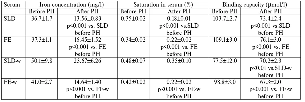
Table 3. Serum iron concentrations, its binding capacity and saturation
The SLD-w group had the highest serum iron and also the highest iron saturation before and even after PH, and binding capacity was the lowest, and had the lowest de-crease after PH. Except for the SLD-w group all groups had a statistical notable decrease in iron in the serum, satura-tion and even binding capacity after PH.
Discussion
Black tea reduces iron absorption in the intestines (23), therefore we reduced the iron absorption from the diet in the SLD and SLD-w groups by black tea – our effort was to minimize iron absorption in the SLD group, when 27 mg/kg of diet was sufficient for hematopoiesis and intensi-fy the differences between groups.
SLD FE SLD-w FE-w
HB (g/l) Before PH 158±2 161±1 158±2 156±4
After PH 143±4 140±3 161±7 148±3
p<0.01 vs. SLD p<0.001 vs. FE
before PH before PH
MCH (pg) Before PH 17.8±0.4 17.1±0.5 19.4±0.2 18.6±0.2
p<0.01 vs. SLD p<0.05 vs. FE
After PH 17.6±0.4 17.0±0.5 19.3±0.3 18.2±0.4
Leuko (109/l) Before PH 10.5±0.7 10.8±0.7 10.7±2.2 9.9±0.4
After PH 6.0±0.2 8.6±1.1 6.7±0.8 6.7±0.7
p<0.001 vs. SLD p<0.001 vs.SLD-w p<0.01 vs.FE-w
before PH before PH before PH
PMN (109/l) Before PH 1.00±0.35 0.85±0.16 1.07±0.31 1.55±0.52
After PH 1.15±0.65 1.33±0.89 3.90±0.62 3.90±0.56
p<0.01 vs.SLD-w p<0.01 vs. FE-w
before PH before PH
Tab. 2: Selected hematological parameters (hemoglobin, MCH, absolute count of leukocytes and polymorphonucleares).
Serum Iron concentration (mg/l) Saturation in serum (%) Binding capacity (μmol/l)
Before PH After PH Before PH After PH Before PH After PH
SLD 36.7±1.7 13.56±0.83 0.35±0.02 0.18±0.01 103.7±2.7 73.4±2.4
p<0.001 vs. SLD p<0.001 vs.SLD p<0.001 vs.SLD
before PH before PH before PH
FE 37.3±1.1 16.45±1.52 0.34±0.02 0.22±0.02 109.1±3.0 76.1±3.0
p<0.001 vs. FE p<0.001 vs. FE p<0.001 vs. FE
before PH before PH before PH
SLD-w 50.1±9.8 23.67±6.26 0.48±0.07 0.35±0.10 77.5±12.0 70.2±2.3
p<0.01 vs.SLD-w before PH
FE-w 41.0±2.7 14.64±1.40 0.42±0.02 0.22±0.02 98.8±3.0 67.3±2.0
p<0.001 vs. FE-w p<0.001 vs. FE-w p<0.001 vs. FE-w
before PH before PH before PH
The parameters of erythrocytes decreased in all groups after PH, especially in FE group, in comparison with the values before PH. The rats fed by the iron enriched diet to-lerated PH poorly. Repeating the blood withdrawals in-fluenced haemapoiesis by increasing hemoglobin content in erythrocytes. The rats of the FE-w group were allowed to waste iron, and did not store it as intensively as the rats of the SLD-w group (s. iron content in liver).
The SLD-w group had the highest serum iron con-centration after PH, although without any statistical signi-ficance. This effect may cause intensive turnover of the iron available during iron insufficiency (14), or temporary is-chemic periods after blood withdrawals. Hypoxia enhanced luminal iron uptake and promoted iron transfer to the blood as well (18). All groups had statistical decreases in iron, saturation and the binding capacity in serum after PH. The SLD-w group had the lowest binding capacity even be-fore PH. We presume that the sufficient proteosynthesis was not supported in the course of blood withdrawals with-out adding the iron into the diet. We suppose that absorbed iron was not stored in serum transport proteins. The highest binding capacity of serum iron was proved in the FE group accord in results in fish living in iron enriched water (7). Iron content in liver tissue was reduced in rats by repeated blood withdrawals, whereas iron supply decreases in organisms have been proved in blood donors (11).
Hepcidin concentrations were about same in all groups before PH, when the iron supply in all rats was sufficient. Different situation occurred after PH. The groups SLD, FE and FE-w showed a decrease in serum hepcidin concentra-tions after PH, whereas the SLD-w group did not. The cause of this decrease may be the diminution of liver iron reserves and the place of hepcidin synthesis. Changes in hepcidin gene expression depend on the type and rate of in-sult. A decrease in hepcidin gene expression was found in rats with alcoholic liver damage (5), within 24 hours after acute anemia in mice (22) and by the third day after intra-vascular haemolysis (12). On the contrary, the SLD-w group, with a lower total iron supply, had a probable ische-mic preconditioning with elevation of hepcidin after PH. This result is in accordance with other authors. The eleva-tion of liver hepcidin gene expression was proved after liver ischemia-reperfusion (13), and from 4 hour to 16 hours after PH in rats (25). Higher serum hepcidin concentration in the SLD-w group was preserved for giving priority to the activity of neutrophils and monocytes, which also produce hepcidin (21). Spontaneous respiratory burst was higher in the SLD-w and FE-w groups before PH, and lower in the FE group. We suppose that an iron enriched diet without blood loss has a negative effect on PMN respiratory burst, as Bergman (3) showed in phagocytosis. Respiratory burst was increased after PH, especially in FE-w. The cause may be the inflammatory increasing of ferritin from the liver, which stimulates the superoxide production by PMN (4). Respiratory burst in SLD-w did not increase enough, parti-cularly after stimulation by PMA, because iron supplies
were low. Li (19) demonstrated that the functions of respi-ratory burst were markedly decreased in iron-deficient rats. Healing processes were studied by means of a quantifi-able marker; i.e., early initiation of liver regeneration. The FE-w group had an earlier start of liver DNA synthesis be-cause the effect of iron enriched diet and blood withdrawals add up. The effect of iron is positive on DNA synthesis of rat hepatocytes (9). Similarly, our unpublished experimen-tal data suggest that drinking black tea did not significantly inhibit liver DNA synthesis 18 hours after PH (1.79±0.18 kBq/mg DNA) vs. drinking water (2.23±0.39 kBq/mg DNA, p=0.325). We assumed that this fact is a result of inhibition of intestinal iron absorption by black tea.
The blood withdrawals before PH induced a “precondi-tioning” state which supported liver regeneration, like hy-perbaric oxygenation (15), sham operation (16), carnitine (8), or ischemic pre-conditioning (2).
Conclusion
An iron enriched diet with stimulation of haematopoesis
led to better response to elective surgery (PH), expressed as earlier initiation of liver regeneration and better PMN func-tions. Our results support the idea of using pre-operation auto-transfusions with iron supplementation to patients.
Sponsored by Research project MZO 00179906.
References
1. Amer J, Fibach E. Chronic oxidative stress reduces the respiratory burst respon-se of neutrophils from beta-thalassaemia patiens. British Journal of Hematology 2005;129:435–41.
2. Bedirli A, Kerem M, Pasaoglu H et al. Effects of ischemic preconditioning on re-generative capacity of hepatocyte in the ischemically damaged rat livers. J Surg Res 2005;125:42–8.
3. Bergman M, Salman H, Pinchasi R et al. Phagocytic capacity and apoptosis of pe-ripheral blood cells from patients with iron deficiency anemia. Biomed Pharmacother 2005;59:307–11.
4. Brailsford S, Lunec J, Winyard P et al. A possible role for ferritin during inflam-mation. Free Radic Res Commun 1985;1:101–9.
5. Bridle K, Cheung TK, Murphy T et al. Hepcidin is down-regulated in alcoholic li-ver injury: implications for the pathogenesis of alcoholic lili-ver disease. Alcohol Clin Exp Res 2006;30:106–12.
6. Burton K. A study of the condition and mechanism of the colorimetric estimati-on of deoxyribestimati-onucleic acid. Biochem J 1956;62:315–23.
7. Carriquiriborde P, Handy RD, Davies SJ. Physiological modulation of iron meta-bolism in rainbow trout (Oncorhynchus mykiss) fed low and high iron diets. J Exp Biol 2004;207:75–86.
8. Červinková Z, Kalinová M, Šimek J. The effect of carnitine on the early phase of liver regeneration after partial hepatectomy in rats with experimental diabetes. Bratisl Lek Listy 1992;93:359–63.
9. Chenoufi N, Loreal O, Drenou B et al. Iron may induce both DNA synthesis and repair in rat hepatocytes stimulated by EGF/pyruvate. J Hepatol 1997;26:650–8. 10. Dahlgren C, Karlsson A. Respiratory burst in human neutrophils. J Immunol
Meth 1999;232:3–14.
11. Fernandez-Real JM, Lopez-Bermejo A, Ricart W. Iron stores, blood donation, and insulin sensitivity and secretion. Clin Chem. 2005;51:1201–5.
12. Frazer DM, Inglis HR, Wilkins SJ et al. Delayed hepcidin response explains the lag period in iron absorption following a stimulus to increase erythropoiesis. Gut 2004;53:1509–15.
13. Goss JA, Seu P, Gao FQ et al. Ischemia-reperfusion of rat liver modulates hepci-din in vivo expression. Liver Transpl 2005;11:800–6.
14. Kolb H, Kolb-Bachofen V. Nitric oxide: a pathogenetic factor in autoimmunity. Immunol Today 1992;13:157–60.
16. Laurent S, Starkel P, Leclercq IA et al. Molecular events associated with accele-rated proliferative response in rat livers when partial hepatectomy is preceded by a sham operation. Eur J Clin Invest 2005;35:140–7.
17. Lee P, Peng H, Gelbart T et al. Regulation of hepcidin transcription by interleu-kin-1 and interleukin-6. Proc Natl Acad Sci USA. 2005;102:1906–10. 18. Leung PS, Srai SK, Mascarenhas M et al. Increased duodenal iron uptake and
transfer in a rat model of chronic hypoxia is accompanied by reduced hepcidin expression. Gut 2005;54:1391–5.
19. Li Q, Liao Q, Luo C et al. Investigation of impairment of neutrophil’s phago-cytosis and bactericidal function in rats with iron deficiency Hua Xi Yi Ke Da Xue Xue Bao 1991;22:274–7.
20. Martin ME, Nicolas G, Hetet G et al. Transferrin receptor 1 mRNA is down-regulated in placenta of hepcidin transgenic embryos. FEBS Lett 2004;574: 187–91.
21. Nemeth E, Rivera S, Gabayan V et al. IL-6 mediates hypo-ferremia of inflamma-tion by inducing the synthesis of the iron regulatory hormone hepcidin. J Clin Invest 2004;113:1271–6.
22. Nicolas G, Viatte L, Bennoun M et al. Hepcidin, a new iron regulatory peptide. Blood Cells Mol Dis 2002;29:327–35.
23. O’Coinceanainn M, Bonnely S, Baderschneider B, Hynes MJ. Reaction of iron (III) with theaflavin: complexation and oxidative products. J Inorg Biochem. 2004;98:657–63.
24. Peyssonnaux C, Datta V, Cramer T, et al. HIF-1 expression regulates the bacteri-cidal capacity of phagocytes. J Clin Invest 2005;115:1806–15.
25. Sheikh N, Batusic DS, Dudas J et al. Hepcidin and Hemojuvelin gene-expression in rat liver damage: In vivo and in vitro studies. Am J Physiol Gastrointest Liver Physiol 2006;291:G482–90.
26. Short J, Zemel R, Kanta J, Lieberman I. Stimulation of deoxyribonucleic acid synthesis in the liver parenchymal cells of the intact rats. Nature 1969;223:956–7. 27. Viatte L, Lesbordes-Brion JC, Lou DQ et al. Deregulation of proteins involved in
iron metabolism in hepcidin-deficient mice. Blood 2005;105:4861–4. 28. Wilhelm J, Frydrychová M, Hezinová A et al. Production of hydrogen peroxide
by peritoneal macrophages from rats exposed to subacute and chronic hypoxia. Physiol Res 1997;46:35–9.
Corresponding author:
Doc. MUDr. Helena Živná, CSc., Charles University in Prague, Faculty of Medicine in Hradec Králové,
Radio-Isotope Laboratory and Vivarium, Šimkova 870, Hradec Kralové, Czech Republic, e-mail: zivna@lfhk.cuni.cz Submitted February 2007.