R E S E A R C H A R T I C L E
Open Access
Double lung, unlike single lung
transplantation might provide a protective
effect on mortality and bronchiolitis
obliterans syndrome
Mohammed Fakhro
1*, Ellen Broberg
2, Lars Algotsson
2, Lennart Hansson
3, Bansi Koul
1, Ronny Gustafsson
1,
Per Wierup
1, Richard Ingemansson
1and Sandra Lindstedt
1Abstract
Background:Survival after lung transplantation (LTx) is often limited by bronchiolitis obliterans syndrome (BOS). Method:Survey of 278 recipients who underwent LTx. The endpoint used was BOS (BOS grade≥2), death or Re-lung transplantation (Re-LTx) assessed by competing risk regression analyses.
Results:The incidence of BOS grade≥2 among double LTx (DLTx) recipients was 16 ± 3% at 5 years, 30 ± 4% at 10 years, and 37 ± 5% at 20 years, compared to single LTx (SLTx) recipients whose corresponding incidence of BOS grade≥2 was 11 ± 3%, 20 ± 4%, and 24 ± 5% at 5, 10, and 20 years, respectively (p> 0. 05). The incidence of BOS grade≥2 by major indications ranked in descending order: other, PF, CF, COPD, PH and AAT1 (p< 0. 05). The mortality rate by major indication ranked in descending order: COPD, PH, AAT1, PF, Other and CF (p< 0. 05).
Conclusion:No differences were seen in the incidence of BOS grade≥2 regarding type of transplant, however, DLTx recipients showed a better chance of survival despite developing BOS compared to SLTx recipients. The highest incidence of BOS was seen among CF, PF, COPD, PH, and AAT1 recipients in descending order, however, CF and PF recipients showed a better chance of survival despite developing BOS compared to COPD, PH, and AAT1 recipients.
Keywords: Lung transplantation, Bronchiolitis obliterans, Graft rejection, Graft survival, Survival rate
Background
Chronic lung allograft dysfunction (CLAD) remains the major barrier to long-term success after lung transplant-ation [1–3]. The primary cause of death after LTx is CLAD. The development of CLAD is rare in the first year after LTx, but the rate increases quickly with cumulative incidence reported to be as high as 40% to 80% within the first five years [4–7]. CLAD that manifests early after transplantation reportedly shows a poorer prognosis than late-onset CLAD. Bronchiolitis obliterans (BO) is the pathologic pattern of injury most commonly seen in lung transplant recipients with progressive loss of lung
function. It is believed to be due to chronic allograft rejec-tion and is characterized by the obliterarejec-tion of small air-ways by fibromyxoid granulation tissue. Distribution is patchy and difficult to detect with transbronchial biopsy [3, 7]. Because BO is difficult to document histologically, the International Society for Heart and Lung Transplant-ation (ISHLT) in 1993 established criteria for its physio-logic counterpart, bronchiolitis obliterans syndrome (BOS). This diagnosis requires a permanent 20% drop in the forced expiratory volume in 1 s (FEV1) not attribut-able to a concurrent process [8]. Since 2014 the diagnosis for chronic rejection was further broadened to CLAD, which includes restrictive allograft syndrome (RAS). CLAD can also be diagnosed by CT scan with visual signs of small airway disease and lung biopsies with severe nar-rowing or complete obstruction of the small airways [8].
* Correspondence:mohammed.fakhro@med.lu.se
1Department of Cardiothoracic Surgery, Skåne University Hospital, Lund University, Lund, Sweden
Full list of author information is available at the end of the article
In Sweden two centers perform lung transplantation for a population of about 10 million, all of whom are covered by national health insurance. Skåne University Hospital is one of these centers. This retrospective re-port reviews the 25-year experience of Skåne University Hospital, Lund University Lung Transplant Program with particular emphasis on chronic rejection between different subgroups of recipients and type of transplant procedure performed. Because of the retrospective na-ture of this study, chronic rejection is referred to as BOS rather than the recently termed CLAD.
Patients and method
Between January 1990 to June 2014, 278 patients under-went lung transplantation at Skåne University Hospital, Lund University. Double lung transplantation (DLTx) was performed in 172 patients, single lung transplantation (SLTx) in 97 patients, and HLTx in 9 patients. Of these, 129 were male and 149 were female. Re-lung transplant-ation (Re-LTx) was performed in 15 patients. Among the Re-LTx recipients, of whom 5 were female and 10 were male, 7 recipients had a DLTx and 8 had a SLTx.
In the present study the median age was 51 years with a range of 12–71 years. The major indications were de-fined as chronic obstructive pulmonary disease (COPD) (n= 67), cystic fibrosis (CF) (n= 54),α1-antitrypsin defi-ciency (AAT1) (n= 55), pulmonary fibrosis (PF) (n= 38), pulmonary hypertension (PH) (n= 39), and a group deemed as “other” (n= 25), which included bronchiec-tasis, sarcoidosis, bronchioalveolary cancer, silicosis, and graft-vs-host disease (GVHD).
HLTx was performed via median sternotomy in 7 pa-tients and via a clamshell (bilateral anterolateral thoracot-omy with a transverse sternotthoracot-omy) incision in the 4th intercostal space in 2 patients. SLTx and DLTx were per-formed in standard fashion. SLTx was perper-formed through a posterolateral thoracotomy in 86 patients, via clamshell in 7 patients, and via median sternotomy in 4 patients. DLTx was performed through a clamshell-incision in 146 patients, via median sternotomy in 17 patients, and via an-terolateral thoracotomy in 9 patients.
Preoperative respiratory support was used in 13 opera-tions (CF 4, PF 5, Re-LTx 3, PH 1). Preoperative ECMO (extracorporeal membrane oxygenation) support was used in 12 operations (CF 6, PF 3, ARDS 1, PH 1, Re-LTx 1).
Intraoperative circulatory support in the form of extra-corporeal circulation (ECC) was used in 105 cases, and intraoperative ECMO was used in 73 cases. Intraopera-tive circulatory support was not used in 115 cases. Re-cipient characteristics are shown in Table 1.
Chronic allograft dysfunction
According to ISHLT guidelines, BOS is defined as more than 20% decline in FEV1from the highest obtained
base-line, [8, 9], and is characterized by perivascular and inter-stitial mononuclear cell infiltrates or chronic rejection characterized by dense scarring and eosinophilic infil-trates. If rapid deterioration of pulmonary function was detected as a sign of chronic allograft dysfunction, bron-choscopies with TBB was conducted and anti-rejection treatment was initiated with pulsed metylprednisolon often together with tacrolimus or everolimus as a replace-ment for cyclosporine. In this study, patients with BOS grade≥2 was included and chosen for analysis.
Statistical methods
The statistical calculations were performed using SPSS Version 19.0. (IBM Corp, Armonk, NY). Primary stratifi-cation of the material was made into two sets of cohorts. The first cohort was based on the main indication for LTx, with the following indicator cohorts: COPD, AAT1, CF, PH, and PF. The second set divided the material
Table 1Recipient characteristics
Baseline Characteristics of the 278 Patients
Variable Median (Range)
or No. (%)
Recipient age, year 51 (12–71)
Recipient primary disease
Cystic fibrosis 54
Pulmonary fibrosis 38
Chronic obstructive pulmonary disease (COPD) 67
α1-antitrypsin deficiency (AAT1) 55
Pulmonary hypertension (PH) 39
Other 25
Transplant type
DLTx 172
SLTx 97
HLTx 9
Re-LTx 15
DLTx 7
SLTx 8
Gender
Female 149
Male 129
Transplant year
1990–2002 126
2003–2014 167
Preoperative ventilator 13
Preoperative ECMO 12
Perioperative ECC 105
Perioperative ECMO 73
based on type of LTx: DLTx or SLTx. The aim of this study was to analyze the occurrence of BOS (grade≥2) after pri-mary LTx. In this analysis, death acted as a competing risk event to BOS. In a competing-risks model, we analyzed in-cidence of BOS grade≥2 and death as two separate out-comes. Specifically, we estimated and compared the cumulative incidence functions for BOS grade≥2 and death using Gray’s test, Gray (1988). All calculations re-garding competing risks were performed using R with the CMPRSK package (available at http://www.r-project.org). For all statistical analyses, a p-value less than 0. 05 was considered significant. All statistical calculations were per-formed by Sidesoft AB, Malmo, Sweden.
Results
Cumulative incidence of BOS grade≥2 and death
Type of transplant
Incidence of BOS (grade≥2) is presented (percentage of probability ± SE) by type of transplant (DLTx and SLTx) in Fig. 1. The incidence of BOS among DLTx-recipients was 16 ± 3% at 5 years, 30 ± 4% at 10 years, 35 ± 5% at 15 years, and 37 ± 5% at 20 years, compared to SLTx-recipients whose incidence of BOS was 11 ± 3% at 5 years, 20 ± 4% at 10 years, 24 ± 5% at 15 years, and 24 ± 5% at 20 years (p> 0. 05). The mortality rate for DLTx recipients was 19 ± 3% at 5 years, 23 ± 4% at 10 years, 28 ± 4% at 15 years, and 43 ± 7% at 20-years compared to the mortal-ity rate of SLTx-recipients, which was 34 ± 5% at 5 years, 55 ± 6% at 10 years, 56 ± 6% at 15 years, and 71 ± 8% at 20 years (p< 0. 05). Kaplan-Meier survival is displayed after development of BOS (grade≥2) until follow-up/ death in Fig. 2. Survival curves are divided into patients that have underwent DLTx vs. SLTx (p> 0. 05).
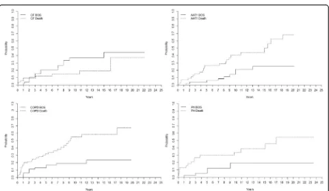
Major indications
The incidence of BOS (grade≥2) is presented by differ-ent diagnostic groups in Fig. 3. The incidence of BOS among AAT1-patients was 4 ± 3% at 5 years, 14 ± 6% at 10 years, 26 ± 8% at 15 years, and 26 ± 8% at 20 years.
For CF-patients it was 20 ± 6% at 5 years, 37 ± 8% at 10 years, 37 ± 8% at 15 years, and 44 ± 10% at 20 years. For COPD-patients it was 13 ± 4% at 5 years, 19 ± 5% at 10 years and 24 ± 7% at 15 years. For PF-patients, the in-cidence of BOS was 25 ± 8% at 5 years, 34 ± 9% at 10 years, 34 ± 9% at 15 years and 34 ± 9% at 20 years while for PH-patients it was 6 ± 4% at 5 years, 19 ± 7% at 10 years, 19 ± 9% at 15 years, and 19 ± 7% at 20 years (p < 0. 05). The mortality rate for AAT1-patients was 26 ± 6% at 5 years, 41 ± 8% at 10 years, 44 ± 8% at 15 years and 68 ± 10% at 20 years. For CF-patients it was 12 ± 5% at 5 years, 15 ± 5% at 10 years, 19 ± 7% at 15 years, and 37 ± 19% at 20 years. For COPD-patients it was 32 ± 6% at 5 years, 55 ± 8% at 10 years, and 58 ± 8% at 15 years. For PF-patients it was 26 ± 8% at five years, 38 ± 9% at 10 years, 38 ± 9% at 15 years, and 52 ± 16% at 20 years, and for PH it was 30 ± 8% at 5 years, 34 ± 8% at 10 years, 39 ± 9% at 15 years, and 54 ± 12 at 20 years (p< 0. 05).
Major indications compared group wise
The patient groups of major indications were compared. The group “other” describes a heterogenic group of pa-tients who underwent LTx due to bronchiectasis, sar-coidosis, bronchioalveolary cancer, silicosis, BOS and graft-vs-host disease (GVHD). The group had a higher incidence of BOS compared to COPD (p= 0. 007), AAT1 (p= 0. 001) and PH (p= 0. 002) patients. The group also showed significant lower risk of death com-pared to COPD (p= 0. 037), AAT1 (p= 0. 281), and PH (p= 0. 300) patients.
Recipients with CF had higher risk of developing BOS compared to AAT1 recipients (p= 0. 048), but AAT1 had a higher mortality (p= 0. 020). Recipients with CF and COPD had the same incidence of developing BOS (p= 0. 164), but COPD recipients had a higher mortality (p= 0. 001). Recipients with CF had higher risk of devel-oping BOS compared to PH recipients (p= 0. 055), but CF and PH recipients had the same mortality (p= 0. 057) (Fig. 3).
Fig. 2Kaplan-Meier figure displaying survival between SLTx and DLTx after development of BOS grade≥2 until death/follow-up (p> 0. 05)
Age and BOS
The incidence of BOS (grade≥2) for patients≤50 years of age was 15 ± 3% at 5 years, 30 ± 5% at 10 years, 35 ± 5% at 15 years, and 38 ± 6% at 20 years. For patients >50 years of age it was 14 ± 3% at 5 years, 22 ± 4% at 10 years, and 26 ± 5% at 15 years (p= 0. 238). The mor-tality rate for patients ≤50 years of age was 20 ± 4% at 5 years, 28 ± 4% at 10 years, 34 ± 5% at 15 years and 41 ± 7% at 20 years. For patients >50 years of age the mor-tality rate was 29 ± 4% at 5 years, 44 ± 5% at 10 years, and 45 ± 5% at 15 years (p= 0. 019) (Fig. 4).
Different time periods and BOS
For the period 1990–2002, the incidence of BOS (grade≥2) in all recipients was 9 ± 3% at 5 years, 23 ± 4% at 10 years, 27 ± 4% at 15 years, and 29 ± 4 at 20 years. The overall mortality rate for the same time period was 24 ± 4% at 5 years, 36 ± 4% at 10 years, 40 ± 5% at 15 years, and 57 ± 6% at 20 years. Between 2003 and 2014, the incidence of BOS was 8 ± 2% at 2 years, 17 ± 3% at 4 years, 21 ± 4% at 6 years, 24 ± 4% at 8 years, and 29 ± 5% at 10 years. The overall mortality rate for the same time period was 14 ± 3% at 2 years, 22 ± 4% at 4 years, 27 ± 4% at 6 years, 32 ± at 8 years, and 36 ± 5% at 10 years (Fig. 5).
Mortality
Post-operative cause of death before and after 12 months is shown in Table 2. The group called “other causes” is defined as mortality caused by myocardial and cerebral ischaemia, and multiple organ failure as well as other causes related to the patient’s age and health status.
Discussion
Lung transplantation is an established treatment for end stage pulmonary disease [10]. The number of clinical lung transplantation is limited by the shortage of organs, which have resulted in a constant searching for new
ways to increase the number of organs [11–14], while survival after lung transplantation mainly limited by CLAD. Survival after lung transplantation has improved significantly over the last decade, however, CLAD, pre-dominantly manifesting as BOS, remains the primary cause of morbidity and mortality after LTx. Although the risk of developing BOS within the first year is low, cu-mulative incidence of BOS quickly increases within the first five years [9, 15, 16].
The risk factors for BOS are still not fully understood [17]. Anti-human leukocyte antigen (HLA) donor-specific antibodies (DSA) have been associated with early and high-grade BOS and death after LTx in some studies but is still controversial [18, 19]. Treatments to remove antibodies or limit antibody-mediated damage using plasmapheresis have been shown to have some effect when DSA are first detected. However, the impact of this treatment on clinical outcome following LTx remains unclear [20]. Bacterial and viral infection has also been identified as a possible trigger of BOS after LTx [21, 22]. Although BOS was generally thought to be irreversible, recent evidence suggests that some patients with BOS may respond to azithromycin with an improvement of their FEV1 with more than 10% [22]. In addition, an-other form of chronic rejection, restrictive allograft syn-drome (RAS), has recently been described, which does not fit the BOS definition but is instead characterized by restrictive functional changes involving peripheral lung pathology, leading to the introduction of the more encompassing term CLAD [5, 23].
The overall cumulative incidence of BOS grade≥2 among our 278 recipients was 15% after 5 years, 26% after 10-years, 30% after 15-years, and 32% after 20 years post-transplant. The incidence of BOS was highest among the group referred to as“other”. This group de-scribes a heterogenic group of patients who underwent LTx due to bronchiectasis, sarcoidosis, bronchioalveolary cancer, silicosis, BOS, and graft-vs-host disease (GVHD)
that might reflect the high incidence of BOS. The group’s heterogenic appearance makes it difficult to draw any conclusions. Besides the group “other,” the highest incidence of BOS was seen among PF recipients followed by CF, COPD, PH, and AAT1 recipients in the described descending order. It has been shown that BOS
and PF respectively exhibit similar disease characteristics with overlapping pathophysiology such as epithelial cell injury and increase in production/deposition of ECM [24]. This patient group is of great interest as the identi-fication of biomarkers in PF could contribute to finding new means of earlier finding BOS [25]. The highest mor-tality was seen among COPD recipients followed by PH, AAT1, PF, and CF recipients in the described descending order, indicating that CF and PF recipients have a better chance of survival despite developing BOS compared to the other major indications such as COPD and PH. However, it should also be acknowledged that patient outcome among LTx-recipients in Sweden might differ in comparison to other countries such as the US due to a significantly higher incidence of COPD/CF vs. intersti-tial lung disease, in combination with Sweden having younger recipients [26]. As well as reports from the ISHLT showing almost double the incidence of intersti-tial lung disease in LTx in comparison to Sweden.
When we compared the different patient groups (pairs of two) of major indications between each other, the group referred to as“other”had significantly higher risk of developing BOS (grade≥2) compared to COPD, AAT1, and PH recipients, keeping in mind that this group of recipients reflects a heterogeneous group of pa-tients where some of the recipients underwent LTx due to BOS and GVHD and where the recipients probably already at the time of transplantation have an immuno-logic response that might lead to the development of BOS in the pulmonary graft.
Recipients with CF had a significantly higher risk of developing BOS compared to AAT1 recipients, but AAT1 had a significantly higher mortality rate, indicat-ing that CF recipients might withstand the development of BOS better than AAT1 recipients. This finding could
Fig. 5Cumulative incidence of bronchiolitis obliterans syndrome (BOS) and mortality after lung transplantation (LTx) for the two different time periods 1990–2002 and 2003–2014. Our findings (Fig. 1) indicate that DLTx and SLTx carried the same risk of developing BOS grade≥2, but DLTx had a significantly lower risk of death. We suspect that these results might reflect a change in postoperative care towards more aggressive infection and rejection therapy in combination with less frequent SLTx in favor of DLTx the last 10–12 years. However, our results could not confirm these suppositions: no difference was found between the risk of developing BOS grade≥2 or death in different time periods. In Fig. 5 we show the results, supporting the claim that DLTx has a significantly lower risk of death compared to SLTx
Table 2Cause of death according to recipient transplantation type and time after transplantation
Tx-type; Cause of Death < 12 months > 12 months p-value
Total: 278
SLTx (n= 97)
Total number of deaths 17 51 0.158
Death from Organ Rejection 2 (12%) 10 (20%)
Death from Infection 4 (23%) 16 (31%)
Death from Malignancy 1 (6%) 10 (20%)
Death from Other Causes 10 (59%) 15 (29%)
DLTx (n= 172)
Total number of deaths 16 47 0.388
Death from Organ Rejection 4 (25%) 20 (42%)
Death from Infection 4 (25%) 6 (13%)
Death from Malignancy 1 (6%) 6 (13%)
Death from Other Causes 7 (44%) 15 (32%)
HLTx (n = 9)
Total number of deaths 1 4 0.576
Death from Organ Rejection 0 (0%) 1 (25%)
Death from Infection 0 (0%) 0 (0%)
Death from Malignancy 0 (0%) 0 (0%)
Death from Other Causes 1 (100%) 3 (75%)
have a positive effect on the clinical implication of favor-ing CF recipients in LTx. Previously the overall survival benefit of LTx in CF has been reported as controversial due to associated risk factors such as CF-related ar-thropathy as well as associated chronic infections with bacterial/fungal agents like B.cepacia, P.Auriginosa and
Aspergillusthat can be serious and life-threatening [27]. Interestingly, recipients with CF had a significantly higher risk of developing BOS compared to PH recipi-ents, however, CF and PH recipients had the same mor-tality, indicating that CF and PH recipients developing BOS have the same chance of survival despite BOS. It has been shown that PH patients undergo extensive re-modeling of the pulmonary arterial walls as part of their pathophysiology, leading to permanent changes of the intima [28]. However, PH is also seen post-LTx among different recipients as bronchiolitis obliterans is often as-sociated with immune-mediated arterio- and venopathy leading to pre- and post-capillary PH [29]. It could be theorized that patients with PH prior to LTx might bet-ter withstand this phenomenon, indicating why PH re-cipients don’t show inferior survival to CF despite BOS. The clinical implication of understanding the develop-ment of BOS in this disease state could be of immense potential. This hypothesis was however not investigated in this study with the need of further data.
Recipients with CF and COPD had the same incidence of BOS, but COPD recipients had a significantly higher mortality, indicating that CF recipients might withstand the development of BOS better than COPD recipients. COPD recipients are often older than CF patients at the time of transplant, and the COPD patients often have comorbidities such as heart and vascular disease that might in part explain these results. We did an expanded analysis to investigate the impact of age on the develop-ment of BOS and the risk of death. We concluded that age had no impact on the development of BOS, but re-cipients 50 years or older had a 9% higher mortality 5 years post-transplant and a 16% increased risk after 10 years post-transplant compared to recipients younger than 50 years (p< 0. 05).
Interestingly, when we compared DLTx to SLTx in the entire cohort we found that DLTx and SLTx recipients had equal risk of developing BOS grade≥2. However, re-cipients receiving DLTx had a significantly better chance of surviving. These results further support a clinical pro-gram favoring DLTx instead of SLTx. We tried to analyze the pattern of DLTx and SLTx among major indications such as COPD, but unfortunately the groups were not big enough to reach statistical evaluation. Our findings indicate that DLTx and SLTx had the same risk of devel-oping BOS, but DLTx had a better chance of survival. We suspect that these results might reflect the fact that during the last 10–15 years, we have initiated early
treatment for viral and bacterial infection, used more ag-gressive therapy at the first sign of rejection, and favored DLTx over SLTx. The frequency of SLTx reached its peak in 2002 at our clinic and has significantly declined since then in favor of DLTx. However, our results did not sup-port these hypotheses as no difference was found be-tween the risk of BOS or mortality in different time periods. In Fig. 5 we show the results for the two differ-ent time periods 1990–2002 and 2003–2014.
Limitations
There have been significant changes in the care of transplant patients over the last 25 years, which affects outcome vari-ables such as survival depending on year of transplantation. Recipient inclusion criteria have broadened over the years and now preoperative ECMO support or ventilator support are no longer contraindications for LTx, representing a com-plex recipient clientele. Several confounding factors have been linked to long-term survival in LTx that could be play-ing a role here. Such variables are recipient/donor age and total lung capacity in addition to recipient kidney function, O2 requirement and allograft ischemic time. In the present study patients diagnosed as BOS grade 1 did not, in the ma-jority, of the cases get a specific treatment or an alternation in the present regime even though the patient was diag-nosed with BOS. It is possible to diagnose BOS through spirometry, where according to ISHLT a drop in FEV1 more than 20% from baseline is associated with BOS grade≥1. Though possible confounders that might affect post-operative pulmonary function besides BOS are recurring in-fections or a decline in FEV1 affected by the natural aging-process or other comorbidities. It should also be acknowl-edged that the definition of FEV1 < 80% from best baseline for BOS is debatable in comparison to current CLAD cri-teria, such as phenotyping into BOS vs. RAS. To differenti-ate those who really have a rejection (BOS) or not, we have in this analysis calculated BOS as BOS grade 2 or more. This can of course be cautious to follow.
Conclusions
Acknowledgements
Lena Mared, Leif Eriksson, Jan-Otto Solem, and Professor Stig Steen. All co-workers at the Department of Cardiothoracic Surgery, Pulmonary Medicine and Thoracic Intensive Care and Anesthesia, Skåne University Hospital, Lund University, Sweden.
Funding None.
Availability of data and materials
The datasets generated and analysed during the current study are not publicly available due to sensitive data based on patient journals from Skåne University Hospital, Lund University.
Authors’contributions
MF conceived of the study, participated in its design, collected the patient data, analyzed the data and drafted the manuscript. EB participated in its design, collected patient data, analyzed the data and helped to draft the manuscript. SL conceived of the study, participated in its design, collected the patient data, analyzed the data and drafted the manuscript. RI conceived of the study, participated in its design and analyzed the data. LA participated in its design and helped to draw the manuscript. LH participated in its design and analyzation of the data. BK participated in its design and analyzation of the data. RG participated in its design and analyzation of the data. PW participated in its design and analyzation of the data. All authors read and approved the final manuscript. The statistical analyzes were performed by Sidesoft AB, Malmo, Sweden.
Ethics approval and consent to participate
This study has been performed in accordance with the Declaration of Helsinki and is approved by the ethic committee in Lund, Lund University with reference number 2016/638.
Consent for publication Not applicable.
Competing interests
The authors declare that they have no competing interests.
Publisher’s Note
Springer Nature remains neutral with regard to jurisdictional claims in published maps and institutional affiliations.
Author details 1
Department of Cardiothoracic Surgery, Skåne University Hospital, Lund University, Lund, Sweden.2Department of Thoracic Intensive Care and Anesthesia, Skåne University Hospital, Lund University, Lund, Sweden. 3Department of Pulmonary Medicine, Skåne University Hospital, Lund University, Lund, Sweden.
Received: 28 June 2017 Accepted: 16 November 2017
References
1. Verleden SE, Vasilescu DM, Willems S, et al. The site and nature of airway obstruction after lung transplantation. Am J Respir Crit Care Med. 2014;189: 292–300.
2. Gauthier JM, Hachem RR, Kreisel D. Update on chronic lung allograft dysfunction. Curr Transplant Rep. 2016;3:185–91.
3. Al-Githmi I, Batawil N, Shigemura N, et al. Bronchiolitis obliterans following lung transplantation. Eur J Cardiothorac Surg. 2006;30:846–51.
4. Vandermeulen E, Lammertyn E, Verleden SE, et al. Immunological diversity in phenotypes of chronic lung allograft dysfunction: a comprehensive immunohistochemical analysis. Transpl Int. 2017;30:134–43.
5. Verleden SE, Vanaudenaerde BM, Vos R, Verleden GM. Phenotypes of chronic lung allograft dysfunction: getting closer step by step? Am J Transplant. 2016; 16:3071–2.
6. Verleden SE, Sacreas A, Vos R, et al. Advances in understanding bronchiolitis obliterans after lung transplantation. Chest. 2016;150:219–25.
7. Royer PJ, Olivera-Botello G, Koutsokera A, et al. Chronic lung allograft dysfunction: a systematic review of mechanisms. Transplantation. 2016;100: 1803–14.
8. Estenne M, Maurer JR, Boehler A, et al. Bronchiolitis obliterans syndrome 2001: an update of the diagnostic criteria. J Heart Lung Transplant. 2002;21: 297–310.
9. Verleden GM, Raghu G, Meyer KC, et al. A new classification system for chronic lung allograft dysfunction. J Heart Lung Transplant. 2014;33:127–33. 10. Fakhro M, Ingemansson R, Skog I, et al. 25-year follow-up after lung
transplantation at Lund University Hospital in Sweden: superior results obtained for patients with cystic fibrosis. Interact Cardiovasc Thorac Surg. 2016;23:65–73.
11. Lindstedt S, Pierre L, Ingemansson R. A short period of ventilation without perfusion seems to reduce atelectasis without harming the lungs during ex vivo lung perfusion. J Transp Secur. 2013;2013:729286.
12. Lindstedt S, Eyjolfsson A, Koul B, et al. How to recondition ex vivo initially rejected donor lungs for clinical transplantation; clinical experience from Lund University Hospital. J Transp Secur. 2011;2011:754383.
13. Machuca TN, Cypel M. Ex vivo lung perfusion. J Thorac Dis. 2014;6:1054–62. 14. Sanchez PG, Bittle GJ, Williams K, et al. Ex vivo lung evaluation of prearrest
heparinization in donation after cardiac death. Ann Surg. 2013;257:534–41. 15. Christie JD, Edwards LB, Kucheryavaya AY, et al. The registry of the
International Society for Heart and Lung Transplantation: twenty-eighth adult lung and heart-lung transplant report–2011. J Heart Lung Transplant. 2011;30:1104–22.
16. Traxler D, Schweiger T, Schwarz S, et al. The lymphatic phenotype of lung allografts in patients with bronchiolitis obliterans syndrome and restrictive allograft syndrome. Transplantation. 2017;101:310–5.
17. Olland A, Reeb J, Leclerq A, et al. Microparticles: a new insight into lung primary graft dysfunction? Hum Immunol. 2016;77:1101–7.
18. Westall GP, Paraskeva MA, Snell GI. Antibody-mediated rejection. Curr Opin Organ Transplant. 2015;
19. Zazueta OE, Preston SE, Moniodis A, et al. The presence of Pretransplant HLA antibodies does not impact the development of chronic lung allograft dysfunction or CLAD related death. Transplantation. 2016;
20. Safavi S, Robinson DR, Soresi S, et al. De novo donor HLA-specific antibodies predict development of bronchiolitis obliterans syndrome after lung transplantation. J Heart Lung Transplant. 2014;33:1273–81. 21. Luckraz H, Sharples L, McNeil K, et al. Cytomegalovirus antibody status of
donor/recipient does not influence the incidence of bronchiolitis obliterans syndrome in lung transplantation. J Heart Lung Transplant. 2003;22:287–91. 22. Vos R, Vanaudenaerde BM, Ottevaere A, et al. Long-term azithromycin
therapy for bronchiolitis obliterans syndrome: divide and conquer? J Heart Lung Transplant. 2010;29:1358–68.
23. Verleden SE, Ruttens D, Vandermeulen E, et al. Restrictive chronic lung allograft dysfunction: where are we now? J Heart Lung Transplant. 2015;34: 625–30.
24. Fernandez IE, Eickelberg O. New cellular and molecular mechanisms of lung injury and fibrosis in idiopathic pulmonary fibrosis. Lancet. 2012;380:680–8. 25. Heijink IH, Rozeveld D, van der Heide S, et al. Metalloproteinase profiling in lung transplant recipients with good outcome and bronchiolitis obliterans syndrome. Transplantation. 2015;99:1946–52.
26. Kistler KD, Nalysnyk L, Rotella P, Esser D. Lung transplantation in idiopathic pulmonary fibrosis: a systematic review of the literature. BMC Pulm Med. 2014;14:139.
27. Lobo LJ, Noone PG. Respiratory infections in patients with cystic fibrosis undergoing lung transplantation. Lancet Respir Med. 2014;2:73–82. 28. Wilkinson M, Langhorne CA, Heath D, et al. A pathophysiological study of
10 cases of hypoxic cor pulmonale. Q J Med. 1988;66:65–85.