S H O R T R E P O R T
Open Access
Transcriptomic changes in the frontal
cortex associated with paternal age
Rebecca G Smith
1, Cathy Fernandes
1, Rachel Kember
1, Leonard C Schalkwyk
1, Joseph Buxbaum
2,
Abraham Reichenberg
1,2and Jonathan Mill
1,3*Abstract
Background:Advanced paternal age is robustly associated with several human neuropsychiatric disorders, particularly autism. The precise mechanism(s) mediating the paternal age effect are not known, but they are thought to involve the accumulation ofde novo(epi)genomic alterations. In this study we investigate differences in the frontal cortex transcriptome in a mouse model of advanced paternal age.
Findings:Transcriptomic profiling was undertaken for medial prefrontal cortex tissue dissected from the male offspring of young fathers (2 month old, 4 sires,n= 16 offspring) and old fathers (10 month old, 6 sires,n= 16 offspring) in a mouse model of advancing paternal age. We found a number of differentially expressed genes in the offspring of older fathers, many previously implicated in the aetiology of autism. Pathway analysis highlighted significant enrichment for changes in functional networks involved in inflammation and inflammatory disease, which are also implicated in autism.
Conclusions:We observed widespread alterations to the transcriptome associated with advanced paternal age with an enrichment of genes associated with inflammation, an interesting observation given previous evidence linking the immune system to several neuropsychiatric disorders including autism.
Keywords:Autism, Advanced paternal age, Gene expression, Transcriptome, Inflammation, Immune response, Brain
Findings
Background
Advanced paternal age is robustly associated with several neuropsychiatric disorders, most notably autism [1]. The mechanism(s) underlying this paternal age effect are not known, although the accumulation of de novo (epi)gen-etic changes over time in the male germ line are thought to be important. Evidence to support this hypothesis comes from preliminary data showing a higher rate ofde novo mutations [2] and altered DNA methylation at birth [3] with increasing paternal age in humans, and in-creased de novo copy number variation (CNV) [4] and altered brain DNA methylation at imprinted loci in ro-dent models of advanced paternal age [5].
Gene expression changes associated with aging in the brain have been investigated largely in terms of
neurodegeneration and dementia [6], although aging per se is associated with widespread transcriptional changes [7]. In a study of aging using mice, for example, brain samples from the cerebellum and neocortex had in-creased expression of genes related to inflammatory and stress responses and decreased expression of genes asso-ciated with growth and trophic factors, protein turnover, DNA synthesis and repair, and neurotransmission [8]. Age-associated changes have been observed in numerous tissue and cell types, including the germ line. Of relevance to paternal age, an expression study of rats of different ages identified over 2,800 loci that are differentially expressed in spermatocytes from older males (18 months) compared to spermatocytes from young males (4 months); of note, many genes associated with base excision repair, nucleotide excision repair, mismatch repair and double strand break repair were altered in spermatocytes from older males [9]. In an analysis of RNA from the spermato-gonial stem cells of mice of four different ages (6 days, 21 days, 60 days and 8 months) using microarrays, 2,819 genes had differential expression between the age groups * Correspondence:[email protected]
1
Institute of Psychiatry, King’s College London, De Crespigny Park, Denmark Hill, London SE5 8AF, UK
3
University of Exeter Medical School, St Luke’s Campus, Magdalen Rd, Exeter EX1 2LU, UK
Full list of author information is available at the end of the article
(P< 0.05, fold change >2) including genes previously iden-tified in gene expression studies of aging in stem cells [10]. Pathway analysis of these genes highlighted an enrichment of genes involved in DNA repair and oxidative stress, which is interesting given the known increase in DNA damage in the spermatozoa of older males. To date, only one study has looked at gene expression changes associ-ated with paternal age in humans [11]. In this study of pa-ternal age and autism, a decrease in the overall variance of gene expression was observed in the offspring of older fa-thers, in addition to a downregulation of genes involved in gene transcription [11]. To date, no research has examined transcriptomic changes in the brain in the context of ad-vanced paternal age.
Methods
C57BL/6J mice were bred and maintained in the Bio-logical Services Unit at the Institute of Psychiatry, Kings
College London, using stocks purchased from Charles River Laboratories. All animal experiments received the approval of the local ethical review panel of King’s College London and were performed in compliance with the UK’s Animals (Scientific Procedures) Act 1986. The work was carried out under licence (PPL 70/7184) and all efforts were made to minimize animal suffering and to reduce the number of animals used. We used tissue collected from an experimentally controlled rodent model of advancing paternal age (described in detail pre-viously [5]) to examine whether the offspring of older fa-thers have altered levels of gene expression. Male offspring of ‘young’ fathers (2 month old, n= 16, four sires with one litter each) and ‘old’ fathers (10 month old, n= 16, six sires with one litter each), with at least two individuals selected from each family, were used in this study (see Additional file 1 for an overview of the samples used). All dissections were performed by a
single individual to ensure topographical similarity be-tween samples. Briefly, brains were removed from the skull and placed dorsal side down on a wetted filter paper on a petri dish kept on ice. The cerebral halves were opened out from the midline, after cutting through the corpus callosum. Approximately 3 mm3 of tissue was cut from the anterior part of the frontal lobes (from bregma 2.46 mm to 1.34 mm [12]), mainly containing the medial prefrontal cortex including some prelimbic cortex, infralimbic cortex, cingulate cortex and motor cortex [13].
High quality RNA (average RNA integrity number (RIN) 8.4) was isolated using the Qiagen AllPrep DNA/ RNA Micro Kit (Qiagen, Manchester, UK). Gene expres-sion was quantified using the Illumina Mouse Ref8 V2 array (Illumina, San Diego, CA, USA), which targets ap-proximately 25,600 transcripts and over 19,100 unique genes. After stringent quality control and preprocessing, raw expression data were batch-corrected using the Com-Bat package [14] before being normalized and analysed using lumi [15] within the R statistical environment. All analysis scripts are available on request from the authors.
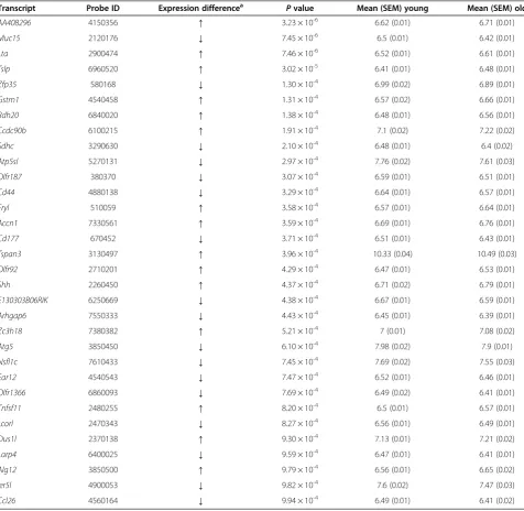
Table 1 Probes showing gene expression differences associated with paternal age (P< 0.001) Transcript Probe ID Expression differencea
Pvalue Mean (SEM) young Mean (SEM) old
AA408296 4150356 ↑ 3.23 × 10⁻⁶ 6.62 (0.01) 6.71 (0.01)
Muc15 2120176 ↓ 7.45 × 10⁻⁶ 6.5 (0.01) 6.42 (0.01)
Lta 2900474 ↑ 7.46 × 10⁻⁶ 6.52 (0.01) 6.61 (0.01)
Tslp 6960520 ↑ 3.02 × 10⁻⁵ 6.41 (0.01) 6.48 (0.01)
Zfp35 580168 ↓ 1.30 × 10⁻⁴ 6.99 (0.02) 6.89 (0.01)
Gstm1 4540458 ↑ 1.31 × 10⁻⁴ 6.57 (0.02) 6.66 (0.01)
Rdh20 6840020 ↑ 1.38 × 10⁻⁴ 6.48 (0.01) 6.56 (0.01)
Ccdc90b 6100215 ↑ 1.91 × 10⁻⁴ 7.1 (0.02) 7.22 (0.02)
Sdhc 3290630 ↓ 2.10 × 10⁻⁴ 6.48 (0.01) 6.4 (0.02)
Atp5sl 5270131 ↓ 2.97 × 10⁻⁴ 7.76 (0.02) 7.61 (0.03)
Olfr187 380370 ↓ 3.07 × 10⁻⁴ 6.59 (0.01) 6.51 (0.01)
Cd44 4880138 ↓ 3.29 × 10⁻⁴ 6.64 (0.01) 6.57 (0.01)
Fryl 510059 ↑ 3.58 × 10⁻⁴ 6.57 (0.01) 6.64 (0.01)
Accn1 7330561 ↑ 3.59 × 10⁻⁴ 6.69 (0.01) 6.76 (0.01)
Cd177 670452 ↓ 3.71 × 10⁻⁴ 6.51 (0.01) 6.43 (0.01)
Tspan3 3130497 ↑ 3.96 × 10⁻⁴ 10.33 (0.04) 10.49 (0.03)
Olfr92 2710201 ↑ 4.29 × 10⁻⁴ 6.47 (0.01) 6.53 (0.01)
Shh 2260450 ↑ 4.37 × 10⁻⁴ 6.71 (0.02) 6.79 (0.01)
E130303B06RIK 6250669 ↓ 4.38 × 10⁻⁴ 6.67 (0.01) 6.59 (0.01)
Arhgap6 7550333 ↓ 4.43 × 10⁻⁴ 6.45 (0.01) 6.39 (0.01)
Zc3h18 7380382 ↑ 5.21 × 10⁻⁴ 7 (0.01) 7.08 (0.02)
Atg5 3850450 ↓ 6.10 × 10⁻⁴ 7.98 (0.02) 7.9 (0.01)
Nsfl1c 7610433 ↓ 7.45 × 10⁻⁴ 7.69 (0.02) 7.55 (0.03)
Ear12 4540543 ↓ 7.47 × 10⁻⁴ 6.52 (0.01) 6.46 (0.01)
Olfr1366 6860093 ↓ 7.69 × 10⁻⁴ 6.49 (0.02) 6.41 (0.01)
Tnfsf11 2480255 ↑ 8.20 × 10⁻⁴ 6.5 (0.01) 6.57 (0.01)
Lcorl 2470343 ↓ 8.27 × 10⁻⁴ 6.56 (0.01) 6.49 (0.01)
Dus1l 2370138 ↑ 9.30 × 10⁻⁴ 7.13 (0.01) 7.21 (0.02)
Larp4 6400025 ↓ 9.59 × 10⁻⁴ 6.47 (0.01) 6.41 (0.01)
Alg12 3850500 ↑ 9.79 × 10⁻⁴ 6.56 (0.01) 6.65 (0.02)
Ier5l 4900053 ↓ 9.82 × 10⁻⁴ 7.6 (0.02) 7.47 (0.03)
Ccl26 4560164 ↓ 9.94 × 10⁻⁴ 6.49 (0.01) 6.41 (0.02)
a
Results and discussion
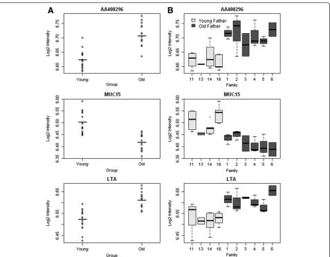
We found numerous differences in gene expression as-sociated with advanced paternal age. Most notably, probes associated with three transcripts were found to be differentially expressed in the offspring of older fa-thers compared to the offspring of younger fafa-thers with a false discovery rate (FDR) < 0.1:AA408296(also known as Diexf) (P= 3.23 × 10⁻⁶, FDR = 0.06) and Lta (P= 3.23 × 10⁻⁶, FDR = 0.06) had elevated expression, whilst Muc15(P= 7.46 × 10⁻⁶, FDR = 0.06) had reduced expres-sion (see Figure 1A). Although the absolute differences in gene expression are small and future work is needed to ascertain their functional significance, for each of these differentially expressed transcripts, consistent pat-terns of altered expression were observed across each of the old-father families (Figure 1B and Additional file 2). The top-ranked differentially expressed transcripts (P< 0.001) are listed in Table 1. Strikingly, almost a quarter of these differentially expressed loci have been previously implicated in the aetiology of autism, including Gstm1 (deletion in GSTM1 observed in autism case-parent trios) [16],Ccdc90b (missense mutation associated with autism in a study of cases and their parents) [17],Cd44 (differential expression in autism discordant monozy-gotic twins) [18], Accn1 (multiple SNPs associated with autism in a familial study) [19], Shh (increased serum SHH expression in autistic patients compared to non-autistic controls) [20],Dus1l(de novomissense mutation observed in autism families) [21] andIer5l(increased ex-pression in lymphoblast cells from autism patients in discordant sibling pairs) [22].
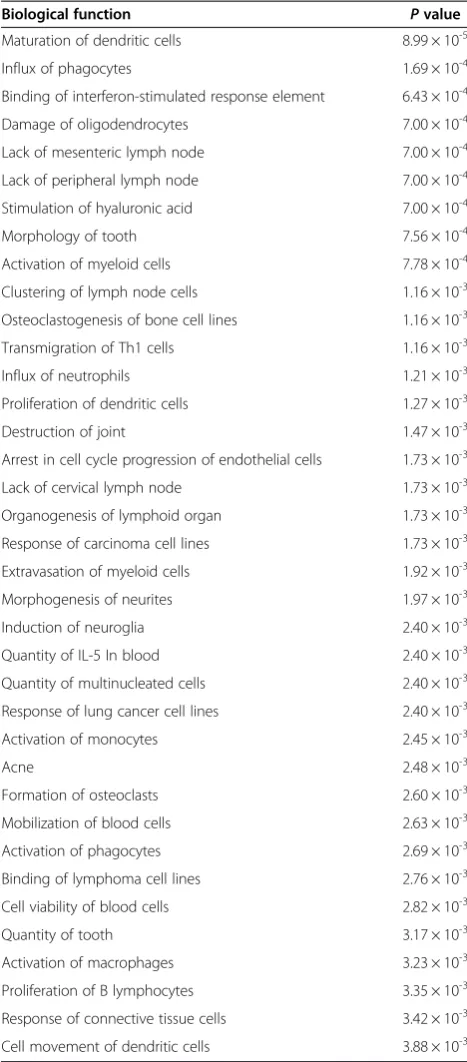
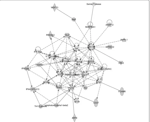
Ingenuity pathway analysis (IPA) [23] and the Database for Annotation, Visualization and Integrated Discovery (DAVID) [24,25] were used to identify gene pathways and functions enriched amongst transcripts differentially expressed in the offspring of old fathers. Of note, IPA iden-tified a significant enrichment for functional pathways in-volved in inflammation and inflammatory disease. Table 2 shows the top-ranked biological functions enriched in probes differentially expressed (P< 0.01) in the frontal cor-tex in the offspring of old fathers. The top-ranked biological network regulates immune cell trafficking and cell-to-cell signalling (Figure 2). Furthermore, the most significantly as-sociated gene ontology term identified by DAVID was im-mune response (14 genes, P= 2.87 × 10⁻⁴) and the second most associated cluster (after cytokine activity) included defence response, response to wounding, inflammatory re-sponse and acute inflammatory rere-sponse. This is interesting given mounting evidence linking the immune system to several neuropsychiatric disorders including autism [26-28]. A recent transcriptomic analysis of post-mortem brains from autistic patients, for example, showed changes in gene networks involved in immune and inflammatory responses in the frontal and temporal cortices [29].
In addition to identifying group-level variation be-tween the offspring of old and young fathers, we also ex-amined family-specific gene expression differences, as many of the genetic (or epigenetic) alterations believed to underlie the paternal age effect in autism are thought likely to be sporadic de novo events occurring in single Table 2 Top-ranked biological functions enriched in probes differentially expressed (P < 0.01) from IPA
Biological function Pvalue
Maturation of dendritic cells 8.99 × 10⁻⁵
Influx of phagocytes 1.69 × 10⁻⁴
Binding of interferon-stimulated response element 6.43 × 10⁻⁴
Damage of oligodendrocytes 7.00 × 10⁻⁴
Lack of mesenteric lymph node 7.00 × 10⁻⁴
Lack of peripheral lymph node 7.00 × 10⁻⁴
Stimulation of hyaluronic acid 7.00 × 10⁻⁴
Morphology of tooth 7.56 × 10⁻⁴
Activation of myeloid cells 7.78 × 10⁻⁴
Clustering of lymph node cells 1.16 × 10⁻³
Osteoclastogenesis of bone cell lines 1.16 × 10⁻³
Transmigration of Th1 cells 1.16 × 10⁻³
Influx of neutrophils 1.21 × 10⁻³
Proliferation of dendritic cells 1.27 × 10⁻³
Destruction of joint 1.47 × 10⁻³
Arrest in cell cycle progression of endothelial cells 1.73 × 10⁻³
Lack of cervical lymph node 1.73 × 10⁻³
Organogenesis of lymphoid organ 1.73 × 10⁻³
Response of carcinoma cell lines 1.73 × 10⁻³
Extravasation of myeloid cells 1.92 × 10⁻³
Morphogenesis of neurites 1.97 × 10⁻³
Induction of neuroglia 2.40 × 10⁻³
Quantity of IL-5 In blood 2.40 × 10⁻³
Quantity of multinucleated cells 2.40 × 10⁻³
Response of lung cancer cell lines 2.40 × 10⁻³
Activation of monocytes 2.45 × 10⁻³
Acne 2.48 × 10⁻³
Formation of osteoclasts 2.60 × 10⁻³
Mobilization of blood cells 2.63 × 10⁻³
Activation of phagocytes 2.69 × 10⁻³
Binding of lymphoma cell lines 2.76 × 10⁻³
Cell viability of blood cells 2.82 × 10⁻³
Quantity of tooth 3.17 × 10⁻³
Activation of macrophages 3.23 × 10⁻³
Proliferation of B lymphocytes 3.35 × 10⁻³
Response of connective tissue cells 3.42 × 10⁻³
litters. We compared the average transcript level for all off-spring within each family to the average across all offoff-spring of young fathers. We found a 1.8-fold enrichment in the number of family-specific significant gene expression differ-ences in the offspring of older fathers (average 426 differen-tially expressed genes) compared to the offspring of young fathers (average 234 differentially expressed genes) (P< 0.001). Significant probes for each of the six old-father fam-ilies are listed in Additional file 3. Of note, a number of probes were differentially expressed in the offspring of more than one advanced-age father including probes repre-sentingMyst1,Tnfsf11andFryl(see Additional file 4).
Conclusions
We present evidence for transcriptomic differences in the medial prefrontal cortex of offspring of old fathers compared to the offspring of young fathers, including
for genes previously implicated in autism, a neuropsychi-atric disease epidemiologically associated with advanced paternal age, and an enrichment of loci involved in the inflammatory response. Previous studies of gene expres-sion changes associated with age in the mouse brain have shown enrichment for genes associated with the in-flammatory response [8], although this is the first study to examine differences associated with advanced paternal age. Future work will examine whether these expression differences result fromde novogenetic (or epigenetic) al-terations occurring in the sperm of older fathers.
Availability of supporting data
The data set supporting the results is available in the Gene Expression Omnibus repository (Currently await-ing upload) or downloaded from our webpage http:// epigenomicslab.com/Paternal Age data.rar.
Additional files
Additional file 1:Overview of the samples used in this study.
Additional file 2:Significant gene expression differences for individual offspring split by sire for‘young’and‘old’fathers were seen for (A)AA408296,(B)Muc15and (C)Lta.
Additional file 3:Top-ranked differentially expressed transcripts (P< 0.001) within each of the six families with an old father.Shown for each transcript is the corresponding rank in the overall old vs young father group comparison.
Additional file 4:Comparison of the top-ranked differentially expressed transcripts (P< 0.001) across each of the six families with an old father.Shown for each transcript is the corresponding rank in the overall old vs young father group comparison. Empty cells indicate a non-significant difference.
Abbreviations
CNV:copy number variation; DAVID: Database for Annotation, Visualization and Integrated Discovery; FDR: false discovery rate; IL: interleukin; IPA: Ingenuity Pathway Analysis; RIN: RNA integrity number; SNP: single nucleotide polymorphism.
Competing interests
The authors declare that they have no competing interests.
Authors’contributions
All authors contributed to the design of the study. RS undertook the lab work. RS and JM drafted the article. RS collected and analysed data, wrote the manuscript, conceived and designed the study, made a critical revision and gave final approval of the manuscript. CF collected data, conceived and designed the study, made a critical revision and gave final approval of the manuscript. RK collected data, made a critical revision and gave final approval of the manuscript. LS analysed the data, made a critical revision and gave final approval of the manuscript. JB and AR conceived and designed the study, provided financial support, made a critical revision and gave final approval of the manuscript. JM wrote the manuscript, conceived and designed the study, made a critical revision and gave final approval of the manuscript. All authors read and approved the final manuscript.
Acknowledgements
This study was supported by the Beatrice and Samuel A Seaver Foundation, by a British Medical Association Margaret Temple Award, and the National Institute of Health Research Biomedical Research Centre for Mental Health at the South London and Maudsley National Health Service Foundation Trust and the Institute of Psychiatry, King’s College London, Pilot Award to Drs Jonathan Mill and Abraham (Avi) Reichenberg.
Author details
1Institute of Psychiatry, King’s College London, De Crespigny Park, Denmark
Hill, London SE5 8AF, UK.2Mount Sinai School of Medicine, Madison Avenue, New York, NY 10029, USA.3University of Exeter Medical School, St Luke’s
Campus, Magdalen Rd, Exeter EX1 2LU, UK.
Received: 10 October 2013 Accepted: 10 March 2014 Published: 23 March 2014
References
1. Lundstrom S, Haworth CM, Carlstrom E, Gillberg C, Mill J, Rastam M, Hultman CM, Ronald A, Anckarsater H, Plomin R, Lichtenstein P, Reichenberg A:Trajectories leading to autism spectrum disorders are affected by paternal age: findings from two nationally representative twin studies.J Child Psychol Psychiatry2010,51(7):850–856.
2. Kong A, Frigge ML, Masson G, Besenbacher S, Sulem P, Magnusson G, Gudjonsson SA, Sigurdsson A, Jonasdottir A, Wong WS, Sigurdsson G, Walters GB, Steinberg S, Helgason H, Thorleifsson G, Gudbjartsson DF, Helgason A, Magnusson OT, Thorsteinsdottir U, Stefansson K:Rate ofde novomutations and the importance of father’s age to disease risk.
Nature2012,488(7412):471–475.
3. Adkins RM, Thomas F, Tylavsky FA, Krushkal J:Parental ages and levels of DNA methylation in the newborn are correlated.BMC Med Genet2011,12:47.
4. Flatscher-Bader T, Foldi CJ, Chong S, Whitelaw E, Moser RJ, Burne TH, Eyles DW, McGrath JJ:Increasedde novocopy number variants in the offspring of older males.Transl Psychiatr2011,1:e34.
5. Smith RG, Reichenberg A, Kember RL, Buxbaum JD, Schalkwyk L, Fernandes C, Mill J:Advanced paternal age is associated with altered DNA methylation at brain-expressed imprinted loci in inbred mice: implications for neuropsychiatric disease.Mol Psychiatry2013,18(6):635–636. 6. Colangelo V, Schurr J, Ball MJ, Pelaez RP, Bazan NG, Lukiw WJ:Gene
expression profiling of 12633 genes in Alzheimer hippocampal CA1: transcription and neurotrophic factor down-regulation and up-regulation of apoptotic and pro-inflammatory signaling.J Neurosci Res
2002,70(3):462–473.
7. Valdes AM, Glass D, Spector TD:Omics technologies and the study of human ageing.Nat Rev Genet2013,14(9):601–607.
8. Lee CK, Weindruch R, Prolla TA:Gene-expression profile of the ageing brain in mice.Nat Genet2000,25(3):294–297.
9. Paul C, Nagano M, Robaire B:Aging results in differential regulation of DNA repair pathways in pachytene spermatocytes in the brown Norway rat.Biol Reprod2011,85(6):1269–1278.
10. Kokkinaki M, Lee TL, He Z, Jiang J, Golestaneh N, Hofmann MC, Chan WY, Dym M:Age affects gene expression in mouse spermatogonial stem/ progenitor cells.Reproduction2010,139(6):1011–1020.
11. Alter MD, Kharkar R, Ramsey KE, Craig DW, Melmed RD, Grebe TA, Bay RC, Ober-Reynolds S, Kirwan J, Jones JJ, Turner JB, Hen R, Stephan DA:Autism and increased paternal age related changes in global levels of gene expression regulation.PLoS One2011,6(2):e16715.
12. Paxinos G:The Mouse Brain in Stereotaxic Coordinates.London: Academic press; 2004.
13. Spijker S:Dissection of rodent brain regions.InNeuroproteomics.New York, NY, USA: Humana Press Inc, Springer; 2011:13–26.
14. Johnson WE, Li C, Rabinovic A:Adjusting batch effects in microarray expression data using empirical Bayes methods.Biostatistics2007,8(1):118–127. 15. Du P, Kibbe WA, Lin SM:Lumi: a pipeline for processing Illumina
microarray.Bioinformatics2008,24(13):1547–1548.
16. Buyske S, Williams TA, Mars AE, Stenroos ES, Ming SX, Wang R, Sreenath M, Factura MF, Reddy C, Lambert GH, Johnson WG:Analysis of case-parent trios at a locus with a deletion allele: association of GSTM1 with autism.
BMC Genet2006,7:8.
17. Neale BM, Kou Y, Liu L, Ma’ayan A, Samocha KE, Sabo A, Lin CF, Stevens C, Wang LS, Makarov V, Polak P, Yoon S, Maguire J, Crawford EL, Campbell NG, Geller ET, Valladares O, Schafer C, Liu H, Zhao T, Cai G, Lihm J, Dannenfelser R, Jabado O, Peralta Z, Nagaswamy U, Muzny D, Reid JG, Newsham I, Wu YMJ,et al:Patterns and rates of exonicde novomutations in autism spectrum disorders.Nature2012,485(7397):242–245.
18. Hu VW, Frank BC, Heine S, Lee NH, Quackenbush J:Gene expression profiling of lymphoblastoid cell lines from monozygotic twins discordant in severity of autism reveals differential regulation of neurologically relevant genes.BMC Genomics2006,7:118.
19. Stone JL, Merriman B, Cantor RM, Geschwind DH, Nelson SF:High density SNP association study of a major autism linkage region on chromosome 17.Hum Mol Genet2007,16(6):704–715.
20. Al-Ayadhi LY:Relationship between Sonic hedgehog protein, brain-derived neurotrophic factor and oxidative stress in autism spectrum disorders.
Neurochem Res2012,37(2):394–400.
21. O’Roak BJ, Vives L, Girirajan S, Karakoc E, Krumm N, Coe BP, Levy R, Ko A, Lee C, Smith JD, Turner EH, Stanaway IB, Vernot B, Malig M, Baker C, Reilly B, Akey JM, Borenstein E, Rieder MJ, Nickerson DA, Bernier R, Shendure J, Eichler EE:Sporadic autism exomes reveal a highly interconnected protein network ofde novomutations.Nature2012,485(7397):246–250. 22. Hu VW, Nguyen A, Kim KS, Steinberg ME, Sarachana T, Scully MA, Soldin SJ,
Luu T, Lee NH:Gene expression profiling of lymphoblasts from autistic and nonaffected sib pairs: altered pathways in neuronal development and steroid biosynthesis.PLoS One2009,4(6):e5775.
23. Ingenuity Pathway Analysis.[http://www.ingenuity.com/products/ipa] 24. Huang DW, Sherman BT, Lempicki RA:Systematic and integrative analysis of large
gene lists using DAVID bioinformatics resources.Nat Protoc2009,4(1):44–57. 25. Huang DW, Sherman BT, Lempicki RA:Bioinformatics enrichment tools:
paths toward the comprehensive functional analysis of large gene lists.
Nucleic Acids Res2009,37(1):1–13.
27. Noriega DB, Savelkoul HF:Immune dysregulation in autism spectrum disorder.Eur J Pediatr2013,173(1):33–43.
28. Michel M, Schmidt MJ, Mirnics K:Immune system gene dysregulation in autism and schizophrenia.Dev Neurobiol2012,72(10):1277–1287. 29. Voineagu I, Wang X, Johnston P, Lowe JK, Tian Y, Horvath S, Mill J, Cantor
RM, Blencowe BJ, Geschwind DH:Transcriptomic analysis of autistic brain reveals convergent molecular pathology.Nature2011,474(7351):380–384. 30. Ingenuity Pathway Analysis legend.[http://www.biolreprod.org/content/
suppl/2011/02/23/biolreprod.110.090019.DC1/90019SupLegend.pdf]
doi:10.1186/2040-2392-5-24
Cite this article as:Smithet al.:Transcriptomic changes in the frontal
cortex associated with paternal age.Molecular Autism20145:24.
Submit your next manuscript to BioMed Central and take full advantage of:
• Convenient online submission
• Thorough peer review
• No space constraints or color figure charges
• Immediate publication on acceptance
• Inclusion in PubMed, CAS, Scopus and Google Scholar
• Research which is freely available for redistribution