Histochemical study of the olfactory rosette of C
yprinus carpio
(Linnaeus, 1758)
Ghosh S. K.
; Chakrabarti P.
*Received: April 2011 Accepted: November 2011
Abstract
The distribution and localization of acid and neutral mucins in various cells lining the olfactory epithelium of Cyprinus carpio have been studied histochemically by employing the PAS-AB technique. Variations in the localization of protein in different cells lining the olfactory epithelium have been correlated with the functional significance of the region concerned. Intense localization of the silver stain in the surface of the olfactory epithelium as well as in the central core confirms the presence of different types of neurons. The localization and functional variations of alkaline phosphatase (ALPase) and adenosine-tri-phosphatase (ATPase) in the cells lining the olfactory epithelium of C. carpio have been discussed.
Keywords: Mucopolysaccharides, Protein, Neuron, ALPase, ATPase, Olfactory Epithelium,
Cyprinus carpio
______________________
Fisheries Laboratory, Department of Zoology, The University of Burdwan, Burdwan-713 104,West Bengal, India.
*Corresponding author’s email: [email protected]
Introduction
The olfactory organ of fishes is of immense importance because of its roles in detection, recognition, selection of food and other activities as a chemoreceptor. A number of workers have studied the histological peculiarities of the olfactory epithelium in fishes, however, there is few information regarding the histochemistry involving identification and localization of the various cellular contents of the cells lining the olfactory epithelium and their role in the sensory reception in teleosts (Ojha and Kapoor, 1972; Datta Munshi and Singh, 1975; Pandey and Mishra, 1984; Belanger et al., 2003; Chakrabarti, 2005a). Still lacuna exists regarding the histochemical studies of the various cells lining the olfactory epithelium of fish. Therefore, an attempt has been made in the present study to examine, more closely, the precise chemical constituents of the cells and correlate them with functional aspects of the olfactory rosette in Cyprinus carpio (Linnaeus).
Materials and methods
Adult healthy fishes of Cyprinus carpio
were collected from the local freshwater body. The fishes were decapitated and the olfactory rosettes were dissected out from the dorsal surface under stereoscopic dissecting binocular microscope. The olfactory rosettes were fixed in 10% neutral formalin. The tissues were then dehydrated through ascending series of ethyl alcohol, cleared in xylene and embedded in paraffin using standard method. Some tissues were also fixed in cold absolute acetone (4C) for enzyme histochemical study. Vertical tissue
sections were cut at 8-10m and were
subjected to following histochemical tests: Periodic Acid Schiff’s (PAS) in combination with Alcian Blue (PAS-AB) for the detection of neutral and acid mucins (Mowry, 1956), Mercury-Bromphenol Blue (MBB) method for detection of basic protein (Bonhag, 1958), Silver impregnation method for axons (Marsland et al., 1954), Calcium-cobalt method for alkaline phosphatase (ALPase) (Gomori, 1951) and Calcium method for Adenosine triphosphatase (ATPase) (Padykula and Herman, 1955).
Results
Detection of mucopolysaccharides
PAS-AB: This combined technique of PAS-AB imparted bright red colour for neutral mucin due to PAS whereas AB produced a blue colour when it reacted with acid mucin. In the present study, the secretory and non-secretory mucous cells in the olfactory epithelium of C. carpio
exhibited purple-bluish colour of varying intensities with PAS-AB histochemical test confirming the presence of mixture of acid and neutral mucopolysaccharide materials in different proportions (Figures 1, 2). On the contrary the mucous mother cells located in the deeper region of the olfactory epithelium exhibited only red colour confirming the presence of neutral mucin exclusively (Figure 2). The supporting cells showed intense PAS-AB reaction due to their mucopolysaccharide content (Figure 2). The intense blue colour in PAS-AB reaction was also discernible in the mast cells located in the deeper region of olfactory epithelium in C. carpio. This indicated that they seemed to have some secretory functions, possibly
the heparin. The connective and collagen tissues in the central core exhibited
moderate reaction to the PAS-AB test.
Figure 1: Localization of acid and neutral mucin (ANM) in the secretory mucous cells (SMC) and non-secretory mucous cells (NMC) of the olfactory epithelium (OEP). Note moderate PAS-AB reaction in the connective tissue of central core (CC) 150.
Figure 2: Localization of ANM in the SMC and supporting cells (SC) of OEP. Note intense neutral mucopolysaccharide reaction in mucous mother cells (MMC) and mast cells (MC) (arrows). Note also moderate PAS-AB reaction in CC 400.
Detection of protein
An intense reaction was discernible in the supporting and mast cells of the olfactory epithelium (Figure 3). The basal cells of olfactory epithelium stained more intensely due to the proteinaceous nature
of these cells. Moreover maximum protein reaction has been encountered in the blood cells and connective tissue of the central core. The weak reaction of protein was discernible in the mucous cells of the olfactory epithelium (Figure 3).
Figure 3: Localization of protein in MC, basal cells (BC) (solid arrows) and SC (broken arrows). Note intense protein reaction in blood cells (arrow head) of CC and weak reaction in SMC 400.
Detection of axons
Different degrees of silver deposition were distributed in e various layers of olfactory lamella. Silver stain was discernible in the apical dendrite process of the olfactory epithelial surface. Maximum localization
of silver stain was discernible in the central core of the olfactory epithelium in
Cyprinus carpio due to the presence of many small and medium sized neurons (Figure 4).
Figure 4: Intense silver stain in the neurons (solid arrows) and synapse of the neurons (broken arrows) in CC 400
Detection of alkaline phosphatase (ALPase)
Intense alkaline phosphatase activity was observed in the olfactory epithelium, especially in the receptor cells and in the basement membrane (Figures 5, 6). The activity of this enzyme was low in the supporting cells (Figures 5, 6). Alkaline phosphatase was found in high
concentration along synaptic connections in primary neurons (Figure 5). A weak ALPase activity was however, detected in the mucous cells (Figure 6). The mast cells as well as the basal cells were also intensely reacted with ALPase (Figures 5, 6).
Figure 5: Intense ALPase reaction in receptor cells (RC) (solid arrows), MC (arrow heads) and BC. Note low reaction in SC (broken arrows) 400.
Figure 6: Intense ALPase activity in the RC (solid arrows), MC (arrow heads), BC and basement membrane (BM). Note weak ALPase activity in SMC and broken arrow indicates low reaction in SC 1000.
Detection of adenosine triphosphatase (ATPase)
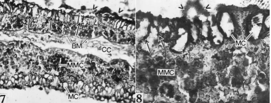
Intense ATPase activity was associated in the receptor cells, mucous mother cell and in the border of mucous cells in the olfactory epithelium of C. carpio. Mucous mother cells were situated just above the basement membrane provided with dense granules. These cells gradually migrated
from deeper to the surface region and transformed into mature mucous cells. The intense activity of this enzyme was also evidenced in the basement membrane and in the luminal secretion of mucin while the central core shows weak ATPase activity (Figures 7, 8).
Figure 7: Intense ATPase activity in the RC (arrows), MMC, BM and border of mucous cells (MC). Note intense ATPase activity in luminal mucin (arrow heads) and weak reaction in CC 400.
Figure 8: Intense ATPase activity in RC (solid arrows), MMC, inner border of MC and luminal secretion (arrow heads) 1000.
Discussion
The conspicuous mucous cells, differing in shape and stages of maturation, i.e.
secretory or peripheral non-secretory discharge their secretion product through
the opening of the extracellular surface coat. The histochemical nature of the
olfactory epithelium mucin in C. carpio
has been studied by employing PAS-AB
histochemical test to establish its chemical nature as well as finding out the
importance of its secretion. It has been noticed during the present investigation
that the content of the mucous cells in the various regions of the olfactory epithelium
in C. carpio differed greatly in chemical nature with regard to their distribution as
well as in their various stages of maturation i.e. secretory or sub-surface
non-secretory mucous cells. Secretory and non-secretory mature mucous cells
situated in the surface of the olfactory epithelium contain a mixture of neutral
and acid mucins as confirmed by the PAS-AB histochemical test. The secretion of mixture of acid and neutral
mucopolysaccharides from the mucous cells probably helps to prevent friction
against microscopic debris and also helps the smooth flow of water in the olfactory
chamber. This is in conformity with the findings of Rahmani and Khan (1980) in
the olfactory mechanism of Anabas testudineus and Chakrabarti (2005b) in the
olfactory epithelium of Puntius javanicus. Zalewsky and Moody (1979) also reported
the presence of the heterogenous nature of mucus and its secretion as a mosaic of
neutral and acidic mucopolysaccharide in the canine gastric mucosa. On the other
hand, the mucous mother cells are provided with exclusively neutral mucin as
advocated by the PAS-AB test. This is probably due to the fact that the mucous
cells are involved in the synthesis of complex glycoprotein macromolecules
during their formation from the multipotent cells. However, Sinha (1975)
reported that during the early stages of development of the mucous cells i.e., in
the mucous mother cells there is a primary synthesis of neutral mucopolysaccharide
granules which in the course of development of the same is either
transformed into acid mucopolysaccharide granules and/or the cells synthesizes new
acid mucopolysaccharides present in the subsequent stages in the mature
non-secretory and non-secretory mucous cells. These polysaccharide granules (acidic and
neutral) ultimately fuse together in the mature non-secretory mucous cells which are gradually pushed towards the periphery
and give rise to a complex mixture of both acidic and neutral mucins (Gona, 1979).
The intense blue colour in PAS-AB reaction in the mast cells in C. carpio is
due to the presence of profuse amount of heparin which is thought to cause
fluctuations in the production of mucus in the olfactory mucosa. Moulton and Beidler
(1967) reported that as the terminal mucus film in the olfactory mucosa is believed to
be an important factor in the olfactory
process this may influence the variations
in the olfactory sensitivity.
The mast cells and the supporting cells of the olfactory epithelium exhibited
intense protein reaction in the present study. The mucopolysaccharide content of
supporting cells and heparin from mast
cells maintains secretion along the surface of the epithelium. Therefore, it is concluded that the secretion is at least in
part, proteinaceous. The basal cells exhibit intense reaction for protein probably for
various metabolic as well as physiological activities.
In the present study, the intense localization of silver reaction in the
olfactory epithelium in C. carpio provides a direct evidence of synaptical connection
of primary and secondary neurons as well as orientation of dendrites of the receptor
cells, along the most superficial layer of the olfactory epithelium. In this fish the
sensory receptor cells in the olfactory epithelium send their axons towards the
central core.
Intense ALPase activity in the
receptor cells and synaptic connection of primary neurons of C. carpio may be
associated with the transport of various nerve impulses during olfactory
transduction mechanism. The intense ALPase activity in nuclei of basal cells may be involved in the process of
regenerating receptor and other cells of the olfactory epithelium. Andres (1966; 1969)
also suggested that basal cells are the precursors of regenerating receptor cells.
Evans et al. (1982) also observed increased
mitotic figures in the basal region in a
constituting epithelium after degeneration. Further, ALPase activity in the mast cells may be involved in secretion and
metabolism of the cell concerned. The ALPase activity in the basement
membrane of the olfactory epithelium
advocates its positive role in the transportation of the various chemicals and nerve impulses through the membrane
during olfactory sensation. Agrawal et al. (1979) reported ALPase activity in the
basement membrane of the skin epidermis of Barbus sophore and confirmed the
positive role of ALPase in the transportation of various chemicals
through the membrane. Intense ALPase activity in the mucous mother cells in the
olfactory epithelium may be related to the synthesis of neutral mucopolysaccharide.
Weinreb and Bilstad (1955) reported that ALPase activity and the occurrence of
neutral mucopolysaccharides occupy the same sites in the digestive tract of rainbow
trout.
Intense ATPase activity of the
border of mucous cells in the olfactory epithelium unequivocally suggests its
secretory nature. The mucin secretion is effected from these cells by exocytosis
which is an active process requiring energy and ATPase activity is the main source of such energy release. Morozov
and Khramtsov (1979) pointed out the role of ATPase in the secretory activity of the
cells of various tissues in different animals. In the mucin component of
non-secretory mucous cells ATPase activity is
observed and probably related to the active
synthesis of carbohydrate and protein, which are the major components of the secreted mucin. The mucin granules of the
mucous mother cells were also positive for ATPase reaction. This reaction is probably
due to the fact that these granules are more
or less directly involved in the synthesis of the mucus component of the mucous mother cells. The role of ATPase in the
biosynthesis of carbohydrates and protein which are the major components of the
secreted mucin at the cytoplasmic area of the mucous cells in the tongue of a rat was
reported by Talesara et al. (1980). On the other hand, ATPase activity in the receptor
cells of the fish studied is related to the transmission of various nerve impulses.
Shantha and Nakajima (1970) have earlier reported that the presence of ATPase in the
olfactory cell axons of the olfactory mucosa of monkey possibly involved in
the process of olfactory sensations is elicited by the contact of odour particles
with these receptor cells. Acknowledgements
The authors are grateful to Dr. G. Aditya, Head of the Department of Zoology, for
providing laboratory facilities and also thankful to the Department of Science and
Technology, New Delhi for providing necessary instrumental facilities for this research work.
References
Agrawal, S. K., Banerjee, T. K. and Mittal, A. K., 1979. Enzyme in the epidermis of a freshwater teleost
Barbus sophore (Cyprinidae, Pisces): a
histochemical investigation.
Mikroskopie, 35, 258-264.
Andres, K. H., 1966. Der Feinball der Regio olfactoria von makro smatikern.
Zeitschrift für Zellforschung und mikroskopische Anatomie, 69,
140-154.
Andres, K. H., 1969. Der Olfaktorische Saum der Katze. Zellforschung und mikroskopische Anatomie, 96,
250-274.
Belanger, R. M., Smith, C. M., Corkum, L. D. and Zielinski, B. S., 2003. Morphology and histochemistry of the
peripheral olfactory organ in the round goby, Neogobius melanostomus
(Teleostei: Gobiidae). Journal of Morphology, 257, 62-71.
Bonhag, P. F., 1958. (vide, Pearse, A. G. E., Vol.1, 1975). Journal of
Morphology, 96, 381.
Chakarbarti, P., 2005a. Histological and histochemical studies on the olfactory rosette of Mugil parsia (Hamilton).
Folia Morphologica, 64, 41-46.
Chakrabarti, P., 2005b. Histoarchitecture and histochemical localization of mucopolysaccharides in the olfactory
epithelium of Puntius javanicus
(Bleeker). Journal of Inland Fisheries
Society of India, 37, 47-52.
Datta Munshi, J. S. and Singh, S. P., 1975. Histochemical observations on the olfactory mucosa of the Indian green snake headed fish, Channa
puncatata (Bloch). Proceedings of the Zoological Society, 28, 1-13.
Evans, R. E., Zielinski, B. and Hara, T. J., 1982. Chemoreception in fishes. In: Ostrander, G. K. (Eds), The laboratory fish, Academic Press, New York,
471-479.
Gomori, G., 1951. Alkaline phosphatase of cell nuclei. Journal of Laboratory
and Clinical Medicine, 37, 526.
Gona, O., 1979. Mucous glycoproteins of teleostean fish: a comparative
histochemical study. Histochemical Journal, 11, 709-718.
Marsland, T. A. Glees, P. and Erikson, L. B., 1954. Modification of the Glees Silver impregnation for paraffin sections. Journal of Neuropathology &
Experimental Neurology, 13, 587.
Morozov, I. A. and Khramtsov, A. V., 1979. Changes in morphological and functional properties of gastric parietal
cells in secretion activation. Fiziol Zh SSSR Im I M Sechenova, 65, 456-461.
Moulton, D. G. and Beidler, L. M., 1967. Structure and function in the peripheral
olfactory system. Physiological Reviews, 47, 1-52.
Mowry, R. W., 1956. Alcian blue techniques for the histochemical study
of acidic carbohydrates. Journal of Histochemistry & Cytochemistry, 6,
82.
Ojha, P. P. and Kapoor, A. S., 1972. Functional anatomy of nose in the
teleost Wallago attu Bl. and Schn.
Archives of Biology, 83, 105-116.
Padykula, H. A. and Herman, E., 1955. The specificity of histochemical
method for adenosine
triphosphatase. Journal of
Histochemistry & Cytochemistry, 3, 170-175.
Pandey, K. C. and Mishra, R. C., 1984. Histochemical studies on the olfactory epithelium of Anabas testudineus
(Bloch). Matsya, 9, 17-24.
Rahmani, A. R. and Khan, S. M., 1980. Histology of the olfactory epithelium and the accessory nasal sacs of an
anabantoid fish, Anabas testudineus
(Bloch). Archives de Biologie, 91,
397-411.
Shantha, T. R. and Nakajima Y., 1970. Histological and histochemical studies on the rhesus monkey (Macaca
mulatta) olfactory mucosa. Zeitschrift für Zellforschung und mikroskopische
Anatomie, 103, 291-319.
Sinha, G. M., 1975. A histochemical study of the mucous cells in the buccopharyngeal region of four Indian
freshwater fishes in relation to their origin, development, occurrence and
probable functions. Acta Histochemica, 53, 217-223.
Talesara, C. L. and Maru, B. K. and Chaudhuri, B. N., 1980. Histochemical distribution and functional role of phosphatase,
cholinesterases and succinic dehydrogenase in the gustatory epithelium and lingual glands of the
rat. Acta Anatomica, 107, 52-59.
Weinreb, E. L. and Bilstad, N. M., 1955. Histology of the digestive tract and adjacent structures of a rainbow trout,
Salmo gairdneri irideus. Copeia, 3,
194-204.
Zalewsky, C. A. and Moody, F. G., 1979. Mechanism of mucus release in
exposed canine gastric mucosa.
Gastroenterology, 77, 719-729.