OPEN A
CCE
SS
JZ
AR R
esearch article
OPEN A
CCE
SS
Facial thermography is not useful in assessing body temperature in
common squirrel monkeys (
Saimiri sciureus
) in comparison to rectal
temperatures
R. Pizzi1*, A. Dowling1, D. Brown1, S. Girling1, S. Pearson1, H. Bacon2 and Y. Martinez Pereira3
1Royal Zoological Society of Scotland, Edinburgh Zoo, 134 Corstorphine Road, EH26 8DF
2Jeanne Marchig International Centre for Animal welfare Education, , Royal (Dick) School of Veterinary Studies, University of Edinburgh, Easter Bush, EH25 9RG 3Hospital for Small Animals, Royal (Dick) School of Veterinary Studies, University of Edinburgh, Easter Bush, EH25 9RG
*Correspondence: [email protected]
Keywords:
infrared, primate, thermal imaging
Article history:
Received: 17 March 2015 Accepted: 9 July 2015 Published online: 23 July 2015
Abstract
A group of 39 captive common squirrel monkeys (Saimiri sciureus) had their body temperature
measurements compared by rectal thermometry and facial infrared thermal imaging (Flir i3, Flir
Systems Inc.). Squirrel monkeys were caught up and manually restrained for examination and temperature determination as part of routine health checks. The mean difference between rectal temperature and maximum facial thermography temperatures was 3.4° C (95% confidence interval = 3.1–3.7° C). The repeatability coefficient of maximum facial temperatures was 3.18° C at a 95% CI. The Pearson correlation coefficient for maximum facial thermography temperatures compared to rectal temperatures was -0.10 (95% CI = -0.27–0.07). This study found no meaningful correlation between facial thermography and rectal temperatures in common squirrel monkeys. Facial thermography had poor accuracy and poor precision compared to rectal temperature measurement. Facial thermography does not appear to be a useful means of detecting altered body temperature in captive common squirrel monkeys.
Introduction
Detection of ill health in an individual captive primate in a large social group can be problematic in zoological and research populations. Capture and restraint are usually needed simply to assess body temperature (Calle and Joslin 2014). Detection
of an elevated or decreased body temperature by means
of a non-contact method that does not require separation or restraint may confer welfare advantages in social captive primates. Catching primates for temperature measurement
can cause stress to the individual animal and group, disrupt
normal behaviours and interactions between group members, interfere with any behavioural research activities being performed on the group, risks injury to animals from catching and manual restraint, and can have an adverse effect on the animal’s welfare when performed unnecessarily or overly frequently (Reichard 2007).
The common squirrel monkey (Saimiri sciureus) is a small
social arboreal primate from South America that is commonly kept in zoological collections, as pets, and in animal research facilities. Conscious restrained squirrel monkeys normally have a rectal temperature of 38 to 39.5° C, while healthy active or
struggling animals may rapidly increase their body temperatures
as high as 41° C (Brady 2000). Body temperatures below 37.7°C may be seen with seriously ill squirrel monkeys (Brady 2000). Elevated body temperature in primates can be associated with a wide range of different health conditions that could require further detailed diagnostic work-up (Knockaert 2007; Varghese et al. 2010; Colvin et al. 2012; Calle and Joslin 2014).
Determination of body temperature in humans and
non-human primates is commonly achieved by means of rectal or oral thermometers, or an infrared thermometer inserted in the ear to measure the temperature of the tympanic membrane
(Davie and Amoore 2010; Sethi et al. 2013). Human infrared thermometers inserted in the ears of squirrel monkeys were not found to be significantly different to rectal temperatures (Long et al. 2011). This method usually requires a primate to be restrained, with the associated disadvantages of this. A hands-off body temperature detection method could potentially be a useful health screening tool in captive primate groups of a social species such as the common squirrel monkey.
Grossbard et al. 2014; Wood et al. 2014; Wilhelm et al. 2015), soft tissue injuries (Stewart et al. 2008), wound healing (Melero et al. 2013), stress determination (Bouwknecht et al. 2007; Ludwig et al. 2007; Travain et al. 2015), reproduction (Talukder et al. 2014), and disease status determination (Biondi et al. 2005; Pérez de Diego et al. 2013; Samara et al. 2013).
Facial infrared thermography has been used as a screening
method for the detection of elevated body temperature in people arriving at airports, as an early detection screening technique during disease outbreaks such as SARS, influenza and Ebola (Nishiura and Kamiya 2011; Kuan and Chang 2012; Bogoch et al. 2015; Gunaratnam et al. 2014). Although surface temperature measurement methods are not always statistically accurate in that they do not measure core body temperature (Ganio et al. 2009), facial thermography can still, with care, be used with precision, and specified maximum facial temperature thresholds can be set. If these are breached, further investigation of a subject’s health status can be initiated (Chan et al. 2003; Bitar et al. 2009; Cho and Yoon 2014).
Thermal imaging cameras detect radiation in the infrared range of the electromagnetic spectrum (approximately 9–14 µm). Thermal images display the amount of infrared energy emitted, transmitted and reflected by an object. The spectrum and quantity of thermal radiation depend on an object’s surface temperature. Other factors however, also influence radiation, such as the emissivity of the measured object, reflected radiation and atmospheric absorption, amongst others. Artefacts may also be caused by factors such as lighting, external heat sources, reflections, moisture and evaporation (Maldague et al. 2001; Hilsberg-Merz 2007; Chiang et al. 2008).
In recent years, the cost of this type of equipment has decreased dramatically, with simple handheld thermal cameras with a sensitivity of 0.1° C ± 2%, and dedicated analysis software, now retailing for under €1000. This is likely to result in increased availability and application of thermography in zoological collections, but the evidence base for many applications in zoological medicine is yet to be developed (Hilsberg-Merz 2007).
The Living Links centre at Edinburgh Zoo is a unique collaboration between the Royal Zoological Society of Scotland (RZSS) and the University of St Andrews. It houses two primate species in a mixed exhibit, common squirrel monkeys and brown capuchins (Cebus apella), in two near identical enclosures for comparative non-invasive behavioural research and display to the visiting public. All research is voluntary, and the monkeys have trained interaction with researchers through research pods, which could allow non-contact temperature measurements via thermography with habituation. As of January 2015 there were 40 common squirrel monkeys and 35 brown capuchins in the population.
The aim of this study was to assess if there was any correlation between rectal temperatures and facial thermography in common squirrel monkeys caught up for routine health examinations, and whether this could be practical and effective as a method for hands-off detection of elevated or depressed body temperatures in individual squirrel monkeys.
Methods
Thirty-nine captive common squirrel monkeys (Saimiri sciureus),
six males and 33 females, in the Living Links exhibit and research facility at the Royal Zoological Society of Scotland’s Edinburgh Zoo, were caught up for routine scheduled physical examination, weighing and replacement of identification neck beads on two separate occasions. Mean body weight was 722 g (95% CI = 664–781 g). The squirrel monkeys were between 1 and 16 years old, with a mean age of 7 years (95% CI = 5.6–8.4 years). Air temperature was 13–15° C on both occasions and humidity 63–
71%. The squirrel monkeys were kept indoors overnight prior to catching, and the housing had subdued fluorescent roof lighting. Squirrel monkeys were caught individually by hand and restrained manually by keeping staff. As soon as an individual was caught and restrained, its rectal temperature was taken with a certified digital thermometer with soft flexible tip (Boots Pharmaceuticals), and four images of the face were captured with the thermal imaging camera (Flir i3, Flir Systems Inc.), from a distance of 50 cm. The camera was held horizontally and vertically perpendicular to the centre of the face, aiming at the nose, with both sides of the face being symmetrical. The monkey then underwent a full veterinary physical examination. Handling and examination took under 5 min in all individuals. Monkeys were then returned to their normal enclosure. Any signs of illness or injuries were recorded over the next six months to rule out possibilities of undetected health problems on the observation dates that could have influenced the body temperature readings. All thermal images were imported into dedicated software for analysis (Flir Tools 4.1 2014, Flir Systems Inc.). Thermal images for inclusion had to meet the criteria of being symmetrical, perpendicular and containing the entire face. The mean and maximum facial temperatures were measured from each individual thermal image. Data were examined for normality. Statistical analysis and reporting was as recommended in statistical guidelines for contributors to medical journals (Altman et al. 1983). Repeatability coefficients were calculated at a 95% confidence level to assess the repeatability of facial thermography measurements under the study conditions. Pearson correlations of both maximum and mean facial thermography temperatures with rectal temperatures were calculated.
Results
One hundred and twenty-nine facial thermal images from 39 individual common squirrel monkeys met the criteria for inclusion in the study. No squirrel monkeys included had any signs of ill health or injury on their veterinary examination, and no squirrel monkeys included developed any signs of ill health in the six months after their examination.
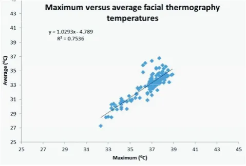
The mean rectal temperature was 40.0° C (95% CI 39.9–40.1° C), and the median was also 40.0° C. The mean maximum facial thermography temperature was 36.6° C (95% CI 36.4–36.9° C) and the median 37° C. The mean of the mean (average) facial thermography temperature was 32.9° C (95% CI 32.6–33.2° C) and the median 33.3° C. The mean difference between rectal temperature and maximum facial thermography temperatures was 3.4° C (95% CI of the difference = 3.1–3.7° C). The mean difference between rectal temperature and mean (average) facial thermography temperatures was 7.1° C (95% CI of the difference = 6.8–7.4° C). The mean difference between maximum and mean (average) facial thermography temperatures was 3.7° C (95% CI of the difference = 3.6–3.9° C). The hottest regions on all facial thermographs were the eyes.
The plot of maximal facial temperatures compared to mean facial temperatures is given in Figure 2, and the plots of maximal
and mean facial temperatures compared to rectal temperatures in
Figures 3 and 4.
The repeatability coefficient of maximum facial temperatures was 3.18° C at a 95% confidence level, and the repeatability coefficient of mean facial temperatures was 3.41° C at a 95%
Figure 2. Correlation between maximum and mean (average) facial
temperatures as determined by thermography in manually restrained
common squirrel monkeys after catching.
Figure 3. Correlation between facial thermography maximum temperatures and rectal temperatures in manually restrained common squirrel monkeys after catching.
Figure 4. Correlation between facial thermography mean (average)
temperatures and rectal temperatures in manually restrained common
squirrel monkeys after catching
confidence level. The Pearson correlation coefficient for maximum versus mean facial thermography temperatures was 0.87 (95% CI = 0.82–0.91), P<0.001. The Pearson correlation coefficient for maximum facial thermography temperatures compared to rectal temperatures was -0.10 (95% CI = -0.27–0.07), P=0.26. The Pearson correlation coefficient for mean facial thermography temperatures compared to rectal temperatures was -0.06 (95% CI = -0.23–0.11), P=0.49.
Discussion
Squirrel monkeys are commonly housed in zoos around the world, used in biomedical research, and kept as pets. However, as with all primates, they have the potential to act as vectors of zoonotic disease (Roberts 1995) both to humans and to other primate species. As a result, strict biosecurity precautions are recommended when handling them (Brady 2000). Obtaining a core body temperature is recognised as being an essential component of a comprehensive physical examination in non-human primates (Long et al. 2011), but handling primates to obtain a core body temperature raises issues of occupational health (Long et al. 2011) and patient welfare, as the capture and handling of
non-human primates not habituated to the process is recognised to be
stressful and potentially detrimental to welfare (Rodas-Martínez et al. 2013). This study attempted to establish the utility of non-invasive facial thermography as a proxy for non-invasive core body temperature measurements, to minimise the potential for human and animal safety and welfare problems.
This study found no meaningful correlation between facial
thermography and rectal temperatures in common squirrel
monkeys. This is evident from the plots in Figures 3 and 4, and the very low correlation coefficients calculated.
The accuracy of facial temperatures, as determined by thermography, was poor, with a mean difference of 3.4° C (95% CI = 3.1–3.7° C) between rectal temperatures and maximum
facial thermography temperatures, and the accuracy of mean
facial temperatures even poorer. This was expected, as it has also been seen in human studies (Sund-Levander et al. 2004; Chiang et al. 2008; Ganio et al. 2009; Sethi et al. 2013), where facial
thermography is not an accurate measure of rectal or core body
temperature, but changes in these may still be reflected in similar changes in facial thermography, enabling its use in screening.
Precision of facial thermography temperature measurements
same animal being taken under identical conditions, only separated by a few seconds. The repeatability coefficients illustrate that one could be 95% certain that the difference between two separate measurements of maximum facial temperatures would be less than or equal to 3.18° C. This is such a wide difference as to make the technique useless in practice for determining if an animal’s temperature was abnormal, even if there had been a reasonable correlation with rectal temperatures. Sikoski et al. (2007) similarly found that in the crab-eating macaque (Macaca fascicularis), thermography of the face, shoulder, axilla and abdomen had no correlation with rectal temperatures, with high between- and within-subject variability in thermography temperatures.
There are limitations to this study that should be considered. While the spectrum and quantity of thermal radiation emitted depend on a subject’s surface temperature, thermal images include infrared energy emitted, transmitted and reflected by an object. Factors such as the emissivity of the measured subject, reflected radiation and atmospheric absorption also influence radiation, and hence thermography temperature readings. Artefacts may be caused by factors such as lighting, external heat sources, reflections, moisture and evaporation (Maldague et al. 2001; Hilsberg-Merz 2007; Chiang et al. 2008). Every effort was
made to minimise these in this study by carrying it out indoors,
under conditions of subdued fluorescent lighting and no air flow. An important limitation to this study was that the squirrel monkeys needed to be initially caught up and restrained for the facial thermography and rectal temperature measurements. Ideally core body temperatures and facial thermograms would
be compared in calm unrestrained animals to eliminate the
possibility of this affecting the results and subsequent correlation calculations (Brady 2000). It is possible that stress associated with capture and restraint resulted in peripheral vasoconstriction and consequently in decreased facial temperatures on thermography. It is alternatively possible that while the rectal temperatures
rose rapidly due to the catch up, the related facial temperatures
rose more slowly, and this difference would not be captured by this study design. These limitations could only be eliminated by implanting validated temperature loggers for comparison with
facial thermography temperatures subsequently obtained from
calm trained animals, or by anaesthetising the monkeys for serial comparative measurements over a period of time (but this may also be affected by anaesthetic drug effects and inability to thermoregulate normally whilst under anaesthesia). Both of these are non-therapeutic interventions, and in the United Kingdom would hence require government licensing and inspection, as for laboratory animals, under the Animals (Scientific Procedures) Act of 1986. This would be unlikely to be acceptable to a zoological collection’s welfare and ethics committee in the United Kingdom.
Infrared thermography has been found to be useful in a variety of human and veterinary health and diagnostic applications (Hilsberg-Merz 2007; Kammersgaard et al. 2013; Melero et al. 2013; Pérez de Diego et al. 2013; Amezcua et al. 2014; Grossbard et al. 2014; Samara et al. 2014; Sanchis-Sánchez et al. 2014; Talukder et al. 2014; Wood et al. 2014; Biondi et al. 2015; Wilhelm et al. 2015). While infrared facial thermography has been widely used for airport arrival screening in an attempt to assess travellers for infectious diseases such as SARS, influenza and Ebola during outbreaks, its actual efficacy in published studies of this use has however been disappointing, with limited correlation and relatively low sensitivity and positive predictive values (Chan et al. 2003; Shu et al. 2005; Bitar et al. 2009; Priest et al. 2011; CADTH 2014). Bitar et al. (2009), reviewing studies using thermal imaging cameras to screen passengers at airports for infectious diseases causing elevated body temperatures, found that in five of seven studies the technique had a positive predictive value of less than 10%, which is extremely poor. This is in part due to
the low incidence of elevated temperatures (1% commonly used in calculations) normally found in passengers in airports during a disease outbreak.
This study cannot demonstrate any value to facial thermography screening for the detection of altered body temperature in common squirrel monkeys being captured and handled for examination, and the associated stress. This is likely to be similar with other similarly sized small primates under typical zoo conditions. With the costs
of basic thermal imaging cameras currently decreasing, their use
is likely to increase in zoological collections, but some caution is needed if their application and subsequent interpretation is to be evidence based. Further research could help determine whether the use of low cost thermal imaging cameras have any clinical application in detecting systemic illness in other larger primate species, which may be more similar to humans.
Acknowledgements
Use of the thermal imaging camera and software was kindly provided by Wildlife Surgery International.
References
Altman D.G., Gore, S.M., Gardner, M.J., Pocock, S.J. (1983) Statistical
guidelines for contributors to medical journal. British Medical Journal
286: 1489–1493.
Amezcua R., Walsh S., Luimes P.H., Friendship R.M. (2014) Infrared
thermography to evaluate lameness in pregnant sows. Canadian
Veterinary Journal 55: 268–272.
Biondi F., Dornbusch P.T., Sampaio M., Montiani-Ferreira F. (2015) Infrared ocular thermography in dogs with and without keratoconjunctivitis
sicca. Veterinary Ophthalmology 18:28–34.
Bitar D., Goubar A., Desenclos J.C. (2009) International travels and fever screening during epidemics: a literature review on the effectiveness and
potential use of non-contact infrared thermometers. Eurosurveillance
14(6):pii=19115. Available at: http://www.eurosurveillance.org/ ViewArticle.aspx?ArticleId=19115
Bogoch I., Creatore M.I., Cetron M.S., Brownstein J.S., Pesik N., Miniota J. (2015) Assessment of the potential for international dissemination of Ebola virus via commercial air travel during the 2014 west African
outbreak. The Lancet 385: 29–35.
Bouwknecht, J.A., Olivier, B., Paylor, R.E. (2007) The stress-induced hyperthermia paradigm as a physiological animal model for anxiety: a review of pharmacological and genetic studies in the mouse.
Neuroscience and Biobehavioral Reviews 31: 41–59.
Brady A.G. (2000) Research techniques for the squirrel monkey (Saimiri
sp.). ILAR Journal, 41: 10–18.
CADTH (Canadian Agency for Drugs and Technologies in Health) (2014)
Mass Thermography Screening for Infection and Prevention: A Review of the Clinical Effectiveness. Available at: http://www.ncbi.nlm.nih. gov/pubmedhealth/PMH0071254/
Calle P.P., Joslin J.O. (2014) New World and Old World monkeys. In: Fowler,
M.E., Miller, R.E.(eds). Zoo and Wild Animal Medicine, 8th edn. St Louis:
Elsevier Saunders, 301–335.
Chan L.S., Lo J.L., Kumana C.R., Cheung B.M. (2003) Utility of infrared
thermography for screening febrile subjects. Hong Kong Medical
Journal 19:109–115.
Chiang M.F., Lin P.W., Lin L.F., Chiou H.Y., Chien C.W., Chu S.F., Chiu W.T. (2008) Mass screening of suspected febrile patients with remote-sensing infrared thermography: alarm temperature and optimal
distance. Journal of the Formosan Medical Association 107: 937–44.
Cho K.S., Yoon J. (2014) Fever screening and detection of febrile arrivals at an international airport in Korea: association among self-reported fever, infrared thermal camera scanning, and tympanic temperature.
Epidemiology and Health 36: e2014004
Colvin J.M., Jaffe D.M., Muenzer J.T. (2012) Evaluation of the precision of
emergency department diagnoses in young children with fever. Clinical
Pediatrics 51:51–57.
Davie A., Amoore J. (2010) Best practice in the measurement of body
temperature. Nursing Standard 24(42): 42–49.
Fonseca B.P.A., Alves A.L.G., Nicoletti J.L.M., Thomassian A., Hussni C.A., Mikail S. (2006) Thermography and ultrasonography in back pain
diagnosis of equine athletes. Journal of Equine Veterinary Science 26:
Ganio M.S., Brown C.M., Casa D.J., Becker S.M., Yeargin S.W., McDermott B.P., Boots L.M., Boyd P.W., Armstrong L.E., Maresh C.M. (2009) Validity
and reliability of devices that assess body temperature during indoor
exercise in the heat. Journal of Athletic Training 44: 124–135.
Grossbard B.P., Loughin C.A., Marino D.J., Marino L.J., Sackman J., Umbaugh S.E., Solt P.S., Afruz J., Leando P., Lesser M.L., Akerman M. (2014) Medical infrared imaging (thermography) of type I thoracolumbar disk
disease in chondrodystrophic dogs. Veterinary Surgery 43: 869-876.
Gunaratnam P.J., Tobin S., Seale H., Marich A., McAnulty J. (2014) Airport arrivals screening during pandemic (H1N1) 2009 influenza in New
South Wales, Australia. Medical Journal of Australia 200: 290–292.
Hilsberg-Merz S. (2007) Infrared thermography in zoo and wild animals. In:
Fowler M.E., Miller R.E. (eds). Zoo and Wild Animal Medicine, 6th edn.
St Louis: Elsevier, 20–31.
Kammersgaard T.S., Malmkvist J., Pedersen L.J. (2013) Infrared thermography – a non-invasive tool to evaluate thermal status of
neonatal pigs based on surface temperature. Animal 7: 2026-2034.
Knockaert D.C. (2007) Recurrent fevers of unknown origin. Infectious
Disease Clinics of North America 21: 1189–1211.
Kuan M.M., Chang F.Y. (2012) Airport sentinel surveillance and entry quarantine for dengue infections following a fever screening program
in Taiwan. BMC Infectious Diseases 12: 182.
Long C.T., Pacharinsak C., Jampachaisri K., Mckeon G.P., Howard A.M., Albertelli M.A., Felt S.A (2011) Comparison of rectal and tympanic
core body temperature measurement in adult Guyanese squirrel
monkeys (Saimiri sciureus sciureus). Journal of Medical Primatology
40: 135–141.
Ludwig N., Gargano M., Luzi F., Carenzi C., Verga M. (2010) Technical note: applicability of infrared thermography as a non-invasive measurement of stress in rabbit. World Rabbit Science 15: 199-206.
Maldague X.P.V., Jones T.S., Kaplan H., Marinetti S. and Prystay M. (2001) Fundamentals of infrared and thermal testing: Part 1. Principles of infrared and thermal testing. In: Maldague X., Moore P.O. (eds).
Nondestructive Handbook, Infrared and Thermal Testing, Volume 3, 3rd
edn. Columbus: American Society for Nondestructive Testing, 718. Melero M., González F., Nicolás O., López I., Jiménez Mde L., Jato-Sánchez
S., Sánchez-Vizcaíno J.M. (2013) Detection and assessment of
electrocution in endangered raptors by infrared thermography. BMC
Veterinary Research 9: 149.
Nishiura H., Kamiya K. (2011) Fever screening during the influenza (H1N1 –
2009) pandemic at Narita International Airport, Japan. BMC Infectious
Diseases 11: 111.
Pérez de Diego A.C., Sánchez-Cordón P.J., Pedrera M., Martínez-López B., Gómez-Villamandos J.C., Sánchez-Vizcaíno J.M. (2013) The use of infrared thermography as a non-invasive method for fever detection
in sheep infected with bluetongue virus. Veterinary Journal 198:
182-186.
Priest P.C., Duncan A.R., Jennings L.C., Baker M.G. (2011) Thermal image scanning for influenza border screening: results of an airport screening
study. PLoS ONE 6(1): e14490. doi:10.1371/journal.pone.0014490
Reichard T.A. (2007) Behavioural training for medical procedures. In:
Fowler M.E., Miller R.E. (eds). Zoo and Wild Animal Medicine, 6th edn.
St Louis: Elsevier Saunders, 66–67.
Roberts J.A. 1995. Occupational health concerns with nonhuman primates
in zoological gardens. Journal of Zoo and Wildlife Medicine 26: 10–23.
Rodas-Martínez A.Z., Canales D., Brousset D.M., Swanson W.F., Romano M.C (2013) Assessment of adrenocortical and gonadal hormones in
male spider monkeys (Ateles geoffroyi) following capture, restraint
and anesthesia. Zoo Biology 32: 641–647.
Samara E.M., Ayadi M., Aljumaah R.S. (2014) Feasibility of utilising an infrared- thermographic technique for early detection of subclinical
mastitis in dairy camels (Camelus dromedarius). Journal of Dairy
Research 81: 38–45.
Sanchis-Sánchez E., Vergara-Hernández C., Cibrián R.M., Salvador R., Sanchis E., Codoñer-Franch P. (2014) Infrared thermal imaging in the diagnosis of musculoskeletal injuries: a systematic review and
meta-analysis. American Journal of Roentgenology 203: 875–882.
Sethi A., Patel D., Nimbalkar A., Phatak A., Nimbalkar S. (2013) Comparison of forehead infrared thermometry with axillary digital thermometry in
neonates. Indian Pediatrics 50:1153–1154.
Shu P.Y., Chien L.J., Chang S.F., Su C.L., Kuo Y.C., Liao T.L.I (2005) Fever
screening at airports and imported dengue. Emerging Infectious
Diseases 11: 460.
Sikoski P., Banks M.L., Gould R., Young R.W., Wallace J.M., Nader M.A. (2007) Comparison of rectal and infrared thermometry for obtaining
body temperature in cynomolgus macaques (Macaca fascicularis).
Journal of Medical Primatology 36: 381–384.
Stewart M., Stafford K.J., Dowling S.K., Schaefer A.L., Webster J.R. (2008) Eye temperature and heart rate variability of calves disbudded with or
without local anaesthetic. Physiology and Behavior 93: 789–797.
Sund-Levander M., Grodzinsky E., Loyd D., Wahren L.K. (2004) Errors in body temperature assessment related to individual variation,
measuring technique and equipment. International Journal of Nursing
Practice 10: 216–223.
Talukder S., Kerrisk K.L., Ingenhoff L., Thomson P.C., Garcia S.C., Celi P. (2014) Infrared technology for estrus detection and as a predictor of time of
ovulation in dairy cows in a pasture-based system. Theriogenology 81:
925–935.
Travain T., Colombo E.S., Heinzl E., Bellucci D., Prato Previde E., Valsecchi P. (2015) Hot dogs: thermography in the assessment of stress in dogs
(Canis familiaris) – a pilot study. Journal of Veterinary Behavior: Clinical Applications and Research 10: 17–23.
Varghese G.M., Trowbridge P., Doherty T. (2010) Investigating and managing
pyrexia of unknown origin in adults. BMJ 341:C5470.
Wilhelm K., Wilhelm J., Fürll M. (2015) Use of thermography to monitor sole haemorrhages and temperature distribution over the claws of
dairy cattle. Veterinary Record 176: 146.
Wood S., Lin Y., Knowles T.G., Main D.C. (2015) Infrared thermography for
lesion monitoring in cattle lameness. Veterinary Record 176: 308–