Chairman
Mohammad Saeid Rahiminejad, MD
Editor-in-Chief
Hassan Abolghasemi, MD
Scientici Editor
Samin Alavi, MD
Iranian Journal of Blood and Cancer
The Official Journal of
Iranian Pediatric Hematology and Oncology Society (IPHOS)
Volume 9, Number 1, March 2017
ISSN: 2008-4595
ناریا ناکدوک ناطرس و نوخ نمجنا
Iranian Pediatric Hematology & Oncology Society
Aggarwal Bharat, India
Alebouyeh Mardawij, Iran
Arzanian Mohammad Taghi, Iran
Biondi Andrea, Italy
Cappellini Maria-Domenica, Italy
Faranoush Mohammad, Iran
Ghavamzadeh Ardeshir, Iran
Khaleghnejad Tabari Ahmad, Iran
Kowsari Farid, Iran
Najmabadi Hosein, Iran
Nakagawara Akira, Japan
Oberlin Odile, France
Pedram Mohammad, Iran
Peyvandi Flora, Italy
Ravindranath Yaddanapudi, USA
Rezvan Houri, Iran
Samiei Farhad, Iran
Schrappe Martin, Germany
Taher Ali, Lebanon
Telfer Paul, UK
Vosough Parvaneh, Iran
Wagner Hans-Peter, Switzerland
Zandian Khodamorad, Iran
EDITORIAL BOARD
“Iranian Journal of Blood and Cancer” is published by “Iranian Pediatric
Hematology and Oncology Society (IPHOS)” in collaboration with
“Iranian Blood Transfusion Organization (IBTO)”
Iranian Journal of Blood and Cancer is Covered in IranMedex
®Editorial Office
Pediatric Hematology and Oncology Society, 1st floor, NO.63, Shahid Toosi
Street, Tohid Square, Tehran, Iran
Postal Code: 1419783311
Tel/Fax: +98(21)66912679
Website: www.ijbc.ir
Email: Info@ijbc.ir
“IJBC” is approved as an “Academic Research Journal” by Medical Journal
Commissions of the “Ministry of Health” and Medical Education of Islamic
Republic of Iran”.
Abolghasemi Hassan
Aghaeipour Mahnaz
Alavi Samin
Alilou Sam
Alizadeh Shaban
Amin Kafiabad Sedigheh
Ansari Shahla
Arjmandi Rafsanjani Khadijeh
Arzanian Mohammad Taghi
Azarkeivan Azita
Bahoosh Gholamreza
Dehghani Fard Ali
Eghbali Aziz
Ehsani Mohammad Ali
Enderami Ehsan
Eshghi Peyman
Faranoush Mohammad
Farshdoosti Majid
Habibi Roudkenar Mehryar
Hadipour Dehshal Mahmoud
Haghi Saba Sadat
Hashemieh Mozhgan
Hedayati Asl Amir Abbas
Honarfar Amir
Ghasemi Fariba
Goudarzipour Kourosh
Jamshidi Khodamorad
Karimi Gharib
Karimijejad Mohammad Hassan
Kariminejad Roxana
Kaviani Saeid
Khaleghnejad Tabari Ahmad
Keikhaei Bijan
Kompany Farzad
Koochakzadeh Leili
Maghsoudlu Mahtab
Mehrvar Azim
Najmabadi Hossein
Naseripour Masood
Nazari Shiva
Rahiminejad Mohammad Saeid
Rahimzadeh Nahid
Ramyar Asghar
Roozrokh Mohsen
Saki Najmaldin
Saki Nasrin
Shamsian Bibi Shahin
Seighali Fariba
Sharifi Zohreh
Tashvighi Maryam
Reviewers
Aim and Scope
The Iranian Journal of Blood and Cancer (IJBC)
is published quarterly in print and
online and includes high quality manuscripts including basic and clinical investigations
of blood disorders and malignant diseases and covers areas such as diagnosis, treatment,
epidemiology, etiology, biology, and molecular aspects as well as clinical genetics of these
diseases editor., as they affect children, adolescents, and adults. The IJBC also includes
studies on transfusion medicine, hematopoietic stem cell transplantation, immunology,
genetics, and gene-therapy. The journal accepts original papers, systematic reviews, case
reports, brief reports and letters to the editor, and photo clinics.
The IJBC is being published since 2008 by the Iranian Pediatric Hematology and
Oncology Society (IPHOS). The contents of the journal are freely available for readers
and researchers and there is no publication or processing fee.
The IJBC has a scientific research rank and is indexed in Directory of Open Access
Journals (DOAJ), Islamic World Science Center (ISC), Index COpernicus (IC), and
Embase. It is also visible in the following databases: Magiran, IranMedex, ISC, Scientific
Information Database (SID), Cambridge Scientific Abstracts (CSA) Academic Search
Complete (ASC), Electronic Journals Library (EJB), CINAHL, GEOBASE, CABI,
Global Health, Open-J-Gate, Excerpta Medica, and Google Scholar.
All Submission should be sent online via our online submission system. For further
inquiries please email the journal directly. The IJBC benefits from editorial freedom.
Our editorial policy is consistent with the principles of
editorial independence presented
by WAME.
http://www.wame.org/resources/policies#independence
Instructions to Authors
Submission Process:
Manuscripts should be sent through the on-line submission system. A submission code is allocated to
each article as well as a short submission ID and all the future contacts should be based on this code
or ID. The articles are primarily evaluated by our internal screeners who check the articles for any
methodological flaws, format, and their compliance with the journal’s instructions. Through a
double-blind review, the articles will be reviewed by at least two external (peer) reviewers. Their comments
will be passed to the authors and their responses to the comments along with the reviewers’ comments
will then be evaluated by the Editor-in-Chief, the Scientific Editor, and a final reviewer who can be a
member of the Editorial Board. The final review process will be discussed in regular editorial board
sessions and on the basis of the comments, and the journal’s scope, the Editors-in-Chief will decide
which articles should be published.
Ethical Considerations:
The journal is a member of the Committee on Publication Ethics (COPE). COPE’s
flowcharts and guidelines are approached in confronting any ethical misbehavior.
The Journal also follows the guidelines mentioned in the
Recommendations for the
Conduct, Reporting, Editing and Publication of Scholarly Work in Medical Journals
issued by the International Committee of Medical Journal Editors (ICMJE)
(
http://www.icmje.org/#privacy
).
The research that involves human beings (or animals) must adhere to the principles of the Declaration
of Helsinki.
(http://www.wma.net/en/30publications/10policies/b3/index.html).
• Informed consent:
All patients and participants of the research should be thoroughly informed about the aims of the
study and any possible side effects of the drugs and intervention. Written informed consent from the
participants or their legal guardians is necessary for any such studies. The Journal reserves the right
to request the related documents.
• Authorship:
Based on the newly released
Recommendations for the Conduct, Reporting, Editing and Publication
of Scholarly Work in Medical Journals
, by the ICMJE, “an Author” is generally considered to be
someone who meets the following conditions 1, 2, 3, and 4.
1-Substantial contributions to the conception or design of the work; or the acquisition, analysis, or
interpretation of data for the work; AND
2-Drafting the work or revising it critically for important intellectual content; AND
3-Final approval of the version to be published; AND
4-Agreement to be accountable for all aspects of the work in ensuring that questions related to the
accuracy or integrity of any part of the work are appropriately investigated and resolved.
• Conflict of Interest:
We request all the authors to inform us about any kinds of “Conflict of Interest” (such as financial,
personal, political, or academic) that would potentially affect their judgment. Authors are preferably
asked to fill the uniform disclosure form available through:
(http://www.icmje.org/coi_disclosure.pdf)
• Plagiarism:
The authors are not allowed to utilize verbatim text of previously published papers or manuscripts
submitted elsewhere.
• Copyright:
If a manuscript contains any previous published image or text, it is the responsibility of the author to
obtain authorization from copyright holders. The author is required to obtain and submit the written
original permission letters for all copyrighted material used in his/her manuscripts.
Retraction Policy:
The IJBC uses the COPE flowchart for retraction of a published article
(http://publicationethics.org/resources/guidelines)
to determine whether a published article should be retracted.
Author Consent Form:
All authors must sign an Author Consent Form and return this form via Email so that the journal
can begin the article’s evaluation process. You hereby warrant that “This article is an original work,
has not been published before and is not being considered for publication elsewhere in its final form
either in printed or electronic form”.
Type of Articles:
Original Articles:
Should contain title page, abstract, keywords, introduction, materials and methods,
results, discussion, conclusion, acknowledgment, references, tables, and figures, enumerated from the
title page. The length of the text should be limited to 3000 words excluding the references and abstract.
Case Reports and Brief Reports:
Should not exceed 1500 words. Both should include abstract,
keywords, introduction, case presentation, discussion, conclusion acknowledgment, and references.
Case reports might have 1 to 4 accompanying figures and/or tables but brief reports should not have
more than one figure or table. Necessary documentations of the case(s) like pathology and laboratory
test reports should be included in the submission package.
Clinical Trials:
should contain patients’ informed consent and the approval of the ethics committee
of the corresponding institution.
Review Articles:
might be requested by the editor, but IJBC will also accept submitted reviews.
Both solicited and unsolicited review articles are subjected to editorial review like the original papers.
Letters to the Editor:
IJBC accepts letters to the editor. Letters should be less than 500 words.
Letters might discuss articles published in the journal during the previous six months or other important
aspects related to the field of hematology. Letters will undergo peer-review processing and will be
edited for clarity.
Photo clinics:
Figures that convey a significant medical point can also be accepted. Photo clinics
should contain one or two high quality figures and a description of the figure no more than 500 words.
24- references should be included.
Paper Preparations:
Cover letter
should contain a statement that you will not resubmit your article to another journal until
the reviewing process will be completed. Also please indicate whether the authors have published or
submitted any related papers from the same study.
Title Page
of the article should include 1) the title of the article; 2) authors’ names; 3) name of the
institution where the work was done; 4) running title (short form of the main title presented on the
top of pages); and 5) complete mailing address, telephone/fax numbers, and email address of the
corresponding author. This page is unnumbered.
Abstract
should be structured for original articles providing background/objective for the study,
methods, results, and conclusion. It should not exceed 250 words altogether. Number this page as page 1.
Abstracts of other types of contributions should be non-structured providing the essential information.
When abstracting a review article a concise summary of the salient points should be addressed.
Preferably, abbreviations should not be mentioned in the abstract.
Keywords
are used for indexing purposes; each article should provide three to five keywords selected
from the Medical Subject Headings (MeSH).
http://www.nlm.nih.gov/mesh/
Introduction
should provide a context or background and specifies the purpose or research objective
of the study or observation.
Method
must indicate clearly the steps taken to acquire the information. Be sure that it includes only
information that was available at the time the plan or protocol for the study was written. It should be
detailed (including: controls, inclusion and exclusion criteria, etc) and may be separated into subsections.
Repeating the details of standard techniques is best avoided.
For reports of randomized controlled trials, authors should refer to the CONSORT statement (http://
www.consort-statement .org/). All randomized clinical trials should be registered in any international
RCT registration centers approved by the WHO. For research conducted in Iran, it is advised to register
at IRCT(www.irct.ir).
Reporting guidelines such as STROBE, STARD, and PRISMA would help you to produce high quality
research and to provide all required information and evidence for related methodology. EQUATOR
Network website would help you in using these guidelines.
The software used for statistical analysis and description of the actual method should be mentioned.
Results
should be presented in chronological sequence in the text, table, and illustration. Organize
the results according to their importance. They should result from your own study.
Tables and illustrations
must
be cited in order which they appear in the text; using Arabic numerals.
Tables should be simple and should not duplicate information in the text of the paper. Figures should be
provided only if they improve the article. For radiographic films, scans, and other diagnostic images,
as well as pictures of pathology specimens or photomicrographs, send the high resolution figures in
jpeg or bitmap format. Color photographs, if found to improve the article, would be published at no
extra-charge at the print version of the journal. Type or print out legends for illustrations on a separate
page, and explain the internal scale and identify the method of staining in photomicrographs.
Discussion
should emphasize the new and important aspects of the study and the conclusions that
follow them. Possible mechanisms or explanations for these findings should be explored. The limitations
of the study and the implications of the findings for future research or clinical practice should be
explored.
Conclusion
should state the final result that the author(s) has (have) reached. The results of other
studies should not be stated in this section.
Supplementary Materials
such as movie clips, questionnaires, etc may be published on the online
version of the journal.
Any technical help, general, financial, and material support or contributions that need acknowledging
but do not justify authorship, can be cited at the end of the text as
Acknowledgments.
References
should be complied numerically according to the order of citation in the text in the
Vancouver style. The numbers of references should not preferably exceed 40 for original articles, 15
for brief, and 8 for case reports.
For the references credited to more than 6 authors please provide the name of the first six authors
and represent the rest authors by the phrase “et al.”
For various references please refer to “the NLM style guide for authors, editors, and publishers”.
(http://www.ncbi.nlm.nih.gov/books/NBK7256/)
Listed below are sample references.
Journal Article:
• Gaydess A, Duysen E, Li Y, Gilman V, Kabanov A, Lockridge O, et al. Visualization of exogenous
delivery of nanoformulated butyrylcholinesterase to the central nervous system. Chem Biol Interact.
2010;187:295-8. doi: 10.1016/j.cbi.2010.01.005. PubMed PMID: 20060815; PubMed Central PMCID:
PMC2998607.
• Javan S, Tabesh M. Action of carbon dioxide on pulmonary vasoconstriction. J Appl Physiol.
In press 2005
Complete Book:
• Guyton AC: Textbook of Medical Physiology. 8th ed. Philadelphia, PA, Saunders, 1996.
Chapter in Book:
• Young VR. The role of skeletal muscle in the regulation of protein metabolism. In Munro HN,
editor: Mammalian protein metabolism. Vol 4. San Diego; Academic; 1970. p. 585-674.
Language and Style:
Contributions should be in either American or British English language. The text must be clear and
concise, conforming to accepted standards of English style and usage. Non-native English speakers
may be advised to seek professional help with the language.
All materials should be typed in double line spacing numbered pages. Abbreviations should be
standard and used just in necessary cases, after complete explanations in the first usage. The editorial
office reserves the right to edit the submitted manuscripts in order to comply with the journal’s style.
In any case, the authors are responsible for the published material.
Correction of Errata:
The journal will publish an erratum when a factual error in a published item has been documented.
For further information please contact the Editorial Office:
Tel: +98 21 66912676
Email: ijbc_iphos@yahoo.com
Website: www.ijbc.ir
Iranian Journal of Blood and Cancer
Volume 9, Number 1, March 2017
Review Article
Leptin in Breast Cancer: Its Relationship with Insulin, Estrogens and Oxidative
Stress ...1
Robab Sheikhpour
Original Articles
Paraoxonase and Arylesterase Activities in Patients with Cancer...5
Mohammad Javad Khodayar, Mohammad Seghatoleslami, Maryam Salehcheh, Fatemeh Jalali
Expression Pattern of Interferon-γ in Human Leukemic T Cell Lines Following
Treatment with Phytoheamagglutinin, phorbol myristate acetate and
Lipopolysaccharide...12
Fatemeh Hajighasemi, Abbas Mirshafiey
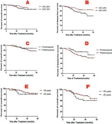
Clinicopathological Features of Non-metastatic Triple Negative Breast Cancer...18
Safa Najafi, Hamid Reza Mozaffari, Masoud Sadeghi
Case Report
Hodgkin’s Lymphoma Occurring Secondary to Autologous Stem Cell Transplantation
in Plasma Cell Leukemia; A Case Report...24
Geetha Narayanan, T Manohar Anoop, Lakshmi Haridas, Lali V Soman
Letter to Editor
Folic Acid Supplementation: to Advise or not to Advise?...27
Mahdi Shahriari
Photo Clinic
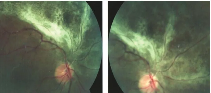
Recurrent Cytomegalovirus Retinitis in a Patient with Leukemia on Maintenance
Chemotherapy...29
Samin Alavi, Kourosh Goudarzipour
Leptin in breast cancer
IJBC 2017; 9(1): 1-4
Leptin in Breast Cancer: Its Relationship with Insulin, Estrogens and
Oxidative Stress
Robab Sheikhpour*
Hematology and Oncology Research Center, Shahid Sadoughi University of Medical Sciences, Yazd, Iran
A R T I C L E I N F O
Review Article
Article History:
Received: 15.12.2016 Accepted: 28.01.2017
Keywords: Leptin Breast cancer Estrogens Insulin Oxidative stress
*Corresponding author: Robab Sheikhpour
Address: Hematology and Oncology Research Center, Shahid Sadoughi University of Medical Sciences, Yazd, Iran
Tel: +98 913 1522462
Email: r.sheikhpour@yahoo.com
ABSTRACT
Breast cancer is the most common cancer in women. Several risk factors such as age, family history of breast cancer, marital status, early menarche and late menopause are related to breast cancer. Obesity is also a main health problem associated with breast cancer incidence and subsequent mortality. Association between obesity and expansion of breast cancer may be due to excessive sex steroid hormone production, particularly estrogen. Moreover, adipose tissue is not only a source of estrogen secretion, but also a producer of certain ‘‘adipocytokines” including leptin. Leptin is a neuroendocrine hormone with 167 amino acid produced predominantly by white adipose tissue. Leptin after binding to receptor activate JAK/STAT/MAP. Leptin also increased expression of cyclin D1 and cdk2 and induces proliferation. It may also develop mammary tumor growth via multiple mechanisms like pro-inflammatory, oxidative, and anti-apoptotic proangiogenic effects. Leptin can increase aromatase activity in MCF-7 cell line which may increase estrogen production and subsequently induce tumor cell growth. Hyperinsulinism through enhanced leptin production by adipose tissue can affect poor breast cancer prognosis.
Introduction
Breast cancer (BC) is the most common cancer in women.1,2 It affects one of every 8 women in the United States. Also, it is one of the most frequent malignancies among Iranian women.3 Several risk factors such as age, family history of breast cancer, marital status, early menarche and late menopause are related to development of breast cancer.4 Obesity, as a main health problem, is associated with increased breast cancer incidence and subsequent mortality.5 However, the mechanism of how obesity relates to the development of breast cancer remains unknown.4 Studies have shown that the association between obesity and breast cancer may be due to excessive sex steroid hormone production, particularly estrogens.6 A group of studies showed that obese individuals have high level of serum leptin that is linked to breast cancer development. In fact, obesity is
characterized as a leptin resistant process.7 Moreover, adipose tissue is not only a source of estrogen secretion, but also a producer of certain ‘‘adipocytokines” including leptins.8 Adipokines, particularly leptin, may have a major role in breast cancer biology.5 It is suggested that leptin could stimulate mammary glands’ growth via multiple mechanisms.4
Leptin
After identification of the obese (OB) gene, “leptin” was discovered and it is now considered as a member of adipokines. It is a 16 KDa neuroendocrine hormone that acts as a multifunctional protein with 167 amino acids, produced predominantly by white adipose tissue.8,9 Leptin is secreted into the blood, where it circulates in both bound and free forms.10 Stomach, placenta, ovary, liver, pituitary and skeletal muscles are among tissues
Iranian Journal of Blood & Cancer
Journal Home Page: www.ijbc.ir
Please cite this article as: Sheikhpour R. Leptin in Breast Cancer: Its Relationship with Insulin, Estrogens and Oxidative Stress. IJBC 2017; 9(1): 1-4.
Sheikhpour R
that expression of leptin mRNA have been reported. Leptin gene expression can be regulated by epigenetic mechanisms. Also there is a reverse relationship between DNA methylation and leptin expression. This relationship was associated with lower methylation density in visceral adipocyte fraction compared to the stromal vascular fraction of white adipose tissue and liver.7 The principal role of leptin is the regulation of energy homeostasis via controlling energy intake and expenditure, by its function on the arcuate nucleus of the hypothalamus.9 Obesity is associated with high levels of leptin. In fact, obesity is associated with leptin resistance.7 However, it is difficult to separate the independent effects of BMI and leptin because of their close biological association.11 There is minimal leptin production in normal conditions which increases in certain pathological processes such as inflammation and malignant transformation.12
Leptin has also contributions to the endocrine and immune systems including reproduction, glucose homeostasis, bone formation, tissue remodelling, inflammation, and angiogenesis.4 Leptin may also play a main role in the growth of mammary tumors via modulation of the extracellular environment, down-regulation of apoptosis and/or up-down-regulation of anti-apoptotic genes.4 It also promotes proliferation and angiogenic differentiation of endothelial cells in vitro and in vivo.7 It is recognized that leptin is expressed in the vicinity of breast cancer cells and leptin receptors are expressed on the cells of ductal and lobular breast carcinomas.6 Breast cancer cell lines MCF-7, T47D and MDA-MB-231 and non-malignant cell line MCF10A also express leptin. It can stimulate the proliferative activity of breast cancer cell lines via the presence of a leptin receptor detected on these cell lines by different signaling pathways.13 Researchers have reported that leptin and its receptors (a member of the cytokine receptor family with two cytokine domains and a single transmembrane domain) are overexpressed in breast tumors.14 In obese mice, the incidence of mammary tumors is correlated with high level of leptin and leptin receptors. Moreover, leptin may exert its capability in breast cancer development via cell proliferation or tumor progression.11
Leptin, Insulin and Breast Cancer
A variety of metabolically active factors such as insulin and glucocorticoids can influence circulating level of leptin.9 Insulin stimulates leptin secretion following meals and leptin is decreased during insulin deficiency.9 Leptin also decreases insulin secretion through direct action on pancreatic beta cells. This finding compared with the fact that insulin is able to increase leptin expression, reveals a negative feedback loop between insulin and leptin.9 A number of studies have shown that there is positive correlation between leptin, obesity and insulin resistance, but other studies could not support this.10 Insulin as a mitogenic agent stimulates the secretion of leptin and hyperinsulinism through enhanced leptin production by adipose tissue can affect poor prognosis of breast cancer patients.9 It is hypothesized that the potential interaction between insulin and metastatic cascade is mediated
through leptin.9
Leptin, Estrogen and Breast Cancer
Numerous studies demonstrated that leptin (OB-R) and estrogen receptors are co-expressed in breast cancers. It seems that interaction between leptin and estrogen promotes breast carcinogenesis.7 Therefore, estrogen as well as other hormones and growth factors can act as intermediates or biological effectors for leptin’s mitogenic activity and stimulates breast cancer.9 Chezet and colleagues reported that leptin can promote breast cancer development in obese women via enhancing estradiol production in situ, not only via adipose tissue but also via epithelial breast cells.6 Another study also reported that estrogen production can be promoted by leptin or follicular estradiol secretion may be limited by it. Leptin can increase aromatase activity in MCF-7 cell line which may increase estrogen production and subsequently induce tumor cell growth. Moreover, leptin receptors expressed in T47D breast cancer cell line induced proliferation of T47D cells by leptin.11 When leptin binds to its receptor Ob-Rl (Obesity receptor), tyrosine phosphorylation and transactivation of signal transducer and activator of transcription 3(STAT 3) were enhanced at the same time with expression of estrogen receptor (ER).15 On the other hand, leptin-induced STAT3 activation acts as a key event in ER α dependent development of malignant diseases and estrogen receptor alpha expression increases the activity of leptin-induced STAT3 in breast cancer cells.15
Impact of Leptin in Angiogenesis
Leptin acts as a positive regulator of vascular endothelial growth factor (VEGF) in breast cancer and blockage of leptin signaling, decreases VEGF expression and tumor growth in mouse xenografts.16 Another study reported that leptin signaling plays a major role in the growth of both ER positive and ER negative breast cancer that is associated with regulation of pro-angiogenic factors (VEGF/VEGF-R2) as a biomarker of poor prognosis in invasive breast cancer and pro- proliferative molecules.9 The data supported potential use of leptin-signaling inhibition as a novel treatment for Breast Cancer.7
Leptin, Oxidative Stress and Breast Cancer
Leptin may play a main role in “reactive oxygen species” (ROS) production. It is interesting that leptin decreases production of mitochondrial ROS; therefore, it can have protective role for cells, but in many cases it increases the oxidative damage in the cells. This mechanism is not clearly understood, but there is some evidence of modulation of the NADPH oxidase enzymes which cause production of several compounds directly involved in cell survival or cycle disruption.13
Impact of Leptin in Apoptosis
Leptin can regulate apoptosis via exerting anti-apoptotic effects. Therefore, it decreases apoptosis through expression of apoptosis inhibitor like survivin and Bcl2 in MCF-7 cells and by inhibiting of pro-apoptotic caspase
Leptin in breast cancer
9 activity.13 Therefore, leptin via pro-inflammatory, oxidative and anti-apoptotic proangiogenic effects can have a main role in the pathogenesis of breast cancer.13
PPAR Ligand and Leptin Signaling in Breast Cancer Peroxisome proliferator-activated receptor (PPAR) is a member of the nuclear receptor family of ligand dependent transcription factor.17 Leptin after binding to receptor activates JAK/STAT/MAPK. It increases GR phosphorylation (pGR) and nuclear translocation. pGR transactivates leptin promoter by binding to GRE motif and activates breast tumor growth. Rosiglitazone (BRL) acts as a new class of antidiabetic drugs and reduces hyperglycemia and hyperinsulinemia in insulin-resistant states. In the presence of BRL, PPAR binds to GRE and as a result GR/PPAR complex is formed, which finally reduce breast tumor growth.17
It has been shown that PPAR ligands suppress ObR mRNA and its promoter activity and block signaling of leptin.18-20 They also reported that PPAR-ligands may show pharmacologic properties and be employed as new therapeutic adjuvant strategies for breast cancer treatment.17
Other Leptin Signaling Pathways in Breast Cancer Another study demonstrated that leptin increases cell proliferation via progression of cell cycle in MCF-7 human breast cancer cells, with up-regulation of “protein kinase C”, PPARc, and PPARa, but others reported that leptin through activation of the mitogen-activated protein kinase (MAP kinase) pathway stimulates proliferation of MCF-7 cell line6 and T47D cell line.20 The effect of leptin on cell proliferation was decreased through inhibition of MAPK pathways, AKT and PI3K activated by leptin.21-26
Conclusion
According to the literature, leptin promotes mammary tumor growth via multiple mechanisms such as inflammatory, oxidative, anti-apoptotic and pro-angiogenic effects. Enhanced leptin production by adipose tissue through hyperinsulinemia can affect poor breast cancer prognosis.
Conflict of Interest: None declared. References
1. Sheikhpour R. New perspective on the role of microRNAs (miRNAs) in breast cancer. BCCR. 2015; 7(1): 2-8.
2. Wang YA, Johnson SK, Brown BL, Carragher LM, Sakkaf KL, Royds JA et al. Enhanced anticancer effect of a phosphati¬dylinositol-3 kinase inhibitor and doxorubicin on human breast epithelial cell lines with different p53 and oestrogen receptor status. Int J Cancer. 2008; 123(7):1536–44.
3. Sheikhpour R, Ghassemi N, Yaghmaei P, Mohiti Ardekani J, Shiryazd M. Immunohistochemical assessment of p53 protein and its correlation with clinicopathological characteristics in breast cancer patients. Indian J Sci Technol. 2014; 4(7): 472-9.
4. Niu J, Jiang L, Guo W, Shao L, Liu Y, Wang L. The Association between leptin level and breast cancer: a meta-analysis. PLoS One. 2013; 8(6):e67349. doi: 10.1371/journal.pone.0067349. PubMed PMID: 23826274. PubMed Central PMCID: PMC3694967. 5. Mohan Reddy N, Kalyan Kumar CH, Kaiser J.
Obesity, an additional burden for breast cancer patients with leptin gene polymorphisms. Columbia International Publishing: AJCRCO. 2013; 1:18-29. doi: 10.7726/ajcrco.2013.1003.
6. Caldefie-Chézet F, Damez M, de Latour M, Konska G, Mishellani F, Fusillier C, et al. Leptin: a proliferative factor for breast cancer? Study on human ductal carcinoma. Biochem Biophys Res Commun. 2005; 334(3):737-41. doi: 10.1016/j. bbrc.2005.06.077. PubMed PMID: 16009333. 7. Gonzalez-Perez RR, Lanier V, Newman G.
Leptin’s pro-angiogenic signature in breast cancer. Cancers (Basel). 2013; 5(3):1140-62. doi: 10.3390/ cancers5031140. PubMed PMID: 24202338.
8. Saxena NK, Vertino PM, Anania FA, Sharma D. Leptin-induced growth stimulation of breast cancer cells involves recruitment of histone acetyltransferases and mediator complex to cyclin d1 promoter via activation of stat3. J Biol Chem. 2007; 282(18):13316-25. doi: 10.1074/jbc.M609798200. PubMed PMID: 17344214.
9. Tourkantonis I, Kiagia M, Peponi E, Tsagouli S, Syrigos KN. The role of leptin in cancer pathogenesis. J Cancer Ther. 2013; 4(2):640-50. doi:10.4236/ jct.2013.42080.
10. Chen DC, Chung YF, Yeh YT, Chaung HC, Kuo FC, Fu OY, et al. Serum adiponectin and leptin levels in Taiwanese breast cancer patients. Cancer Lett. 2006; 237(1): 109–14. doi: 10.1016/j.canlet.2005.05.047. PubMed PMID: 16019138.
11. Harris HR, Tworoger SS, Hankinson SE, Rosner BA, Michels KB. Plasma leptin levels and risk of breast cancer in premenopausal women. Cancer Prev Res (Phila). 2011; 4(9):1449-56. doi: 10.1158/1940-6207.CAPR-11-0125. PubMed PMID: 21680707. 12. Polyzos SA, Mantzoros CS. Leptin in health and
disease: facts and expectations at its twentieth anniversary. Metabolism. 2015; 64(1):5-12. doi: 10.1016/j.metabol.2014.10.017. PubMed PMID: 25467841.
13. Delort L, Rossary A, Farges MC, Vasson MP, Caldefie-Chézet F. Leptin, adipocytes and breast cancer: focus on inflammation and anti-tumor immunity. Life Sci. 2015; 140:37-48. doi: 10.1016/j.lfs.2015.04.012. PubMed PMID: 25957709.
14. Anuradha C, Madanranjit P, Surekha D, Raghunadharao D, Santhoshi Rani N, Vishnupriya S. Association of leptin receptor (LEPR) Q223R polymorphism with breast cancer. Glob J Med Res. 2012; 12(1): 20-31.
15. Binai NA, Damert A, Carra G, Steckelbroeck S, Löwer J, Löwer R, et al. Expression of estrogen receptor alpha increases leptin-induced STAT3 activity in breast cancer cells. Int J Cancer. 2010;
Sheikhpour R
127(1):55-66. doi: 10.1002/ijc.25010. PubMed PMID: 19876927.
16. Alshaker H, Krell J, Frampton AE, Waxman J, Blyuss O, Zaikin A, et al. Leptin induces upregulation of sphingosine kinase 1 in oestrogen receptor-negative breast cancer via Src family kinase-mediated, janus kinase 2-independent pathway. Breast Cancer Res. 2014; 16(5):426. doi: 10.1186/s13058-014-0426-6. PubMed PMID: 25482303.
17. Catalano S, Mauro L, Bonofiglio D, Pellegrino M, Qi H, Rizza P, et al. In vivo and in vitro evidence that PPAR ligands are antagonists of leptin signaling in breast cancer. Am J Pathol. 2011; 179(2):1030-40. doi: 10.1016/j.ajpath.2011.04.026. PubMed PMID: 21704006 .
18. Mantzoros CS, Magkos F, Brinkoetter M, Sienkiewicz E, Dardeno TA, Kim SY, et al. Leptin in human physiology and pathophysiology. Am J Physiol Endocrinol Metab. 2011; 301(4):E567-84. doi: 10.1152/ ajpendo.00315.2011. PubMed PMID: 21791620. 19. Bluher S, Shah S, Mantzoros CS. Leptin deficiency:
clinical implications and opportunities for therapeutic interventions. J Investig Med. 2009; 57(7):784-8. doi: 10.2310/JIM.0b013e3181b9163d. PubMed PMID: 19730134.
20. Iciek R, Wender-Ozegowska E, Zawiejska A, Mikolajczak P, Mrozikiewicz PM, Pietryga M, et al. Placental leptin and its receptor genes expression in pregnancies complicated by type 1 diabetes. J Physiol Pharmacol. 2013; 64(5):579-85. PubMed
PMID: 24304572.
21. Lorincz AM, Sukumar S. Molecular links between obesity and breast cancer. Endocr Relat Cancer. 2006; 13(2):279-92. . doi: 10.1677/erc.1.00729. PubMed PMID: 16728564.
22. Chen C, Chang YC, Liu CL, Chang KJ, Guo IC. Leptin-induced growth of human ZR-75-1 breast cancer cells is associated with up-regulation of cyclin D1 and c-Myc and down-regulation of tumor suppressor p53 and p21WAF1/CIP1. Breast Cancer Res Treat. 2006; 98(2):121-32. doi: [PubMed - indexed for M. PubMed PMID: 16752079. 23. Laud K, Gourdou I, Pessemesse L, Peyrat JP, Djiane
J. Identification of leptin receptors in human breast cancer: functional activity in the T47-D breast cancer cell line. Mol Cell Endocrinol. 2002; 188(1-2):219-26. PubMed PMID: 11911959.
24. Ray A, Nkhata KJ, Cleary MP. Effects of leptin on human breast cancer cell lines in relationship to estrogen receptor and HER2 status. Int J Oncol. 2007; 30(6):1499-509. PubMed PMID: 17487372.
25. Soma D, Kitayama J, Yamashita H, Miyato H, Ishikawa M, Nagawa H. Leptin augments proliferation of breast cancer cells via transactivation of HER2. J Surg Res. 2008; 149(1):9-14. doi: 10.1016/j. jss.2007.10.012. PubMed PMID: 18262553.
26. Frankenberry KA, Skinner H, Somasundar P, McFadden DW, Vona-Davis LC. Leptin receptor expression and cell signaling in breast cancer. Int J Oncol. 2006; 28(4):985-93. PubMed PMID: 16525650.
PON1 activity in cancer
IJBC 2017; 9(1): 5-11
Paraoxonase and Arylesterase Activities in Patients with Cancer
Mohammad Javad Khodayar1, Mohammad Seghatoleslami2, Maryam Salehcheh3*, Fatemeh Jalali4
1Department of Toxicology and Pharmacology Research Center, School of Pharmacy, Jundishapur University of Medical Sciences, Ahvaz, Iran 2Department of Hematology-Oncology, School of Medicine, Ahvaz Jundishapur University of Medical Sciences, Ahvaz, Iran
3Department of Medical Laboratory Sciences, Para-Medical Faculty, Ahvaz Jundishapur University of Medical Sciences, Ahvaz, Iran 4School of Pharmacy, Ahvaz Jundishapur University of Medical Sciences, Ahvaz, Iran
A R T I C L E I N F O
Original Article
Article History:
Received: 05.10.2016 Accepted: 28.12.2016
Keywords: Paraoxonase Arylesterase Malondialdehyde
Lipid profile
Cancer
*Corresponding author: Maryam Salehcheh
Address: Department of Medical Laboratory Sciences, Para-Medical Faculty, Ahvaz Jundishapur University of Medical Sciences, Ahvaz, Iran
Tel: +98 916 6075337
Fax: +98 61 33738330
Email: maryamselehcheh@ajums.ac.ir maryamsalehcheh@gmail.com
ABSTRACT
Background: Cancer has the highest disease-related mortality rate in Iran. Reduced activity of paraoxonase reported in patients with cancer may be due to a reduction in its antioxidant properties and a subsequent increased risk of developing cancer. We aimed to assess antioxidant and oxidative status in patients with cancer through measuring the activity of PON1 as an antioxidant enzyme and determining MDA as a marker of oxidative stress.
Methods: This case-control study was conducted on 50 patients with colon, lung, blood or breast cancer and 50 age- and sex-matched healthy controls matching during 2014-2015. Paraoxonase-1 and arylesterase activities were measured with paraoxon and phenylacetate substrates and their malondialdehyde levels and serum lipid profile were determined through spectrophotometry.
Results: Serum paraoxonase activity was lower in patients with cancer (28.52±2.77 IU/L) compared with the healthy subjects (96.57±1.49 IU/L; P<0.0001). Similarly, serum arylesterase activity was lower in patients with cancer (49.27±2.90) than the controls (66.91±2.47; P<0.0001). MDA levels were higher in patients with cancer (1.3166±0.0876) than the healthy controls (0.9008±0.0452). The Mann-Whitney U-Test showed significant differences between the two groups in terms of their triglyceride levels (P<0.05). Although serum HDL levels were higher in the control group compared with the cases, the difference was not statistically significant (P>0.05). Serum VLDL, LDL and total cholesterol levels differed significantly between the two groups (P<0.05).
Conclusion: The results obtained showed a reduction in paraoxonase activity and an increased lipid oxidation in the patients with cancer and thereby reduced the antioxidant power of paraoxonase and weakened the body’s antioxidant system.
Introduction
In many countries, cancer is the second cause of mortality.1,2 in Iran, it is the third leading cause of death after cardiovascular diseases and accidents.3 The etiology of cancer remains unknown and different factors have been proposed to cause it. Some studies propose genetic factors as the fundamental causes of cancer,4,5 and other have proposed environmental factors,6,7 nutrition,8,9 and infections, smoking and alcohol.10-12 Nevertheless, the most fundamental cause of cancer is
known to be oxidative stress, which is inevitable for aerobic organisms,13 and can be a major mediator in the damage of cell structures such as proteins, membranes, lipids and the DNA,14 as an excessive reactive oxygen species (ROS). Increased oxidative stress and oxygen free radicals increase the risk of developing different types of cancer.15 Low antioxidant levels that increase free radical activities significantly increase the risk of cancer.16 Reactive oxygen metabolites play a major role in the pathogenesis of gastric and intestinal mucosal
Iranian Journal of Blood & Cancer
Journal Home Page: www.ijbc.ir
Please cite this article as: Khodayar MJ,Seghatoleslami M, Salehcheh M, Jalali F. Paraoxonase and Arylesterase Activities in Patients with Cancer. IJBC 2017; 9(1): 5-11.
Khodayar MJ et al.
inflammation and cancer.17 Lipid peroxidation is a known indicator of free radical activity16 and malondialdehyde (MDA) is one of the final products of lipid peroxidation with a higher rate in patients with cancer.18-21 The antioxidant system is a set of enzymes and antioxidants that act against free radicals and oxidants; paraoxonase 1 (PON1) appears to be one of these antioxidant enzymes. Some in-vitro studies have proposed paraoxonase as a strong oxidation inhibitor that causes H2O2 hydrolysis.22 Moreover, PON1 prevents LDL oxidation by removing the oxidized phospholipids.23
Paraoxonase 1 is synthesized in the liver and binds to HDL (an active site) in the blood and improves the antioxidant properties of HDL.24 This enzyme has several different catalytic activities, including paraoxonase, arylesterase, diazoxonase and lactonase activities.25-27 PON1 is the best and most known PON and is able to destroy lipid peroxides before their accumulation on LDL.28 The physiological substrates of the enzyme are still unknown, but paraoxonase and arylesterase activities are both performed on single PON1 and paraoxon and phenylacetate are their synthetic substrates, respectively.17 Oxidizers may be produced or restored in the body through the oxidation metabolic pathways or due to the consumption of oxidized fats. Given that this enzyme binds to HDL, its activity levels were measured along with the lipid profile. Changes in size significantly affect the shape of the binding and the stability of PON1 and reduce its antioxidant capacity.29 The oxidation of low-density lipoproteins (LDL) in the artery walls is responsible for the initiation and progression of atherosclerosis. High-density lipoproteins (HDL) prevent atherosclerosis and can reduce LDL oxidation. HDLs have a variety of functions that help its protective effect against atherosclerosis, including antioxidant, anti-fibrinolytic and anti-inflammatory properties, and also inhibit matrix metalloproteinase and help keep the endothelial plaques normal and intensify endothelial restoration. As LDLs and their oxidized species are associated with the failure of various tissues followed by different diseases such as cerebral, cardiac, hepatic and renal diseases, diabetes and cancer, most recent studies has been dedicated to the antioxidant properties of paraoxonase.30 Different studies have also confirmed the role and significance of PON1 in the pathogenesis of different diseases such as diabetes, chronic renal failure, obesity, and the metabolic syndrome, cardiovascular diseases, Alzheimer’s disease, HIV infection, chronic hepatic disorder and cancer.31,32 As a result, paraoxonase 1 is likely to reduce the risk of cancer through its antioxidant properties. Given the limited number of studies on the role of PON1 in cancer, we aimed to assess antioxidant and oxidative status in patients with cancer through measuring the activity of PON1 as an antioxidant enzyme and determining MDA as a marker of oxidative stress.
Materials and Methods
The present case-control study was conducted on 50 patients from Khuzestan province with colon, lung, blood, or breast cancer admitted to the Adult Hematology
Division of Shafa Hospital in Ahvaz, Iran, during 2014-2015. The patients were diagnosed with these cancers through blood tests and histopathological findings.
A total of 50 age- and sex- matched healthy individuals were selected as the control group. The controls lacked underlying diseases, diabetes, kidney or liver failure and blood diseases and were selected from the healthy subjects referring to the hospital’s medical laboratory. Fasting venous blood samples were obtained and after being transferred to the laboratory, their serum was centrifuged at 3000 rpm for 15 minutes and immediately frozen at -80 °C for the tests.
Paraoxonase Activity Measurement
Paraoxonase activity was measured by adding 20 µl of the serums (dilution 1:10) to 180 µl of paraoxon (1.2 mmol paraoxon in 1 M Tris-Hcl and 1 M NaCl buffer containing 1 M CaCl2 and PH=8.5) at 37 °C and with a wavelength of 405 nm.33 Paraoxonase activity was expressed in nmolmin-1ml-1 in serum.
Arylesterase Activity Measurement
Arylesterase activity was measured with phenyl acetate (Fluka) according to the method proposed by Gan and colleagues using the synthetic substrate of paraoxonase 1. Phenyl acetate was purchased from the Merck Group in Germany along with some other necessary substances. The substrate solution was prepared fresh every day and stored in a closed container and shaken intensely before each use. 10 µl of the serum was then added to the reaction mixture containing 2 mmol of phenyl acetate and 2 mmol of CaCl2 in 100 mmol of Tris-HCl buffer (pH=8). The substrate hydrolysis rate was measured using a spectrophotometer at 37 °C and with a wavelength of 270 nm with UV 1250 (made by Shimadzu in Japan). The enzyme activity was calculated with an extinction coefficient of 1310 M-1cm-1 mol/liter and the results were reported in mol/min/ml of serum.34
Oxidative Status Measurement
Serum levels of MDA were measured as a marker of lipid peroxidation using Yagi’s method, 35 and based on their reaction with thiobarbituric acid and a measurement of the solution absorption and its mixture with n-butanol by mixing 125 μl of its serum with 5.1 ml of phosphoric acid in a test tube and by adding 0.5 ml of thiobarbituric after stirring. The tube containing the mixture was then placed in boiling water for 45 minutes. After cooling, 1 ml of n-butanol was added and centrifuged for 10 minutes; the mixture’s pink supernatant was then separated and its absorption was measured at a wavelength of 532 nm and the standard curve solution of MDA formed from tetraethoxypropane was thus obtained.35
Lipid Profile Measurement
Triglycerides (TG), Total Cholesterol (CH) and High-Density Lipoproteins (HDL) were measured with standard biochemical methods using commercial laboratory kits (made by Pars Azmoon Co., Tehran, Iran) and also with enzymatic methods using autoanalyzer BT3000; LDL
PON1 activity in cancer
levels were calculated using Friedewald’s formula or through electrophoresis.36
Friedwald’s Formula
LDL=Total cholesterol-[HDL+TG/K], where k=5. Very low-density lipoprotein cholesterol, VLDL-C The samples’ VLDL-C was calculated using the following equation:
VLDL-C (mg/dl)=TG (mg/dl)/5
Data Analysis
Data analysis was done using SPSS software, version 22. The quantitative variables were expressed as mean ± standard deviation and the qualitative variables as a percentage. The t-test was used to compare the groups and assess their differences. Pearson’s correlation coefficient was used to assess the dependence between the variables. Non-parametric tests were used for the non-normally-distributed variables. The level of statistical significance was set at P≤0.05.
Results
Of the 50 patients examined, 14 (28%) were women and 36 (72%) were men with a mean±SD age of 54.22±13.99 years (range: 25-88 years). Of the total of 50 healthy subjects examined, 17 (34%) were women and 33 (66%) were men with a mean±SD age of 42.22±11.96 years (range: 25 to 70 years) (table 1).
Table 2 presents a comparison of the paraoxonase and arylesterase activities, the MDA levels and the lipid profile. These results suggest a significant reduction in paraoxonase and arylesterase activities in the patients and a significant increase in serum levels of MDA and triglyceride in the group of patients compared to the control group. An inverse correlation was therefore
observed between paraoxonase and arylesterase activities and MDA levels (r=-0.457, P≤0.0001 and r=-0.303, P<0.002, respectively) and a direct correlation was also observed between paraoxonase activity and HDL cholesterol levels (r=0.213, P=0.039).
Discussion
Few studies have been conducted to measure PON1 activity in patients with cancer. The present study is one of the few in which paraoxonase, MDA and arylesterase levels are simultaneously measured in patients with blood, colon, lung, or breast cancer. Oxidative stress damages the biological membranes, the intracellular organelles and macromolecules such as proteins and DNA, and can lead to the production of active compounds such as aldehydes, ketones, and hydroxy acids. These radicals are produced in the body as a result of oxidation and restoring reactions within the body or else as a result of environmental factors outside the body. An imbalance in the formation and removal of these radicals, including reactive oxygen species (ROS), can cause genetic damage, interfere with cellular signals and cause neurodegenerative diseases, aging and metastasis. Cardiovascular diseases such as atherosclerosis and coronary artery disease are their long-term pathological presentation.37 Oxidant-antioxidant balance appears to be important in the initiation and progression of cancer.38 This study therefore measured PON1 in patients with cancer relying on the enzyme’s antioxidant properties and found the enzyme’s activity to be significantly lower in patients with cancer compared with the controls. Previous studies have reported similar findings. For example, Akçay and colleagues examined patients with gastric cancer and found PON1 activity to be significantly lower in them compared to in the controls.23 Baskül19 also obtained similar results. In line with the present findings, another study examined
Table 1: Mean age and BMI of the case and control group
Variables Patients group Control group
Men Women Men Women
Sex 36 14 33 17
Age (year) 12.718±55.72 50.35±16.735 46.909±11.601 33.11±6.009
(kg/m2) *BMI 28.84±0.257 28.35±0.201 26.306±0.208 26.55±0.325
*BMI, body mass index
Table 2: Comparison of Paraoxonase, Arilesterase enzymes activity, Lipid peroxidation and lipid profile in two groups of the study
(individuals suffering from cancer and healthy ones)
Variable Patients group Control group P value
Paraoxonase )U/L) 28.52 ±2.77** 96.57±1.49 0.000
Aril Esteraz )U/L) 2.9**±49.27 66.91±2.47 0.000
MDA(nmol/L) 1.316±0.087 ** 0.9008±0.0452 0.000
HDL-C (mg/dl) 48.91±2.72 53.98±1.56 0.281
Triglyceride (mg/dl) 137.17±12.92 * 105.12±5.0 0.02
LDL-C (mg/dl ) 58.28±2.59 ** 88.96±2.63 0.000
Cholesterol)mg/dl) 139.93±5.09 ** 168.1±3.29 0.000
VLDL-C (mg/dl ) 27.11±16.56* 21.02±7.085 0.02
TG: Triglycerides; LDL: Low-density lipoprotein; CHO: Cholesterol; HDL: High-density lipoprotein; VLDL: Very low density lipoprotein; PON: Serum paraoxonase; ARE: Arylesterase; MDA: Malondialdehyde; Results have been stated as±standard deviation average (values are mean±SD). *Significant difference with control group approximate to 0.05; **Significant difference with control
group approximate to 0.0001
Khodayar MJ et al.
patients with esophageal and gastric malignancies and found a significant reduction in arylesterase and paraoxonase in them.17 Another study observed a significant reduction in PON1 in patients with lung cancer.15 Oxidative stress is also one of the main risk factors for cancer35 that sometimes occurs in the body due to disrupted mitochondrial function or inadequate defense mechanisms.36,37 The present study used MDA as a measure of serum lipid peroxidation. Low antioxidant enzyme activities reduce the antioxidant capacity and increase lipid peroxidation and its metabolic product, MDA, while increased antioxidant enzyme activities inhibit lipid peroxidation and thus reduce MDA production.39 As expected, MDA levels were significantly higher in cancer patients compared to in the healthy controls, suggesting an increased lipid peroxidation in these patients. Previous studies also confirm this finding.19,21,39,40 One of the main capabilities of HDL is that it functions as a depository of antioxidant enzymes that can reduce ample levels of oxidized phospholipids from the blood. Paraoxonase is one of the main blood plasma antioxidants that limit the accumulation of oxidized phospholipids in plasma lipoproteins.41 Although some other HDL-binding proteins, such as apolipoprotein A1, lecithin cholesterol acyl transferase and platelet-activating factor acetyltransferase, also have antioxidant properties, the antioxidant activity of PON1 appears to be the most significant.42
Measuring the lipid profile yielded the following results: Triglyceride and VLDL levels increased significantly in the group of patients compared to in the controls; however, HDL cholesterol levels were lower in the patients than in the controls, although the difference was not statistically significant. A significant reduction was also observed in LDL and total cholesterol levels in the patients compared to in the controls. Similar results were reported in the study by Akçay et al.23 Previous studies have compared PON1 and lipid peroxidation (MDA) levels in patients with gastric cancer and hepatitis and have found a significant inverse relationship between them.19,43 In another study, PON1, ARE and MDA levels were measured in those exposed to ionizing radiation and a significant inverse relationship was observed, although not between ARE and MDA.44 A negative correlation appears to exist between PON activity and MDA levels due to the extensive oxidative damage to PON. The binding of PON to HDL confirms the dependence of this enzyme on lipids. The hydrophobic environment of HDL is necessary to paraoxonase activity. Phospholipids, especially those with long fatty acid chains, stabilize PON and are essential to its binding on lipoprotein surfaces.45 The mechanism of reduced PON1 activity in cancer is not yet identified. The reduced paraoxonase activity in the group of cancer patients compared to in the healthy controls could be due to several factors, including enzyme inactivation. In this process, PON1 free sulfhydryl group reacts with specific oxidized lipids and ultimately becomes inactive.46 The attack of free radicals (ROS) on the enzyme may be responsible for its inactivation. Moreover, the reduction in paraoxonase activity may be caused by the increased oxidative stress in the patients.47 The present study found no significant reductions in HDL
levels in the group of patients compared to in the control group. Previous studies have shown that changes in HDL structure can lead to the non-binding of PON1 to HDL, thereby reducing serum PON1 levels. Another mechanism associated with the reduction of PON1 activity could be due to the suppression of the enzyme due to genetic defects46 or perhaps due to the down regulation of its transcription in the liver. Another reason for the reduction in paraoxonase may be the disrupted liver structure and function. Since the liver is the largest and most important organelle in the body and since colon, lung and breast cancer commonly metastasize to the liver, patients with cancer tend to also develop liver damage. Since PON1 is produced in the liver, an impaired liver function can reduce the production of PON1 or cause the production of impaired HDL.48
Overall, paraoxonase has been shown to become inactive after the hydrolysis of lipid peroxides in patients with high levels of lipid peroxidation.47 Moreover, paraoxonase is an HDL-dependent enzyme and any changes in the metabolism and structure of HDL can reduce paraoxonase activity.48 Moreover, since PON1 is reduced as an antioxidant enzyme in the body, the oxidant-antioxidant balance is impaired and oxidative stress thus increases. The oxidant, inflammatory and angiogenic environments then lead to carcinogenesis and enable the progression of cancer.23 As PON1 is an antioxidant factor, using factors that increase its activity may help with the treatment of cancer. Overall, the present study shows that paraoxonase levels reduce in patients with cancer and consequently increase oxidative stress and lipid peroxidation. This finding suggests the positive effects of antioxidants on cancer. Nevertheless, the high levels of MDA and the reduced PON1 activity suggest an impaired oxidant-antioxidant balance in patients with cancer, suggesting oxidant-antioxidant balance to have a major role in the pathogenesis of cancer.
One of the limitations of the present study is that it did not assess the many different elements involved in oxidant-antioxidant balance, thereby making the generalization of the results to the entirety of oxidant-antioxidant balance irrespective of the other factors at play a matter that should be pursued with extreme caution; the following measures are therefore recommended: To better understand the antioxidant properties of paraoxonase in patients with cancer, antioxidant factors such as vitamins, including vitamin E and C, and antioxidant enzymes such as catalase, glutathione and peroxidase are recommended to be studied along with paraoxonase. Dietary antioxidants are also recommended to be administered to patients with cancer either in combination or separately and their effects to be assessed on PON1.
Conclusion
In patients with cancer, reduced paraoxonase activity accompanied by reduced arylesterase activity indicates weak antioxidant activities in the body. Increased MDA levels as a marker of lipid peroxidation suggest the lower oxidative status potentially caused by oxidant-antioxidant imbalance (including reduced antioxidant power, more
PON1 activity in cancer
oxidized substances or both). These findings demonstrate oxidative stress or its aggravation in patients with cancer.
Acknowledgements
This paper is the result of a thesis approved by the Faculty of Pharmacy, Ahvaz Jundishapur University of Medical Sciences, under the approval number D-91/019 and the code of ethics (IR.AJUMS.REC.1395.219) Hereby, the authors would like to express their gratitude to the university faculty members and personnel and all the affiliated professors and healthcare personnel of Shafa Hospital in Ahvaz.
Conflict of Interest: None declared. References
1. King JC, Cousins RJ. Zinc. In: Shils ME, Shike M, Ross AC, Caballero B, Cousins RJ, editors. Modern Nutrition in Health and Disease. 10th ed. Baltimore: Lippincott Williams & Wilkins; 2006: 271–85. 2. Díaz MeP, Osella AR, Aballay LR, Muñoz SE,
Lantieri MJ, Butinof M, et al. Cancer incidence pattern in Cordoba, Argentina. Eur J Cancer Prev. 2009; 18(4): 259-66. doi: 10.1097/CEJ.0b013e3283152030. PubMed PMID: 19404198.
3. Mousavi SM, Gouya MM, Ramazani R, Davanlou M, Hajsadeghi N, Seddighi Z. Cancer incidence and mortality in Iran. Ann Oncol. 2009; 20(3): 556-63. doi: 10.1093/annonc/mdn642.
4. Palli D, Galli M, Caporaso NE, Cipriani F, Decarli A, Saieva C, et al. Family history and risk of stomach cancer in Italy. Cancer Epidemiol Biomarkers Prev. 1994; 3(1): 15–8. PubMed PMID: 8118379.
5. Inoue M, Tajima K, Yamamura Y, Hamajima N, Hirose K, Kodera Y, et al. Family history and subsite of gastric cancer: data from a case-referent study in Japan. Int J Cancer. 1998; 76(6):801-5. PubMed PMID: 9626344. doi:10.1002/(SICI)1097-0215(19980610)76:6<801::AID-IJC6>3.0.CO;2-1. 6. Haenszel W, Kurihara M, Segi M, Lee RK. Stomach
cancer among Japanese in Hawaii. J Natl Cancer Inst. 1972; 49(4): 969–88. PubMed PMID: 4678140. 7. Kamineni A, Williams MA, Schwartz SM, Cook
LS, Weiss NS. The incidence of gastric carcinoma in Asian migrants to the United States and their descendants. Cancer Causes Control. 1999; 10(1): 77–83. doi: 10.1023/A:1008849014992. PubMed PMID: 10334646.
8. Tsugane S, Sasazuki S. Diet and the risk of gastric cancer: review of epidemiological evidence. Gastric Cancer. 2007; 10(2): 75–83. doi: 10.1007/s10120-007-0420-0. PubMed PMID: 17577615.
9. Rocco A, Nardone G. Diet. H. pylori infection and gastric cancer: evidence and controversies. World J Gastroenterol. 2007; 13(21): 2901–12. PubMed PMID: 17589938. PubMed Central PMCID: PMC4171140. 10. Sitas F, Urban M, Bradshaw D, Kielkowski D, Bah
S, Peto R. Tobacco attributable deaths in South Africa. Tob Control. 2004; 13(4): 396–9. doi:10.1136/ tc.2004.007682.
11. Chow WH, Swanson CA, Lissowska J, Groves FD, Sobin LH, Nasierowska-Guttmejer A, et al. Risk of stomach cancer in relation to consumption of cigarettes, alcohol, tea and coffee in Warsaw, Poland. Int J Cancer. 1999; 81(6): 871–6. doi: 10.1002/(SICI)1097-0215(19990611)81:6<871::AID-IJC6>3.0.CO;2-#. PubMed PMID: 10362132. 12. Vineis P, Alavanja M, Buffler P, Fontham E,
Franceschi S, Gao YT, et al. Tobacco and cancer: recent epidemiological evidence. J Natl Cancer Inst. 2004; 96(2): 99–106. PubMed PMID: 14734699. 13. Benz CC, Cristina Y. Ageing, oxidative stress and
cancer: paradigms in parallex. Nat Rev Cancer. 2008; 8(11): 875-9. doi: 10.1038/nrc2522. PubMed PMID: 18948997. PubMed Central PMCID: PMC2603471. 14. Djordjević VB, Zvezdanović L, Cosić V. [Oxidative
stress in human disease]. Srp Arh CelokLek. 2008; 136(suppl 2): 158-65. PubMed PMID: 18924487. 15. Elkiran ET, Mar N, Aygen B, Gursu F, Karaoglu
A, Koca S. Serum paraoxonase and arylesterase activities in patients with lung canser in a turkish population. BMC Canser. 2007; 7: 48. doi: 10.1186/1471-2407-7-48.
16. Barber DA, Harris SR. Oxygen free radicals and antioxidants: a review. Am Pharm. 1994; 34(9): 26-35. PubMed PMID: 7977023.
17. Krzystek-Korpacka M, Boehm D, Matusiewicz M, Diakowska D, Grabowski K, Gamian A. paraoxonase1 (PON1) status in gastroesophageal malignancies and associated paraneoplastic syndromes- Connection with inflammation. Clin Biochem. 2008; 41(10-11): 804-11. doi: 10.1016/j.clinbiochem.2008.03.012. PubMed PMID: 18423402.
18. Bekerecioğlu M, Aslan R, Uğras S, Kutluhan A, Şekeroğlu R, Akpolat N, et al. Malondialdehyde levels in serum of patients with skin cancer. Eur J Plast Surg. 1998; 21(5): 227- 9. doi:10.1007/s002380050076. 19. Başkol M, Başkol G, Koçer D, Artış T, Yılmaz
Z. Oxidant - antioxidant parameters and their relationship in patients with gastric cancer. Journal of Turkish Clinical Biochemistry. 2007; 5(3):83-9. 20. Kaynar H, Meral M, Turhan A, Keles M, Celik
G, Akcay F. Glutathione peroxidase, glutathione-S-transferase, catalase, xanthine oxidase, Cu–Zn superoxide dismutase activities, total glutathione, nitric oxide, and malondialdehyde levels in erythrocytes of patients with small cell. Cancer Lett. 2005; 227(2): 133-9. doi: 10.1016/j.canlet.2004.12.005. PubMed PMID: 16112416.
21. Torun M, Yardim S, Gönenç A, Sargin H, Menevşe A, Símşek B. Serum β–carotene, vitamin E, vitamin C and malondialdehyde levels in several types of cancer. J Clin Pharm Therapeut. 1995; 20(5):259-63. doi: 10.1111/j.1365-2710.1995.tb00660.x. PubMed PMID: 8576292.
22. Sözmen EY, Sözmen B, Girgin FK, Delen Y, Azarsiz E, Erdener D, et al. Antioxidant enzymes and paraoxonase show a coactivity in preserving low-density lipoprotein from oxidation. Clin Exp Med. 2001; 1(4): 195- 9. doi: 10.1007/s102380100003.
Khodayar MJ et al.
PubMed PMID: 11918278.
23. Akcay MN, Yilmaz I, Polat MF, Akcay G. serum paraoxonase levels in gastric canser. Hepatogastroenterology. 2003; 50 (suppl2): cclxxiii-cclxxv. PubMed PMID: 5244199.
24. Juretic D, Tadijanovic M , Rekic B, Simeon-rudolf V, Rainer E, Baricic M. Serum paraoxonase activities in hemodialysed uremic patients: Cohort Study. Croate Med J. 2001; 42(2):146-50. PubMed PMID: 11259735. 25. Furlong CE, Li WF, Brophy VH, Jarvik GP, Richter
RJ, Shih DM, et al. The PON 1 gene and detoxication. Neurotoxicology. 2000; 21(4): 581-7. PubMed PMID: 11022865.
26. Ferre N, Tous M, Paul A, Zamora A, Vendrell JJ, Bardaji A, et al. araoxonase Gln–Arg(192) and Leu– Met (55) gene polymorphisms and enzyme activity in a population with a low rate of coronary heart disease. Clin Biochem. 2002; 35(3):197–203. doi: 10.1016/ S0009-9120(02)00295-3. PubMed PMID: 12074827. 27. Billecke S, Draganov D, Counsell R, Stetson P,
Watson C, Hsu C, et al. Human serum paraoxonase (PON 1) isozymes Q and R hydrolyze lactones and cyclic carbonate esters. Drug Metab Dispos. 2000; 28(11): 1335-42. PubMed PMID: 11038162.
28. Marsillach J, Parra S, Ferré N, Coll B, Alonso-Villaverde C, Joven J, et al. Paraoxonase-1 in chronic liver diseases, neurological diseases and HIV infection. In: The Paraoxonases: Their Role in Disease Development and Xenobiotic Metabolism. Netherlands: Springer; 2008. p. 187-98. doi: 10.1007/978-1-4020-6561-3_12.
29. Li HL, Liu DP, Liang CC. Paraxonase gene polymorphisms, oxidative stress, and disease. J Mol Med. 2003; 81(12): 766-79. PubMed PMID: 14551701. 30. Blatter MC, James RW, Messmer S, Barja F, Pometta
D. Identification of a distinct human high-density lipoprotein subspecies defined by a lipoprotein-associated protein, K-45. Identity of K-45 with paraoxonase. Eur J Biochem. 1993; 211: 871-9. doi: 10.1111/j.1432-1033.1993.tb17620.x. PubMed PMID: 8382160.
31. Goswami B, Tayal D, Gupta N, Mallika V. Paraoxonase: a multifaceted biomolecule. Clinica Chimica Acta. 2009; 410(1):1-12. doi: 10.1016/j. cca.2009.09.025. PubMed PMID: 19799889. 32. Camps J, Marsillach J, Joven J. The paraoxonases: role
in human diseases and methodological difficulties in measurement. Crit Rev Clin Lab Sci. 2009; 46(2):83-106. doi: 10.1080/10408360802610878. PubMed PMID: 19255916.
33. Wiley InterScience. Current Protocols in Toxicology. John Wiley & Sons, Inc; 2005.
34. Cole TB, Li WF, Richter RJ, Furlong CE, Costa LG. Inhibition of paraoxonase (PON1) by heavy metals. Toxicol Sci. 2002; 66(1-S):312.-Yagi, Kunio. A simple fluorometric assay for lipoperoxide in blood plasma. Biochemical medicine. 1976; 15(2): 212-216. 35. Armstrong D. Free radicals in diagnostic medicine:
A Systems Approach to Laboratory Technology, Clinical Correlations, and Antioxidant Therapy. New
York: Springer; 1994. doi: 10.1007/978-1-4615-1833-4. 36. Friedewald WT, Levy RI, Fredrickson DS. Estimation
of the concentration of low-density lipoprotein cholesterol in plasma, without use of the preparative ultracentrifuge. Clin Chem. 1972; 18 (6): 499–502. PubMed PMID: 4337382.
37. Allen RG. Oxidative stress and superoxide dismutase in development, aging and gene regulation. Age. 1998; 21(2): 47-76. doi: 10.1007/s11357-998-0007-7. PubMed PMID: 23604352. PubMed Central PMCID: PMC3455717.
38. Oliveira CP, Kassab P, Lopasso FP, Souza HP, Janiszewski M, Laurindo FR, et al. Protective effect of ascorbic acid in experimental gastric cancer: reduction of oxidative stress. World J Gastroenterol. 2003; 9(3): 446-8. PubMed PMID: 12632494. PubMed Central PMCID: PMC4621558.
39. Aviram M, Rosenblat M, Bisgaier CL, Newton RS, Primo - Parmo SL, La Du BN. Paraoxonase inhibits high-density Lipoprotein oxidation and preserves its functions: a possible peroxidative role for paraoxonase. J Clin Invest. 1998; 101(8):1581-90. doi: 10.1172/JCI1649. PubMed PMID: 9541487. 40. Batcioglu K, Mehmet N, Ozturk IC, Yilmaz M,
Aydogdu N, Erguvan R, et al. Lipid peroxidation and antioxidant status in stomach cancer. Cancer Invest. 2006; 24(1): 18-21.doi: 10.1080/07357900500449603. PubMed PMID: 16466987.
41. Cabana VG, Catherine CA, Feng N, NeathS, Lukens J, Getz GS. Serum paraoxonase: effect of the apolipoprotein composition of HDL and the acute phase response. J Lipid Res. 2003; 44(4): 780-92. doi: 10.1194/jlr.M200432-JLR200. PubMed PMID: 12562837.
42. Mackness MI, Hallam SD, Peard T, Warner S, Walker CH. The separation of sheep and human serum A-esterase activity into thelipoprotein fraction by ultracentrifugation. Comp Biochem Physiol B. 1985; 82(4):675-7. PubMed PMID: 3004805.
43. Ali EM, Shehata HH, Ali-Labib R, Esmail, Zahra LM. Oxidant and antioxidant of arylesterase and paraoxonase as biomarkers in patients with hepatitis C virus. Clin Biochem. 2009; 42(13- 14): 1394-400. doi: 10.1016/j.clinbiochem.2009.06.007. PubMed PMID: 19538950.
44. Serhatlioglu S, Gursu MF, Gulcu F, Canatan H, Godekmerdan A. Levels of paraoxonase and arylesterase activities and malondialdehyde in workers exposed to ionizing radiation. Cell Biochem Funct. 2003; 21(4): 371-5.
45. Dirican M, Akca R, Sarandol E, Dilek K. Serum paraoxonase activity in uremic predialysis and hemodialysis patients. J Nephrol. 2004; 17(6): 813-8. PubMed PMID: 15593056
46. Baskol G, Karakucuk S, Oner AO, Baskol M, Kocer D, Mirza E, et al. Serum paraoxonase 1activity and lipid peroxidation levels in patients with age-related macular degeneration. Ophthalmologica. 2006; 220(1): 12–6. doi: 10.1159/000089269. PubMed PMID: 16374043.
PON1 activity in cancer
47. Aviram M, Rosenblat M, Billecke S, Erogul J, Sorenson R, Bisgaier CL, et al. Human serum paraoxonase (PON 1) is inactivated by oxidized low density lipoprotein and preserve by antioxidants. Free Radical Biol Med. 1999; 26(7-8): 892-904. PubMed PMID: 10232833.
48. James RW, Deakin SP. The importance of high-density lipoproteine for paraoxonase (pon-1) secretion,stability, and activity. Free Radic Biol Med. 2004; 37(12):1986-94.doi: 10.1016/j. freeradbiomed.2004.08.012 . PubMed PMID: 15544917.

