*Corresponding author: Pooneh Rahimi, Department of Hepatitis and AIDS, Pasteur Institute of Iran, Pasteur street, Tehran, IR Iran.
Tel: +98-2166403496 Fax: +98-2166480777 E-mail: [email protected]
Designing a recombinant Bacmid construct of HCV core+1 in Baculovirus
expression system
Fahimeh Safarnezhad Tameshkel1, Pooneh Rahimi2*, Mohammad Reza Khataminejad1
1Department of Biology, Islamic Azad University, Tonekabon branch, Tonekabon, IR Iran.
2Department of Hepatitis and AIDS, Pasteur Institute of Iran.
Received: May 2015, Accepted: July 2015
ABSTRACT
Background and Objectives: Hepatitis C virus (HCV) chronically infects around 200 million people worldwide and fre-quently causes liver cirrhosis and hepatocellular carcinoma. Rapid detection of this virus results in decreasing the distance between infection and initiation the anti-viral treatment, and may prevent most of the undesirable consequences. The new detected HCV protein "Core+1" made from the ribosomal frame shift in Core region is an important candidate for diagnostic tools. This study was conducted to design a recombinant Bacmid plasmid expressing the HCV 1a Core+1 sequence in the Baculovirus expression system for further diagnostic applications.
Materials and Methods: The HCV Core +1 gene was amplified by PCR using the pcDNA-HAF recombinant vector that contained the Core+1 sequence from HCV genotype 1a as a template, and the specific primers with 2 restriction sites for Nco I and Xba I restriction enzymes. The PCR product was cloned in XbaI/NcoI restriction sites of the linearized pFastBac-HTB vector and evaluated by using those restriction enzymes and sequencing. Then the recombinant pFastBac-HTB vector was transformed in DH10Bac and the result was screened and confirmed by X-Gal discrimination and PCR.
Results: The HCV 1a Core+1 was successfully amplified and the PCR product was confirmed by using the related restriction enzymes and sequencing. Cloning of pFastBac vector with the purified PCR product of HCV Core+1 was confirmed. Finally, the recombinant Bacmid was successfully transformed in DH10Bac.
Conclusion: The recombinant Bac-Core+1 expression vector is considered as an important tool to transfect the sf9 cell line and expression the Core+1 protein.
Keywords: Hepatitis C Virus, HCV Core+1, pFastBac vector, Bacmid expression vector, Baculovirus expression system.
stranded positive RNA genome with a single open
reading frame (1, 2). This virus is a major cause of chronic liver diseases, including steatosis, cirrho-sis and hepatocellular carcinoma (1, 3). It has been proposed that HCV infection is also associated with insulin resistance and type 2 diabetes mellitus (1, 3). This small enveloped virus encoding a polyprotein as a precursor which is processed by host and viral proteases into at least 10 different proteins (1, 3). There is a second functional Open Reading Frame (ORF), within the core gene, encoding an addition-al protein named addition-alternative reading frame protein
ORIGINAL
AR
TICLE
INTRODUCTION
http://ijm.tums.ac.ir (ARFP), frameshift (F) or Core+1 (to indicate its
po-sition) (3, 4). Several studies have shown that HCV F protein has been produced from reading frame by more than one type of coding event (4, 5). The effect of this viral protein on virus replication re-mains elusive. However there are some evidences about immune response against this protein through measuring the anti- Core+1 antibody in HCV in-fected patients such as hepatocellular carcinoma (6, 7). Core+1 is produced through the ribosomal frame shift in core region so, these two viral proteins have the overlapped ORFs (5, 8). Since HCV core protein is a multifunctional protein, Core+1 may contribute to some functions attributed to the core protein (9). Several studies reported the presence of anti-F pro-tein antibodies in some HCV-positive patients (7, 10, 11). However, the exact role of this virus product in induction of immune response and virus – associated pathogenesis remains controversial (6, 10, 12).
Con-sideration of different expression systems, to find the most efficient and safest with lower costs and labor
intensive, is an important context in modern biotech-nology. The most described organism in this content is E.coli expression system with quick growth in simple media, distinct biological process and high yield of recombinant protein expression (13, 14). At this point, some enzyme-linked immunoabsorbent assays has been developed using E.coli expressed
purified recombinant HCV Core+1 protein (10, 15). Lack of post -transcriptional modifications in eu -karyotics proteins results in designing other expres-sion systems such as Baculovirus expresexpres-sion system (8, 16). The Baculovirus system has become one of the most powerful eukaryotic systems for recombi-nant protein expression since 1983. This expression system provides the post translational processing, and folding of recombinant proteins that are highly similar to those occurred in natural infection in a eu-karyotic host (16-18). This study was conducted to design a recombinant Bacmid plasmid expressing the HCV 1a Core+1 sequence in Baculovirus expression system for further diagnostic applications.
MATERIALS AND METHODS
PCR. The HCV Core +1 gene was amplified by
PCR in 50 μl reaction mixture using the pCDEF-HAF recombinant vector (kindly gifted from Professor Jing-hsiung Ou) containing the Core+1 sequence
from HCV genotype 1a as a template. 1 μl of this
tem-plate DNA was added to the PCR mixture which con -tained 1 μ of 20 pmol of each forward
5´-ATCCAT-GGGCACGAATCCTAAACCTC-3´ and reverse 5´- CAT C TAG AT TAT CAC G C C G T C T T C
-CAGAAC-3´ primers, (the underlined nucleotides
demonstrate Nco I and Xba I digestion sites).
Five μl of 10X PCR reaction buffer, 1 μl dNTP mix
and 0.5 μl of PFU DNA polymerase (Fermentas Life Sceince, Lithuania), and 40.5 μl dDW. This
reac-tion was amplified in a Master cycler Gradient (Ep
-pendorf, Germany) using the following conditions:
initial denaturation at 94°C for 5 minutes, followed by 35 cycles at 94°C for 30 second, 55°C for 30
sec-ond and 72°C for 35 secsec-ond and a final extension
phase at 72°C for 5 minutes. The PCR products were run on agarose gel electrophoresis for further
analy-sis and seubsequently purified and sequenced using
ABI Prism automated sequencer Pairwise alignment was done by comparing the sequence of PCR product with Core+1 sequence of the pCDEF-HAF recombi-nant vector.
Cloning of the Core+1 genes. The Core+1 seg-ment was gel-extracted using the PCR product
puri-fication kit (QIAquick Gel Extraction Kit, QIAGEN GmbH) and was cloned through the XbaI and NcoI sites into the pFastBacHTB donor vector
(Fermen-tas Life Science, Lithuania), making the recombinant pFastBac-Core+1 plasmid. The fidelity of cloning
was evaluated using the relevant restriction enzymes and sequencing of PCR product as described in pre-vious section.
Construction of recombinant Bacmid. E. coli strain DH10Bac was transformed using the pBac-Core+1 recombinant donor plasmid. To perform
site-specific transposition of the Core+1 frag
-ment from the donor plasmid to the Bacmid DNA
which already available in DH10Bac E. coli cells, the transformed DH10Bac cells were transferred to
the Luria-Bertani broth (LB) agar plate containing Gentamicin (7 µg/ml), Kanamycin (50 μg/ml), Tet -racycline (10 μg/ml), X-gal (100 μg/ml) and isoprop
-ylthio-β-galactoside (IPTG, 40 µg/ml) and incubated
at 37°C for 48 h.
Afterward, the largest white colonies were
re-streaked on the new LB agar plates containing the mentioned reagents to reconfirm their white phe
-notype. Finally, the confirmed white colonies were
overnight cultivated in LB broth medium containing
the mentioned antibiotics and subjected to Bacmid extraction using plasmid extraction mini kit accord-ing to the manufacturer's instructions (AccuPrep R
Plasmid Extraction Kit, Bioneer Coroperation, Ko
-rea). In order to confirm the extracted recombinant
Bacmid, PCR was performed using universal M13 forward and reverse primers and the recombinant Bacmid contains pFastBac-HCV Core+1, the recom-binant Bacmid - pFastBac and the non-recomrecom-binant Bacmid without pFastBac (as separate templates re-spectively) as instructed by the manufacturer
(Fer-mentas Life Science, Lithuania).
RESULTS
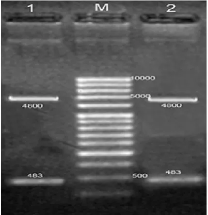
Core+1 amplification, cloning and confirma
-tion. HCV Core+1 sequence from genotype 1a was
amplified successfully through PCR by using specif
-ic primers and the amplified fragment (483bp) was
cloned into pFastBacHTB plasmid. The fidelity of the cloning was confirmed by restriction digestion and
sequencing (Fig. 1).
Construction of the recombinant Bacmid DNA. E-coli DH10Bac strain was transformed using the
re-combinant pFastBac- HCVCore+1 vector and Blue/
white screening was performed. The extracted
re-combinant Bacmid was verified by PCR using the M13 universal primers and a 2856 bp amplified prod -uct was observed (Fig. 2). There was a recombinant Bacmid –pFastBac (Lane 3,4) as the negative control (2373), and also, a 273 bp fragment(Lane C) repre -sented a PCR control for the non-recombinant Bacmid alone (without pFastBac) (Fig. 2).
Fig. 1. Digestion of HCV (1a) Core+1 PCR product. Lane M: 1Kb DNA Ladder, Lane (1,2): pFastBacHTB donor plas-mid :(4800bp), Core+1 fragment: (483bp).
Fig. 2. Confirmation of the recombinant Bacmid. Lane M: 1Kb DNA Ladder, Lane C: Non- recombinant Bacmid alone without pFastBac (273bp); Lane (1,2): recombinant Bacmid contains pFastBac-HCV Core+1 (2856bp); Lane (3,4): re-combinant Bacmid - pFastBac (2373bp).
DISCUSSION
HCV is classified into seven major genotypes, with
more than 80 subtypes that amongst them subtypes 1a, 1b, 2a, 2b, 2c, and 3a represent a worldwide epidemic including Iran (1, 3). HCV has a single ORF and its
ssRNA genome encodes three structural (Core, E1, and E2) and seven non-structural (P7, NS2, NS3, NS4A, NS4B, NS5A, and NS5B) proteins (1, 2). An additional HCV protein, the F protein results from a -2/ +1 frameshift at the HCV AUG initiator codon of the polyprotein so, this overlapping ORF lacks an AUG
http://ijm.tums.ac.ir coding sequence is independent of the downstream E1
region so, it is proposed that it is a truncated core pro-tein (9). Shorter forms of it ranging from 126 to 155 codons in length and are also conserved. However it has been revealed that the length of the F protein is
genotype specific (4, 9). According to these findings
expression an intact form of F protein is very import-ant especially when it used to develop the laboratory
diagnostic methods such as ELISA. There are some
reports of expressing the Core+1 protein in different expression systems other than Baculovirus expression system such as E. coli and mammalian expression sys-tems (9, 20). Also, in some of these investigations the expressed Core+1 proteins were used in
immunoper-cipitation /ELISA tests to study its antigenic proper -ties (9). Recombinant Baculovirus expression systems
are one of the important tools for highly efficient gene
expression through infection of insect cells (16, 17). This system is used for expression of many
mamma-lian recombinant proteins with high efficiency. It has some significant advantages like the modifications,
processing and other features of natural mammalian host cells with high yield of products.
This study was conducted to design and make a re-combinant Baculovirus vector (Bacmid) contains the HCV Core+1 sequence derived from the genotype 1a for further investigations. There are some studies that HCV structural genes were expressed in Baculovirus expression system (21).
In conclusion, the HCV Core+1 candidate to Bac-to-Bac system and successfully cloned. It was trans-ferred to Bacmid by transposition mechanism. Final-ly, we suggest the use of this Bacmid to express the Core+1 protein for further studies.
ACKNOWLEDGMENTS
Our great appreciate goes to Professor Jing-hsiung Ou, Department of Molecular Microbiology and
Im-munology, University of Southern California, Keck School of Medicine, Los Angeles, California
90033-1054. We wish to thank Mrs. F. Motevalli and Dr.
R.Vahabpour, and Miss G. BahramAli for their great
technical advice. Also, we would like to appreciate all staffs of Department of Hepatitis and AIDS, Pas-teur Institute of Iran for collaborating in this research project.
This project was financially supported by Pasteur
Institute of Iran under the project number 683.
REFERENCES
1. Mauger DM, Golden M, Yamane D, Williford S, Lem-on SM, Martin DP, et al. FunctiLem-onally cLem-onserved archi-tecture of hepatitis C virus RNA genomes. Proc Natl Acad Sci U S A 2015; 112: 3692-3697.
2. Shakeri MT, Nomani H, Ghayour Mobarhan M, Sima HR, Gerayli S, Shahbazi S, et al. The prevalence of hepatitis C virus in mashhad, iran: a population-based study. Hepat Mon 2013; 13: e7723.
3. Li HC, Ma HC, Yang CH, Lo SY. Production and pathogenicity of hepatitis C virus core gene products. World J Gastroenterol 2014; 20: 7104-7122.
4. Kotta-Loizou I, Vassilaki N, Pissas G, Kakkanas A, Bakiri L, Bartenschlager R, et al. Hepatitis C virus core+1/ARF protein decreases hepcidin transcription through an AP1 binding site. J Gen Virol 2013; 94: 1528-1534.
5. Komurian-Pradel F, Rajoharison A, Berland JL, Khouri V, Perret M, Van Roosmalen M, et al. Antigen-ic relevance of F protein in chronAntigen-ic hepatitis C virus infection. Hepatology 2004; 40: 900-909.
6. Vassilaki N, Mavromara P. The HCV ARFP/F/core+1 protein: production and functional analysis of an un-conventional viral product. IUBMB Life 2009; 61: 739-752.
7. Rehermann B. Pathogenesis of chronic viral hepati-tis: differential roles of T cells and NK cells. Nat Med 2013; 19: 859-868.
8. Jackson RJ, Hellen CU, Pestova TV. Termination and post-termination events in eukaryotic translation. Adv Protein Chem Struct Biol 2012; 86: 45-93.
9. Niepmann M. Hepatitis C virus RNA translation. Curr Top Microbiol Immunol 2013; 369: 143-166.
10. Hashempour T AM, Bamdad T, Merat S, Zaer-Rezaee H, Fakharzadeh E, et al. Development of a recombi-nant based ELISA using specific antibodies to F pro-tein in HCV chronically infected patients-A seroprev-alence study. Iranian Journal of Virology 2010; 4: 1-6. 11. Cohen M, Bachmatov L, Ben-Ari Z, Rotman Y,
Tur-Kaspa R, Zemel R. Development of specific an-tibodies to an ARF protein in treated patients with chronic HCV infection. Dig Dis Sci 2007; 52: 2427-2432.
12. Yue M, Deng X, Zhai X, Xu K, Kong J, Zhang J, et al. Th1 and Th2 cytokine profiles induced by hepatitis C virus F protein in peripheral blood mononuclear cells from chronic hepatitis C patients. Immunol Lett 2013; 152: 89-95.
13. Assenberg R, Wan PT, Geisse S, Mayr LM. Advances in recombinant protein expression for use in pharma-ceutical research. Curr Opin Struct Biol 2013; 23: 393-402.
14. LaVallie ER, McCoy JM. Gene fusion expression sys-tems in Escherichia coli. Curr Opin Biotechnol 1995; 6: 501-506.
15. Gao DY, Zhang XX, Hou G, Jin GD, Deng Q, Kong XF, et al. Assessment of specific antibodies to F pro-tein in serum samples from Chinese hepatitis C pa-tients treated with interferon plus ribavarin. J Clin Microbiol 2008; 46: 3746-3751.
16. Kost TA, Condreay JP, Ames RS. Baculovirus gene delivery: a flexible assay development tool. Curr Gene Ther 2010; 10: 168-173.
17. Trowitzsch S, Bieniossek C, Nie Y, Garzoni F, Berger I. New baculovirus expression tools for recombinant protein complex production. J Struct Biol 2010; 172: 45-54.
18. Chuang WC, Allain JP. Differential reactivity of
pu-tative genotype 2 hepatitis C virus F protein between chronic and recovered infections. J Gen Virol 2008; 89: 1890-1900.
19. Firth AE, Brierley I. Non-canonical translation in RNA viruses. J Gen Virol 2012; 93: 1385-1409. 20. Baghbani-arani F, Roohvand F, Aghasadeghi MR,
Eidi A, Amini S, Motevalli F, et al. Expression and characterization of Escherichia coli derived hepatitis C virus ARFP/F protein. Mol Biol (Mosk) 2012; 46: 251-259.