N.V.L Suvarchala Reddy V Asian Journal of Pharmacology and Toxicology, 03(10), 2015,13-18
Introduction
Hypertension is a cardiovascular disease where elevated arterial pressure causes pathological changes in the vasculature and hypertrophy of the left ventricle. Hypertension is considered a state of oxidative stress that can contribute to the development of
atherosclerosis 1 and other hypertension-induced
organ damage 2. Assessment of antioxidant activities
and lipid peroxidation by-products in hypertensive’s indicates an excessive amount of Reactive Oxygen Species (ROS) and a reduction of antioxidant mechanism activity in blood as well as in several other
cellular systems, 3including not only vascular wall cells,
4but also those found in circulating blood 5, 6. Chronic
intake of glucocorticoids, especially at
supraphysiological doses, leads to elevated systolic
blood pressure7. Increased vascular sensitivity to
glucocorticoids has been also demonstrated in patients
with essential hypertension8.
Angiotensin converting enzyme, is a zinc
metallopeptidases that converts the angiotensin I (inactive decapeptide) to angiotensin II (a potent vaso-constrictor), and bradykinin (a hypotensive peptide) to inactive components. High ACE activity leads to increased concentration of angiotensin II and hypertension. Therefore, development of agents that
hinder the conversion of angiotensin I to angiotensin II, and brady-kinin to inactive components began as a therapeutic strategy to treat hypertension. ACE inhibitors such as captopril and lisinopril play key roles in treating hypertension and maintaining the
electrolyte balance. But captopril 9 has been reported
to have side effects on prolonged use. The oxidative stress in cardiac and vascular myocytes caused by ROS has been noted to induce cardiovascular tissue injury and to sustain homeostasis of the vascular wall, a balance between the endogenous transmitter’s angiotensin II, nitric oxide, and ROS is of great value. It has been clearly noted that hypertension caused by chronically increased levels of angiotensin II is
mediated in part by superoxide anions 10.
The cardiovascular diseases caused by increased levels of angiotensin II are found to be mediated by vasoconstriction and thus decreased concentration of vascular nitric oxide seems to promote the angiotensin
II dependent cardiovascular diseases 11. It is claimed
that phenolic compounds are powerful chain breaking
antioxidants 12. The scavenging activity of phenolic
group is due to its hydroxyl group 13. Superoxide
radicals can react with water to form hydrogen peroxide. Hydrogen peroxide is scavenged by Received on: 02/07/2015
Accepted on: 05/08/2015 Published on: 22/08/2015
Corresponding Author N. V.L Suvarchala reddy V Sr. Assistant Professor, Department of pharmacology, Gokaraju Rangaraju College of Pharmacy
Email:suvarchalakiran@gmail.com
QR Code for Mobile users
Antihypertensive, ACE Inhibitory and Antioxidant
Activity of Whole Plant of
Rhynchosia beddomei
N. V.L Suvarchala reddy V*a, d, S.J. Anarthe, C.V.S Subrahmanyamb and N.M. Raghavendrac
a Department of Pharmacology, Gokaraju Rangaraju College of Pharmacy, Osmania
University, Hyderabad.
b Department of Pharmaceutics, Gokaraju Rangaraju College of Pharmacy, Osmania
University, Hyderabad
c Department of Pharmaceutical Chemistry, Gokaraju Rangaraju College of Pharmacy,
Osmania University, Hyderabad
dJawaharlal Nehru Technololgical University, Hyderabad
ABSTRACT
Pharmacological investigation of methanolic extract of Rhynchosia
beddomei whole plant (MERB) for its antihypertensive activity, ACE inhibition, antioxidant activity via radical scavenging activity. Albino Wistar rats were treated with dexamethasone (30 μg/kg/day s.c) or saline for 14 days. MERB (300 mg/kg b.w., p.o.) was administered from day 8 to 14 day of study. Chronic fructose treatment in rats has repeatedly been shown to elevate blood pressure in association with insulin resistance. MERB (300 mg/kg b.w, p.o) was able to prevent the establishment of hypertension by decreasing the elevated blood pressure levels. The reduction in blood pressure is attributed to the inhibition of ACE by 49.6%. The preliminary phytochemical investigation suggests that the MERB possesses flavonoids, phenolics and steroids. MERB exhibited 1,
1-diphenyl-2-picrylhydrazyl radical-scavenging activity with IC50 value of
7.4 µg/ml as well as superoxide ion extinguishing ability with IC50 value of 12.5 µg/ml. MERB exhibits antihypertensive activity by inhibiting angiotensin converting enzyme and antioxidant activity by radical scavenging property. These findings reveal the presence of potential
active constituents of MERB.
N.V.L Suvarchala Reddy V Asian Journal of Pharmacology and Toxicology, 03(10), 2015,13-18
flavonoids. Flavonoids are a group of polyphenolics compounds, which have been reported to possess ACE
inhibitory activity 14. The activity of flavonoids and
other polyphenols may be due to the formation of chelate complexes with the zinc atom within the active
centre of zinc-dependent metallopeptidases 15.
The genus Rhynchosiabelongs to the family Fabaceae
(Leguminosae). Rhynchosia beddomei is an endemic
medicinal plant from the Eastern Ghats of India. The plant was used in various human ailments by the tribal
people of the Eastern Ghats. Rhynchosia beddomeiwas
found to have abortifacient, antibacterial, antifungal,
diabetic and hepatoprotective properties 16, 17. The
leaves are also used for wounds, cuts, boils and
rheumatic pains by tribal people in India 18, 19. It is also
observed that MERB has a variety of anticancer effects such as cell growth and kinase activity inhibition, apoptosis induction, suppression of the secretion of matrix metalloproteinases and tumor invasive
behavior 20. The objectives of the present study were to
explore the effects of the methanolic extract of
Rhynchosia beddomei for anti-hypertensive action and pharmacological effects on hypertensive related abnormalities induced by dexamethasone and high fructose diet in normal rats and also to investigate probable antioxidant activity of MERB.
Materials and Methods
Animals: Healthy male rats (Albino wistar strain) weighing 200-250gm was selected for the study. The rats were kept in a laboratory animal unit with a 12-h light/dark cycle. Throughout the experiment, room temperature was maintained at 25 °C. The rats were maintained on a standard chow diet and water ad libitum prior to dietary manipulation. They were trained for the first week to become acclimated to the procedure of indirect blood pressure measurement. Preparation of plant extract: Whole plant of
Rhynchosia beddomei Baker was collected from seshachalam hills in tirupathi, chittoor district of Andhra Pradesh. The plant was identified and authenticated by Dr. K. Madhava Chetty. The coarse powdered whole plant (1Kg) was extracted using reflux with methanol for 1h to obtain methanol extract. Acute toxicity: Acute toxicity of Rhynchosia beddomei
was determined using female albino mice. The animals were fasted 3 h prior to the experiment according to the procedure (OECD guideline no. 423) and were observed for 48 h following oral administration of
different doses of Rhynchosia beddomei, as per the
guidelines.
In-Vitro Antioxidant Assays
DPPH radical scavenging activity: The hydrogen donating ability of extracts was examined in the presence of DPPH stable radical. One milliliter of 0.3 mM DPPH methanolic solution was added to 2.5 mL of test solution of different concentrations and allowed to react at room temperature. After 30 minutes the absorbance values were measured at 517 nm. Methanol (1.0 mL) and plant extract solution (2.5 mL)
was used as blank, DPPH solution (1.0 mL, 0.3 mM) and methanol (2.5 mL) served as negative control. Ascorbic acid was used as standard.
NBT reduction assay: A reaction mixture (3mL) per tube was prepared with 1.4 mL of 50 mM KH2PO4-KOH pH 7.4 containing 1mM EDTA, 0.5 mL of 100 µM hypoxanthine, 0.5mL of 100µM NBT. The reaction was started by adding 0.066 units per tube of xanthine oxidase freshly diluted in 100µL of phosphate buffer and 0.5 mL of test extract in saline. The subsequent rate of NBT reduction was determined by spectrophotometric method at 560nm. Ascorbic acid was used as standard. The results were expressed as the percentage inhibition of NBT.
In vitro ACE-inhibitory activity: ACE-inhibitory
activity was measured in vitro using the
spectrophotometric assay 21, 22. The substrate, hippuryl
histidil-leucine (HHL) and angiotensin converting enzyme (ACE) from rabbit lung (EC 3.4.15.1). Testing solutions (40 µl) were incubated with 100 µl of 0.1 M borate buffer (pH 8.3) containing 5 mM HHL and 0.3 M
NaCl and with 20 µl of ACE (2 mU) at 370C for 30 min.
The reaction was stopped with 150 µl of 1 M HCl. The hippuric acid formed was extracted with ethyl-acetate (1000 µl), centrifuged at 1500 rpm for 10 min and 750 µl of the organic phase were evaporated. The residue was dissolved in 800 µl of distilled water and the absorbance was measured at 228 nm. Triplicate tests were performed for each sample. Inhibitory activity was expressed as the protein concentration-determined by the bicinchoninic acid assay (Pierce, Rockford, IL, USA) using bovine serum albumin as standard- needed to inhibit 50% of ACE activity (IC50). Dexamethasone induced Hypertension: The Wistar albino rats were fed with pellet feed and water ad libitum. The behavior of rats was examined daily. On the day 1 the animals were weighed individually and
the weights were noted down 23,. Administration of
positive control (dexamethasone) and vehicle was done for 13 days consecutively in such a way that the rats would be in hypertensive stage. From 8th day the
treatment was done with the Rhynchosia beddomei
extracts (300 mg/kg) and standard amlodipine (3
mg/kg b.w, p.o.). Blood Pressure (BP), Systolic blood pressure (SBP), Diastolic blood pressure (DBP), Beats per minute (BPM), was measured by the tail–cuff method.
Fructose-induced hypertension and effect of
Rhynchosia beddomei: At age of 6 weeks, the
experimental rats were randomly assigned to two
groups. The Group-I of rats received the control diet containing 60% vegetable starch, 11% fat, and 29%
protein throughout the experiment 24. The second
N.V.L Suvarchala Reddy V Asian Journal of Pharmacology and Toxicology, 03(10), 2015,13-18
Pulse rate and systolic blood pressure were measured every 3 days. At the end of the experiment, the animals were sacrificed by decapitation and blood samples were collected for biochemical determinations. Plasma glucose and triglycerides levels were determined using a Hitachi 704 automatic plasma analyzer. Plasma insulin was determined by the radio immunological method with rat insulin as standard.
Results and Discussion:
In acute toxicity study of Rhynchosia beddomei the
maximum accepted dose was found to be 3000 mg/kg
b.w. (p.o). The MERB was subjected for in vivo
antihypertensive activity with dexamethasone induced
hypertension model. Dexamethasone induced
hypertension group showed a significant increase in BP, SBP, DBP and moderate decrease in BPM when results were compared to control group (Table 1). Further rats treated with MERB (300 mg/kg b.w, p.o) and standard amlodipine (3 mg/kg b.w, p.o) showed a significant decrease in the BP, SBP, DBP & BPM when compared with dexamethasone induced group (Table 1). The increased BP by the administration of dexamethasone may be due to salt retention and
glucocorticoid actions of dexamethasone. MERBlowers
the elevated blood pressure levels. This is followed by multifaceted metabolic changes ensuing in decline in food consumption; reduction in body weight, profound
obesity often accompanied by diabetes and
development of insulin resistance with enhanced blood glucose and triglyceride levels. MERB prevented the rise in triglyceride, glucose in blood and also prevented the progressive reduction in body weight caused by dexamethasone (Table 2.1).
Fructose induced hypertension group showed a highly significant increase in BP, SBP DBP; Also the present study revealed that the rats treated with MERB (300 mg/kg b.w, p.o) and standard Amlodipine (3 mg/kg b.w, p.o) showed a significant decrease in the BP, SBP, DBP, Mean BP and Heart rate when compared with fructose induced hypertension group in table 2. Fructose induced hypertension group showed a significant increase in the triglyceride, insulin levels and no change in the glucose level respectively when results were compared to control group. Rats were treated with MERB (300 mg/kg b.w, p.o) and standard amlodipine showed significant decrease in the triglyceride, insulin and no change in the glucose levels was observed when results were compared with fructose induced hypertension group shown in table 2.1.
In Fructose induced hypertension model, rats treated with MERB (300 mg/kg b.w, p.o) and standard Amlodipine (3 mg/kg b.w, p.o) showed a significant decrease in the body weight at week 2 and 6 respectively when compared with fructose induced group (Table 2.2). Animals also showed a significant reduction in the feed intake at week 6 when compared with the control group, which resulted in significant
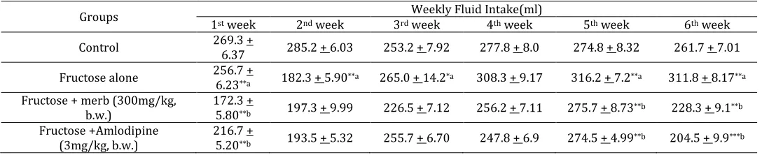
decrease in body weight with MERB and amlodipine treated (table 2.2 and 2.3-supplementary information). In addition, Fructose induced hypertension group showed a significant reduction in the fluid intake at week 1 & 2 followed by the significant increase the fluid intake at week 5 & 6 when compared with control group. Whereas, MERB and amlodipine treated groups showed significant decrease in the fluid intake on the week 1, week 5 week 6 respectively. There is no significant change in the fluid intake found in the remaining weeks which were shown in table no 2.4 of supplementary information).
DPPH radical scavenging activity and NBT reduction assay showed that MERB possess antioxidant activity. DPPH radical scavenging activity IC50 value for Vitamin C and MERB were 8.4 µg/ml and 7.4 µg/mL
respectively. In NBT inhibition assay, IC50 value for
MERB was 12.5 µg/ml against the standard Vitamin C
(8.4 µg/ml). The whole plant of Rhynchosia beddomei
demonstrated ACE inhibitory activity at a
concentration of 800 µg/ml, showing an inhibition of 72.52 %. The IC50 value of MERB extract was 385 µg/ml and standard captopril, was 0.3 ng/ml (Table 3). MERB inhibited ACE in a concentration dependent manner. The antihypertensive, ACE inhibition and antioxidant properties are possibly due to the presence of phenolic and flavonoid content in the MERB.
From the Histopathological studies of myocardium shows intact arrangement of the cardiac muscle fibers where as the in the induced group, myocardium shows partially haphazard arrangement of the cardiac muscle fibers. MERB and standard treated shows its restoration to normal.
GROUPS Dexamethasone induced hypertension. BP SBP DBP BPM Control 110.0 +
0.705
114.9 + 1.679
92.48 + 1.709
329.9 + 3.794 Dexamethasone
(0.2 ml s.c)
121.1 + 0.965
126.0 + 1.641***a
117.9 + 1.231***a
321.8 + 1.265*b
Dexamethasone + MERB(300 mg/kg, b.w.)
90.92 + 2.302
98.73 + 3.405***b
79.33 + 1.929***b
289.5 + 5.607***b
Dexamethasone + Amlodipine (3mg/kg, b.w.)
86.92 + 0.830
86.88 + 0.861***b
85.88 + 0.511***b
280.1 + 5.464***b
Table 1: Effect of MERB on Dexamethasone induced hypertension in rats
Values are expressed as Mean ±SEM, n=6, analysed in graph pad prism version 5.04 by one way ANOVA followed by Tukey’s multiple comparision test. Where * represents significant at P ≤
0.05, ** represents highly significant at P≤0.01, ***represents very
significant at P≤0.001.a control vs Dexamethasone induced b
N.V.L Suvarchala Reddy V Asian Journal of Pharmacology and Toxicology, 03(10), 2015,13-18
Parameters Control Fructose
Fructose+MERB (300 mg/kg, b.w.)
Fructose + Amlodipine (3 mg/kg, b.w.)
BP 103.3 ± 3.34 157.0 ± 2.29***a 107.4 ± 3.5***b 101.0 ± 1.34***b
SBP 105.0 ± 2.5 144.2 ± 3.54***a 116.2 ± 4.04***b 116.1 ± 1.42***b
DBP 72.8 ±1.78 126.0 ± 1.43**a 105.2 ± 3.25**b 92.3 ± 2.5**b
Mean BP 101 ± 1.31 122.5 ± 2.20***a 100.7 ± 3.00***b 96.3 ± 2.00***b
Heart rate 294.8 ± 4.3 337.4 ± 7.75***a 292 ± 4.27**b 304.3 ± 1.79**b
Table 2: Antihypertensive effect of MERB on fructose fed albino wistar ratsa
aValues are expressed as Mean ±SEM, n=6, analysed in graph pad prism version 5.04 by one way ANOVA followed by Tukey’s comparision
test Where * represents significant at P ≤ 0.05, ** represents highly significant at P≤0.01, ***represents very
significant at P≤0.001.a control vs fructose induced group, b fructose induced group vs drug treatment groups.
GROUPS Glucose (gm/dL) Triglycerides (gm/dL) Insulin (IU/ml) Control 86.69 + 0.068 1.352 + 0.135 2.843 + 0.218 Fructose 167.54 + 0.327 3.277 + 0.415**a 4.452 + 0.196***a
Fructose + MERB (300 mg/kg, b.w.) 89.46 + 0.369 1.725 + 0.386**b 3.423 + 0.382**b
Fructose + Amlodipine (3 mg/kg, b.w.)
82.32 + 0.542 1.665 + 0.289**b 3.235 + 0.208**b
Table 2.1: antihypertensive effect of MERB on biochemical changesa
aValues are expressed as Mean ±SEM, n=6, analysed in graph pad prism version 5.04 by one way ANOVA followed by Tukey’s comparision
test. Where * represents significant at P ≤ 0.05,** represents highly significant at P≤0.01, ***represents very significant at P≤0.001.a control vs
fructose induced group, b fructose induced group vs. drug treatment groups.
Groups Weekly body weight changes (gms)
0 day 1st week 2nd week 3rd week 4th week 5th week 6th week
Control 157.2 ± 5.45 166.9 ± 4.1 195.4 ± 6.5 205.8 ± 7.1 217.6 ± 8.7 224.8 ± 9.2 236.6 ± 9.2 Fructose 192.5 ± 6.26 217.4 ±
5.1**a 228.9 ± 8.4*a 236.3 ± 9.1*a 240.3 ± 10*a 245.5 ± 10.9*a
256.3 ± 10.8*a
Fructose + MERB
(300mg/kg b.w.) 218.0 ± 8.08 229.3 ± 6.7
215.8 ±
8.3**b 246.5 ± 12.2 249.5 ± 12.4 225.9 ± 13.6*b
233.1 ± 12.1**b
Fructose + Amlodipine
(3 mg/kg, b.w.) 213.1 ± 5.06 220.3 ± 5.1 229.1 ± 5.7 237.31 ± 6.2 232.1 ± 6.2 231.13 ± 6.2*b
224.11 ± 6.9**b
Table 2.2: Effect of drug treatment on body weight changesa
aValues are expressed as Mean ±SEM, n=6, analysed in graph pad prism version 5.04 by one way ANOVA followed by Tukey’s comparision
test. Where * represents significant at P ≤ 0.05, ** represents highly significant at P≤0.01, ***represents very significant at P≤0.001. a control vs
fructose induced group, b fructose induced vs MERB and amlodipine treated groups.
Groups Weekly Feed Intake(gms)
1st week 2nd week 3rd week 4th week 5th week 6th week
Control 60.83 + 1.352 58.97 + 1.349 52.07 + 2.063 52.67 + 1.169 49.78 + 1.169 55.20 + 0.735 Fructose alone 54.00 + 1.732*a 45.38 + 1.411*a 43.20 + 1.652 40.17 + 1.338 41.97 + 1.228 38.88 + 0.749**a
Fructose
+MERB 35.67 + 1.626**a 39.58 + 1.42*b 32.08 + 1.154*b 35.47 + 0.914 35.10 + 2.173 33.37 + 1.265*b Fructose +
Amlodipine 30.17 + 1.27**a 37.56 + 1.45*b 28.77 + 1.672*b 27.52 + 1.146**b
23.03 +
1.320**b 25.58 + 0.479**b
Table 2.3: Effect of drug on weekly feed intake.
Values are expressed as Mean ±SEM, n=6, analysed in graph pad prism version 5.04 by one way ANOVA followed by Tukey’s comparision test, Where * represents significant at P ≤ 0.05, ** represents highly significant at P≤0.01, ***represents very significant at P≤0.001. a control vs
fructose induced group, b fructose induced vs MERB and amlodipine treated groups.
Groups Weekly Fluid Intake(ml)
1st week 2nd week 3rd week 4th week 5th week 6th week
Control 269.3 +
6.37 285.2 + 6.03 253.2 + 7.92 277.8 + 8.0 274.8 + 8.32 261.7 + 7.01 Fructose alone 256.7 +
6.23**a 182.3 + 5.90**a 265.0 + 14.2*a 308.3 + 9.17 316.2 + 7.2**a 311.8 + 8.17**a
Fructose + merb (300mg/kg, b.w.)
172.3 +
5.80**b 197.3 + 9.99 226.5 + 7.12 256.2 + 7.11 275.7 + 8.73**b 228.3 + 9.1**b
Fructose +Amlodipine (3mg/kg, b.w.)
216.7 +
5.20**b 193.5 + 5.32 255.7 + 6.70 247.8 + 6.9 274.5 + 4.99**b 204.5 + 9.9***b
Table 2.4: Effect of drug treatment on weekly fluid intake.
Values are expressed as Mean ±SEM, n=6, analysed in graph pad prism version 5.04 by one way ANOVA followed by Tukey’s comparision test, Where * represents significant at P ≤ 0.05, ** represents highly significant at P≤0.01, ***represents very significant at P≤0.001. a control vs
N.V.L Suvarchala Reddy V Asian Journal of Pharmacology and Toxicology, 03(10), 2015,13-18
Extract / Standard Concentration % Inhibition IC50
MERB
100 21.10 ± 0.38
385 ± 1.32 (µg/ml)
200 34.90 ± 0.33
400 56.04 ± 0.49
800 72.52 ± 0.54
Captopril
0.1 35.12 ± 0.23
0.3 ± 0.02 (ng/ml)
0.2 45.38 ± 0.32
0.4 68.20 ± 0.45
0.8 78.01 ± 0.56
Table 3. In vitro ACE inhibitory activity of MERB and Captopril MERB- Rhynchosia beddomei bark methanolic extract Values are expressed as mean ± SEM of three parallel measurements. *P < 0.01 when compared with standard.
Test extract DPPH radical scavenging activity IC50 (µg/mL)
NBT inhibition assay IC50 (µg/mL)
MERB 7.4 12.5
Vitamin C 8.4 8.4
Table 4. NBT and DPPH radical scavenging activity of Rhynchosia beddomei extracts
Values are expressed as mean ± SEM of three parallel measurements. *P < 0.01 when compared with standard.
Figure 1: Histopathology report of Myocardium: In the histological report for the myocardium A. Control the myocardium shows intact arrangement of the cardiac muscle fibers, B. Induced group the myocardium shows partially haphazard arrangement of the cardiac muscle fibers, C. MERB treated, the myocardium shows intact arrangement of the cardiac muscle fibers, which shows its restoration to normal. D. Standard the myocardium shows intact arrangement of the cardiac muscle fibers.
Conclusion
Hypertension is a risk factor for apoplectic stroke. ACE regulates blood pressure and ACE inhibition will help reduce hypertension, cardiovascular disease and other related ailments. Antihypertensive activity of MERB in dexamethasone and fructose treated rats was confirmed by decreased BP, ACE inhibition; and antioxidant activity by radical scavenging property. At firstly the phytochemical constituents of MERB extract were found to be triterpenoids, steroids, flavonoids and saponins. These results may lend further support to mount up evidence that the MERB extract containing compounds which, if taken in sufficient quantities, could conceivably be beneficial in the
attenuation and prevention hypertension and
hyperinsulinemia. The current study has demonstrated
that the whole plant extract of Rhynchosia beddomei is
capable of inhibiting angiotensin converting enzyme, quenching free radicals and acting as reducing and chelating agents. Further studies are required to
investigate the antihypertensive properties of
individual components of extract of R. beddomei.
References
1. Romero JC, Reckelhoff JF. Role of angiotensin and oxidative stress in essential hypertension. Hypertension 1999;34:943–949.
2. Raij L. Nitric oxide in hypertension: relationship with renal injury and left ventricular hypertrophy. Hypertension 1998; 31: 189–193.
3. McIntyre M, Bohr DF, Dominiczak AF. Endothelial function in hypertension, the role of superoxide anion. Hypertension 1999; 34:539–545.
4. Orie NN, Zidek W, Tepel M. Reactive oxygen species in essential hypertension and non-insulin-dependent diabetes mellitus. Am J Hypertension1999; 12:1169– 1174.
5. Yasunari K, Maeda K, Nakamura M, Yoshikawa J. Oxidative stress in leukocytes is a possible link between blood pressure, blood glucose, and C-reactive protein. Hypertension 2002; 39:777–780.
6. Redon J, Oliva MR, Tormos C, Giner V, Chaves FJ, Iradi A, Saez GT. Antioxidant activities and oxidative stress byproducts in human hypertension. Hypertension 2003; 41: 1096–1101.
7. Saruta T. Mechanism of glucocorticoid-induced hypertension. Hypertension Res 1996; 19: 1–8.
8. Ullian ME. The role of corticosteriods in the regulation of vascular tone. Cardiovas Res 1999;41:55–64.
9. John T, Groel MD, Samir S, Tadros MD, Gerald R, Dreslinski MD, Alan C, Jenkins MD. Long-Term Antihypertensive Therapy with Captopril. Hypertension 1983;5(5):145-151.
10. Zhang GX, Kimura S, Murao K, Shimizu J, Matsuyoshi H, Takaki M. Role of neuronal NO synthase in regulating vascular superoxide levels and mitogen-activated protein kinase phosphorylation Cardiovas Res 2009; 81:389. 11. DeGasparo M. Angiotensin II and nitric oxide
interaction. Heart Failure Reviews 2002; 7:347–358. 12. Hateno T, Edamatsu R, Mari A. Effects of interactions of
N.V.L Suvarchala Reddy V Asian Journal of Pharmacology and Toxicology, 03(10), 2015,13-18
14. Ojeda D, Enrique J, Alejandro Z, Herrera-Arellano A, Jaime T, Laura A. Inhibition of angiotensin convertin enzyme (ACE) activity by the anthocyanins delphinidin- and cyanidin-3-O-sambubiosides from Hibiscus sabdariffa. J Ethnopharmacol 2010;127(1): 7-10.
15. Loizzo MR, Said A, Tundis R, Rashed K, Statti AG, Hufner A, Menichini F. Inhibition of angiotensin converting enzyme (ACE) by flavonoids isolated from Ailanthus excelsa (Roxb) (Simaroubaceae). Phytotherapy Res 2007; 21(1): 32-36.
16. Chetty KM, Sivaji K, Rao KT. Flowering plants of Chittoor district Andhra Pradesh India. 1st ed. Tirupati: Student
offset printers 2008; 98-99.
17. Bakshu LMD, Raju RRV. Antimicrobial activity of
Rhynchosia beddomei. Fitoter 2001;72:579-582.
18. Bakshu LMD, Raju RRV. Chemical characterization of Essential oil of Rhynchosia beddomei. Journal of Applied Biological Sciences 2009; 3(1): 31-32.
19. Woods M, Key J. The genus Rhynchosia (Fabaceae) in Alabama. Phytologia 2009; 91(1): 3–17.
20. Chouhan A, Iqbal S, Maheshwari RS, Bafna A. Effect of Leaf Extracts of Rhynchosia Beddomeion Ehrlich Ascites Carcinoma in Mice. The International Journal of Advanced Scientific Research and Review 2011; 1(4): 29-36 21. Cushman DW, Cheung HS. Spectrophotomeic assay and
properties of the angiotensin converting enzyme of rabbti lung. Biochem Pharmacol 1971;20: 1637–1648.
22. Miguel M, Recio I, Gomez-Ruiz JA, Ramos M, Lopez-Fandino R. Angiotensin-I-converting enzyme inhibitory activity of peptides derived from egg white proteins by enzymatic hydrolysis. J Food Prot 2004; 67:1914–1920. 23. Mahendran P, Shyamala Devi CS. Effect of Garcinia
cambogia extract on lipids and lipoprotein composition in dexamethasone administered rats. Indian Journal of Physiol Pharmacol 2001;45:345-50.
24. Vikrant V, Grover JK, Tandon SS, Rathi SS, Gupta N. Treatment with extracts of Momordica charantia and
Eugenia jambolana prevents hyperglycemia and hyperinsulinemia in fructose fed rats. J Ethnopharmacol 2001; 76:139-43.