R E S E A R C H
Open Access
HLA and killer cell immunoglobulin-like
receptor (KIRs) genotyping in patients with
acute ischemic stroke
Antonino Tuttolomondo
1*†, Domenico Di Raimondo
1†, Rosaria Pecoraro
6,7, Alessandra Casuccio
2,
Danilo Di Bona
5, Anna Aiello
3, Giulia Accardi
3, Valentina Arnao
4, Giuseppe Clemente
1, Vittoriano Della Corte
8,
Carlo Maida
1, Irene Simonetta
1, Calogero Caruso
3, Rosario Squatrito
6, Antonio Pinto
1and on behalf of KIRIIND (KIR
Infectious and Inflammatory Diseases) Collaborative Group
Abstract
Introduction:In humans, a major component of natural killer (NK) and T cell target recognition depends on the surveillance of human leukocyte antigen (HLA) class I molecules by killer immunoglobulin-like receptors (KIRs).
Aims:To implement the knowledge about the immunological genetic background of acute ischemic stroke susceptibility in relation to the frequency of the KIR genes and HLA alleles.
Methods:Subjects with acute ischemic stroke and subjects without stroke were genotyped for the presence of KIR genes and of the three major KIR ligand groups, HLA-C1, HLA-C2, and HLA-Bw4, both HLA-B and HLA-A loci.
Results:Between November 2013 and February 2016, consecutive patients with acute ischemic stroke were recruited. As healthy controls, we enrolled subjects without acute ischemic stroke. Subjects with acute ischemic stroke in comparison with controls showed a higher frequency of 2DL3, 2DL5B, 2DS2, and 2DS4 KIR genes and a lower frequency of HLA-B-Bw4Ialleles. Subjects without acute ischemic stroke showed a higher frequency of interaction between KIR 2DS2 and HLAC2. We also observed a higher frequency of 2DL3 and 2 DL4 KIR genes in subjects with atherosclerotic (LAAS) subtype. Multiple logistic regression analysis showed a protective effect towards stroke of HLA-B-Bw4Iand interaction between KIR 2DL2 and HLAC1 and 2DS2-HLAC2 and a detrimental effect of 2DL2-HLA-C1_A interactions.
Conclusion:Our findings of a higher frequency of activating KIR genes seem to be consistent with findings previously reported patients with coronary syndrome. This higher frequency of“proinflammatory”genes in subjects with ischemic stroke could also explain the immunoinflammatory activation of the acute phase of stroke.
Keywords:Stroke, Killer immunoglobulin-like receptors (KIRs), HLA
Background and purpose
Evidence for the contribution of T cell-mediated immunity to cardiovascular diseases has only recently emerged [1,2]. T cells infiltrate atherosclerotic plaques, and in these sites, proinflammatory immune responses mediated by CD4+
T-helper type 1 (TH1) and CD8+cytotoxic T lymphocytes are preponderant [1–4]. Natural killer (NK) cells activating or inhibition pathways mediated by human leukocyte anti-gen (HLA) expression could have a potential detrimental or protective role towards inflammation-mediated acute com-plications of atherosclerosis such as acute ischemic stroke.
In humans, a major component of NK and T cell tar-get recognition depends on the surveillance of human leukocyte antigen (HLA) class I molecules by killer immunoglobulin-like receptors (KIRs), a family of di-verse activating or inhibitory receptors that are expressed on the surface of NK cells and T cell subsets
© The Author(s). 2019Open AccessThis article is distributed under the terms of the Creative Commons Attribution 4.0 International License (http://creativecommons.org/licenses/by/4.0/), which permits unrestricted use, distribution, and reproduction in any medium, provided you give appropriate credit to the original author(s) and the source, provide a link to the Creative Commons license, and indicate if changes were made. The Creative Commons Public Domain Dedication waiver (http://creativecommons.org/publicdomain/zero/1.0/) applies to the data made available in this article, unless otherwise stated. * Correspondence:bruno.tuttolomondo@unipa.it
†Antonino Tuttolomondo and Domenico Di Raimondo contributed equally
to this work.
1Internal Medicine and Stroke Care Ward, Department of Health Promotion
[5]. Different KIRs can transmit inhibitory or activating signals to the cell, and combinations of KIR genes synergize to generate genes with widely differing balance between activating and inhibitory types.
KIR receptors have been reported as involved in the susceptibility to several diseases, including celiac disease, rheumatoid arthritis, systemic lupus erythematosus, in-fectious diseases, and cancer [4,5]. Our group previously reported [8] that immunocompetent subjects carrying the homozygous A haplotype or the HLABw4Tallele are at higher risk of developing symptomatic disease after primary cytomegalovirus (CMV) infection.
Although HLA genes, as well as the innate immune genes KIR, are considered to be important determinants of T cell involvement in inflammation, few studies, to our knowledge, evaluated their role in the clinical setting of susceptibility of acute complications of atherosclerotic disease [5,6].
One study [7] reported that CD4+CD28nullT cell clones from patients with acute coronary syndrome (ACS) expressed a broader spectrum of KIRs with preference for the stimulatory variant, CD158j, and that CD4+ T cell clones from patients acquired de novo expression of the DAP12 molecule, an adapter chain that transmits CD158j-derived signals. Consequently, these T cells ac-quire cytolytic capability that may suggest T cell involve-ment also in acute complication of atherosclerosis also indicating a possible role of KIR genetic background in regulation of inflammatory cell involvement in acute car-diovascular event.
No study, to the best of our knowledge, analyzed the relationship between KIR aplotype and HLA allele and the risk of developing an acute ischemic stroke.
Study hypothesis
The principal hypothesis of our study was that a higher risk of developing an ischemic stroke may be related to a higher frequency of some KIR genes and their HLA ligands and of their interactions in comparison with subjects without ischemic stroke. An additional hypothesis was that a higher risk of
developing certain subtypes of ischemic stroke may be due to a higher frequency of some genetic variants of KIR and their HLA ligands or of their interactions.
Aim of the study
The aim of this analytical cross-sectional study was:
– To assess if genetic variants of KIR and their HLA ligands may influence susceptibility to ischemic stroke and its TOAST subtype and to implement the knowledge about the immunological and genetic
background of acute ischemic stroke susceptibility in connection with the expression of the KIR and HLA alleles
– To further characterize immune-inflammatory de-gree of the acute phase of ischemic stroke by means evaluation of peripheral frequency of CD28nullcells in a sample of patients with acute stroke in compari-son with subjects without ischemic stroke
Materials and methods
Between November 2013 and July 2016, consecutive pa-tients with acute ischemic stroke were recruited from Internal Medicine with Stroke Care ward of Policlinico “P. Giaccone”, University of Palermo and Pronto Soc-corso Unit of Giuseppe Giglia Hospital, Cefalù (Pa-lermo). As controls, we enrolled consecutive healthy subjects admitted to our ward in the same recruitment period and without acute ischemic stroke and without exclusion criteria (see above).
Ischemic stroke has been defined as: “An episode of neurological dysfunction caused by focal cerebral, spinal, or retinal infarction lasting more than 24 hours”[8].
For stroke patients, we evaluated medical history, 12-lead ECG, 24-h electrocardiography monitoring, trans-thoracic echocardiography, carotid ultrasound, brain CT, or MRI at admission.
Patients and controls were excluded if they had one of these exclusion criteria: rheumatologic disorders, chronic inflammatory disease, acute systemic infections, recent venous thrombosis, recent acute myocardial in-farction (AMI) (within 3 months), and recent cerebro-vascular event (TIA or stroke within 6 months).
Type 2 diabetes mellitus was determined using a clin-ically based algorithm that considered age at onset, pre-senting weight and symptoms, family history, onset of insulin treatment, and history of ketoacidosis [9]. Hyper-tension was defined according to the 2013 ESH/ESC guidelines [10]. Hypercholesterolemia was defined as total serum cholesterol ≥200 mg/dL, and hypertriglyc-eridemia as total serum triglyceride ≥150 mg/dL on the basis of the National Cholesterol Education Program– Adult Treatment Panel III reports [11,12]. Coronary ar-tery disease has been evaluated by means the presence of clinical symptomatology of angina, myocardial infarc-tion, or any previous coronary revascularization.
Cerebrovascular disease (TIA/ischemic stroke) has been evaluated by means patient history, neurological exam, and hospital records.
Ethical approval and consent to participate
study, as well as for sampling and banking of the bio-logical material.
Stroke subtype evaluation
The type of acute ischemic stroke was classified ac-cording to the TOAST classification [13]: (1) large ar-tery atherosclerosis (LAAS), (2) cardioembolic infarct (CEI), (3) lacunar infarct (LAC), (4) stroke of other determined etiology (ODE), and (5) stroke of undeter-mined etiology (UDE).
Cell isolation, staining, and flow cytometry
Peripheral blood has been drawn at 48 h after symptom onset and after informed consent had been obtained from the patient or his/her authorized representative. Peripheral blood mononuclear cells (PBMCs) have been obtained by density gradient centrifugation using the lymphocyte separation medium (ICN Pharmaceutical, Costa Mesa, CA). This protocol yields an average PBMC composition of 60% T cells, 15% monocytes/macro-phages, 10% B cells, and 15% natural killer cells [29]. White cells were obtained from 2 ml of peripheral EDTA-anticoagulated venous blood. Cells were labeled with human monoclonal anti-CD4 antibodies conjugated with fluorescein isothiocyanate and CD28 anti-bodies conjugated with phycoerythrine (Becton Dickin-son). Mouse IgG1 antibodies conjugated with fluorescein isothiocyanate (IgG1-FITC) and IgG2a conjugated with phycoerythrine IgG2a-PE (Becton Dickinson) were used as the isotype controls. The samples were incubated for 30 min in the dark at ambient temperature, washed with 5% saline, and centrifuged. The pellet was suspended in 1% formalin. Expression of the CD28 receptor on lym-phocytes was studied with a FACSCalibur flow cyt-ometer (FACSCalibur/Sysmex XT1800i dual platform) operating with CellQuest OS2 software. The population of CD4+CD28nullwas expressed as a percentage of CD4 + cells (CD4+CD28nulland CD4+CD28+).
HLA and KIR genotyping
Peripheral whole blood samples were collected as indi-cated, and genomic DNA was extracted from leukocytes by a commercial kit (PureLink® Genomic DNA, Thermo-Fisher Scientific, Waltham, MA, USA). Using the poly-merase chain reaction sequence-specific primer (PCR-SSP) technique, the DNA subjects with acute is-chemic stroke and subjects without stroke were geno-typed for the presence of the three major KIR ligand groups, HLA-C1, HLA-C2, and HLA-Bw4, both HLA-B and HLA-A loci (Epitop-TYPE kit; BAG Health Care GmbH, Lich, Germany). HLA-C and Bw4 KIR ligand groups were assigned directly by using specific oligo-nucleotide primers to type the codon corresponding to amino acid 80 for HLA-C (HLA-C1, Cw alleles with
asparagine at position 80; HLA-C2, Cw alleles with ly-sine at position 80) and for HLA-Bw4 (Bw4-I, Bw alleles with isoleucine at position 80; Bw4-T, Bw alleles with threonine at position 80). KIR genotyping was per-formed for both inhibitory and activating KIR using the KIR-TYPE kit (BAG Health Care GmbH).
The following KIR genes were analyzed, which in-cluded the inhibitory receptors KIR2DL1, 2DL2, 2DL3, 2DL5, 3DL1, 3DL2, and 3DL3; the activating receptors 2DL4, 2DS1, 2DS2, 2DS3, 2DS4, 2DS5, and 3DS1; two pseudogenes (2DP1 and 3DP1) and the common vari-ants of KIR2DL5 (KIR2DL5A, KIR2DL5B); the KIR2DS4 alleles; and KIR3DP1 alleles. KIR gene profiles were de-termined by the presence or absence of each KIR gene.
The detection of individual receptors Kir/HLA ligands was performed with the sequence-specific primer method. Therefore, the amplification occurs only if the primers are complementary to the target sequence and is then highlighted to agarose gel electrophoresis. The specific primers were selected so as to detect single KIR/ HLA ligand genes.
Statistical analysis
Statistical analysis of quantitative and qualitative data, including descriptive statistics, was performed for all items. Continuous data are expressed as mean ± SD, un-less otherwise specified. Baseline differences between groups were assessed by the chi-square test or Fisher exact test, as needed for categorical variables, and by the independent Student ttest for continuous parameters if the data were normally distributed. If the data did not show a normal distribution, a non-parametric test was used (Mann Whitney’s U test). Logistic regression ana-lysis examined the correlation between patient charac-teristics (independent variables) and patient groups (dependent variable) in simple and multiple regression models. An overall sample size of 124 patients (62 for each study group) has been calculated to document a difference of at least 20% in the prevalence of KIR genes and HLA alleles between the patient groups. The sample sizes took into account a significance level of 5% and a power of 80%.
Data were analyzed by IBM SPSS Software 22 version (IBM Corp., Armonk, NY, USA). All p values were two-sided, and p< 0.05 was considered statistically significant.
Results
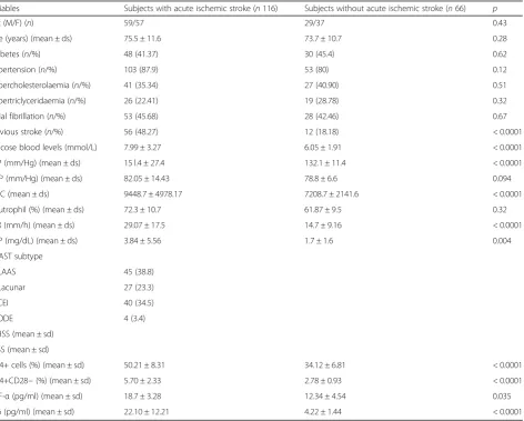
General and clinical characteristics of patients with acute ischemic stroke and of subjects without acute is-chemic stroke are listed in Table1.
The mean age of patients with acute ischemic stroke was 75.5 ± 11.6 years in subjects with stroke and 73.7 ± 10.7 years in subjects without acute ischemic stroke.
Subjects with acute ischemic stroke showed in com-parison with subjects without stroke a similar prevalence of hypertension, diabetes, hypercholesterolaemia, and atrial fibrillation, whereas subjects with ischemic stroke showed in comparison with subjects without acute stroke a higher prevalence of previous stroke (48.3% vs 18.2%;p< 0.0001) and higher mean glucose blood levels (7.99 ± 3.27 mg/dl vs 6.05 ± 1.91 mg/dl; p< 0.0001), higher mean SBP values (151.4 ± 27.4 mm/Hg vs 132.1 ± 11.4 mm/Hg;p< 0.0001), higher SBP values (15 l.4 ± 27.4 vs 132.1 ± 11.4/Hg;p< 0.0001), higher mean WBC count (9448.7 ± 4978.17 vs 7208.7 ± 2141.6; p< 0.0001), and
higher ESR (29.07 ± 17.5 mm/h vs 14.7 ± 9.16;p< 0.0001) and CRP (3.84 ± 5.56 vs 1.7 ± 1.6;p= 0.004) mean values. Subjects with acute ischemic stroke showed in com-parison with subjects without ischemic stroke higher peripheral percentage of CD4+ (50.21 ± 8.31% vs 34.12 ± 6.81%; p< 0.0001) and CD28null (5.70 ± 2.33% vs 2.78 ± 0.93%; p< 0.0001) cells; furthermore, subjects with stroke showed higher serum levels of TNF-α (18.7 ± 3.28 pg/ml vs 12.34 ± 4.54 pg/ml; p= 0.035) and IL-6 (22.10 ± 12.21 pg/ml vs 4.22 ± 1.44 pg/ml; p< 0.0001) (see Table1).
KIR genotype analysis of patients with acute ischemic stroke in comparison with subjects without ischemic stroke showed a higher frequency of 2DL3 (74.1% vs 42.4% p< 0.0001), 2DL5B (33.6% vs 18.8%; p= 0.03), 2DS2 (37.9% vs 16.6%;p= 0.003), 2DS4 (41.3 vs 16.6;p < 0.0001), and 3DP1 (14.6% vs 0; p< 0.0001) KIR genes (see Table2 and Fig. 1) and a lower frequency of 3DL1
Table 1General, demographic, and clinical findings in subjects with acute ischemic stroke and in subjects without ischemic stroke
Variables Subjects with acute ischemic stroke (n116) Subjects without acute ischemic stroke (n66) p
Sex (M/F) (n) 59/57 29/37 0.43
Age (years) (mean ± ds) 75.5 ± 11.6 73.7 ± 10.7 0.28
Diabetes (n/%) 48 (41.37) 30 (45.4) 0.62
Hypertension (n/%) 103 (87.9) 53 (80) 0.12
Hypercholesterolaemia (n/%) 41 (35.34) 27 (40.90) 0.51
Hypertriclyceridaemia (n/%) 26 (22.41) 19 (28.78) 0.32
Atrial fibrillation (n/%) 53 (45.68) 28 (42.46) 0.67
Previous stroke (n/%) 56 (48.27) 12 (18.18) < 0.0001
Glucose blood levels (mmol/L) 7.99 ± 3.27 6.05 ± 1.91 < 0.0001
SBP (mm/Hg) (mean ± ds) 15 l.4 ± 27.4 132.1 ± 11.4 < 0.0001
DBP (mm/Hg) (mean ± ds) 82.05 ± 14.43 78.8 ± 6.6 0.094
WBC (mean ± ds) 9448.7 ± 4978.17 7208.7 ± 2141.6 < 0.0001
Neutrophil (%) (mean ± ds) 72.3 ± 10.7 61.87 ± 9.5 0.32
ESR (mm/h) (mean ± ds) 29.07 ± 17.5 14.7 ± 9.16 < 0.0001
CRP (mg/dL) (mean ± ds) 3.84 ± 5.56 1.7 ± 1.6 0.004
TOAST subtype
LAAS 45 (38.8)
Lacunar 27 (23.3)
CEI 40 (34.5)
ODE 4 (3.4)
NIHSS (mean ± sd)
SSSS (mean ± sd)
CD4+ cells (%) (mean ± sd) 50.21 ± 8.31 34.12 ± 6.81 < 0.0001
CD4+CD28−(%) (mean ± sd) 5.70 ± 2.33 2.78 ± 0.93 < 0.0001
TNF-α(pg/ml) (mean ± sd) 18.7 ± 3.28 12.34 ± 4.54 0.035
IL-6 (pg/ml) (mean ± sd) 22.10 ± 12.21 4.22 ± 1.44 < 0.0001
(67.2% vs 89.3%; p< 0.0001), 3DL2 (67.2% vs 100%;p< 0.0001), 3DL3 (62.9% vs 100%;p< 0.0001), 2DS5 (14.6% vs 51.5%; p < 0.0001), and 2DP1 (77.5% vs 100%) KIR genes in comparison with subjects without stroke. No difference was reported in the frequency of KIR haplo-types between the groups.
With regard to HLA alleles, subjects without ische-mic stroke showed in comparison with stroke sub-jects a significantly higher frequency of HLA-B-Bw4I alleles (65.1% vs 8.6%; p< 0.0001) (see Table 2 and Fig. 1).
With regard to KIR-HLA ligand interactions, sub-jects with acute ischemic stroke showed in comparison with subjects without acute stroke a higher frequency of interaction between KIR2DS2 and HLAC2 (25% vs 6.06%; p= 0.001) (see Table 3 and Fig.1) and a lower frequency of interaction between KIR2DL2 and HLA-C1 (21.55% vs 40.9%;p= 0.007).
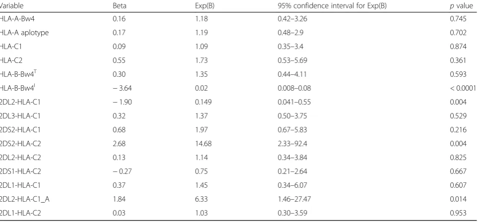
Multiple logistic regression analysis considering vari-ables predictive of ischemic stroke showed the protective effect of HLA-B-Bw4I alleles (B=−3.649; p< 0.0001) and of interaction between KIR2DL2 and HLA-C1 (B= −1.901;p= 0.004), whereas a detrimental effect was ob-served with regard to interaction between KIR2DS2 and HLAC2 (B= 2.687; p= 0.004) and between KIR2DL2 and HLA-C1_A (B= 1.847;p= 0.014) (see Table4).
General, demographic, and clinical findings in subjects with acute ischemic stroke in relation to TOAST sub-type are listed in Table5.
With regard to KIR gene frequency in relation to TOAST subtype, we observed a higher frequency of 2DL3 (86.3% vs 62.9% vs 72.5%;p= 0.033), 2DL4 (88.8% vs 59.25% vs 65%; p= 0.010), 3DL2 (82.2% vs 51.8% vs 62.5%;p= 0.016), and 3DL3 (80% vs 48.14% vs 55%;p= 0.015) KIR genes in subjects with LAAS subtype in com-parison with subjects with other subtypes, whereas with Table 2Frequencies of KIR haplotypes and HLA alleles among individuals acute ischemic stroke and in subjects without stroke
Aplotipo KIR Subjects with acute ischemic stroke (n116) Subjects without acute ischemic stroke (n66) p
2DL1 (n/%) 64 (55.17) 43 (65.16) 0.21
2DL2 (n/%) 41 (35.34) 26 (39.3) 0.63
2DL3 (n/%) 86 (74.1) 28 (42.4) < 0.0001
2DL4 (n/%) 84 (72.4) 66 (100) < 0.0001
2DL5A (n/%) 14 (12.06) 12 (18.8) 0.27
2DL5B (n/%) 39 (33.6) 12 (18.8) 0.03
2DS1 (n/%) 24 (20.6) 18 (27.2) 0.36
2DS2 (n/%) 44 (37.9) 11 (16.6) 0.003
2DS3 (n/%) 31 (26.7) 17 (25.7) 1
2DS4 (n/%) 48 (41.3) 11 (16.6) 0.001
2DS5 (n/%) 17 (14.6) 34 (51.5) < 0.0001
3DL1 (n/%) 78 (67.2) 59 (89.3) 0.001
3DL2 (n/%) 78 (67.2) 66 (100) < 0.0001
3DL3 (n/%) 73 (62.9) 66 (100) < 0.0001
3DS1 (n/%) 60 (51.7) 37 (56.06) 0.64
2DP1 (n/%) 90 (77.5) 66 (100) < 0.0001
3DP1 (n/%) 17 (14.6) 0 < 0.0001
3DP1*003 (n/%) 73 (62.9) 66 (100) < 0.0001
KIR-aplotypes
HLA-A (n/%) 52 (44.8) 30 (45.4) 1.0
HLA-A+B (n/%) 62 (53.4) 37 (56.0) 0.76
HLA alleles
HLA-Bw4 T (n/%) 31 (26.7) 13 (19.6) 0.36
HLA-A-BW4 (n/%) 31 (26.7) 16 (24.2) 0.85
HLA-B-Bw4I(n/%) 10 (8.6) 43 (65.1) < 0.0001
HLA-C1 (n/%) 38 (32.7) 26 (39.3) 0.420
HLA-C2 (n/%) 36 (31.03) 19 (28.7) 0.86
regard to interactions between KIR and HLA alleles, we observed only a higher frequency of the interaction be-tween KIR2DL1 and HLA-C1 alleles in subjects with cardioembolic stroke subtype (22.5% vs 4.4% vs 14.81%;
p= 0.047) (see Table5and Fig.2).
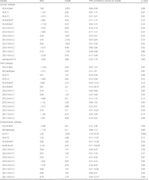
Multiple logistic regression analysis considering vari-ables predictive of TOAST subtype of ischemic stroke showed a significant association between 2DL5A (B= 2.81; p= 0.03) and 3DL1 (B= 2.17; p= 0.02) KIR genes and cardioembolic stroke, whereas with regard to HLA alleles, only HLA-C1 allele (B= 2.36; p= 0.004) was sig-nificantly associated with cardioembolic stroke (see Ta-bles6and7).
At multiple regression analysis, we also observed a possible trend toward significance with regard to the protective role of HLA-A-Bw4 HLA alleles (p= 0.05) and of interaction between KIR 2DS2 and HLA-C2 alleles (p= 0.05) towards LAAS subtype of stroke (see Table 7).
Discussion
Our study showed that subjects with acute ischemic stroke in comparison with subjects without stroke were more likely to have a higher frequency of 2DL3, 2DL4, 2DL5B, 2DS2, 2DS4, and 3DP1 KIR genes and a signifi-cantly lower frequency of HLA-B-Bw4Iallele.
The definition of an immunological genetic profile in patients with acute ischemic stroke could better characterize the pathogenesis of the disease, offering new perspectives of diagnostic and therapeutic evalu-ation. Considering the complexity of genetic studies, we hypothesized that due to the high polymorphism of KIR genes by contributing to the variability of the immune response both innate and adaptive, these genes could be a suitable subject of a study addressed to explain genetic basis of inflammatory pathogenesis of ischemic stroke and of its subtype.
Several studies have reported that susceptibility to is-chemic stroke and its functional prognosis seems to be Fig. 1frequency of KIR genes and of interaction KIR-HLA in subjects with ischemic stroke
Table 3Frequencies of KIR-HLA combinations among individuals with acute ischemic stroke and in subjects without stroke
HLA and KIR interaction Subjects with acute ischemic stroke Subjects without acute ischemic stroke p
2DL2-HLA-C1 (n/%) 25 (21.55) 27 (40.9) 0.007
2DL3-HLA-C1 (n/%) 38 (32.75) 22 (33.3) 0.93
2DS2-HLA-C1 (n/%) 37 (31.89) 23 (34.48) 0.94
2DS2-HLA-C2 (n/%) 29 (25.0) 4 (6.06) 0.001
2DL2-HLA-C2 (n/%) 26 (22.41) 11 (16.67) 0.34
2DS1-HLA-C2 (n/%) 15 (12.93) 5 (7.57) 0.42
2DL1-HLA-C1 (n/%) 16 (13.79) 7 (10.6) 0.52
2DL1-HLA-C2 (n/%) 17 (14.65) 13 (13.79) 0.41
influenced by systemic inflammatory processes and that stroke patients with systemic inflammation exhibit clin-ically poorer outcomes [14].
The KIRs (also known as CD158) are a family of re-ceptors encoded by 14 polymorphic genes [15] seven of which are inhibitory and seven of which are activating. Although the current view focuses primarily on the pacts of KIR expression on NK cell function, it is im-portant to note that KIR are also expressed on subsets of T cells, including invariant NKT cells, and can also directly influence their function.
Furthermore, some authors [16–19] have shown that the relative response of CD56brightand CD56dimNK cells is associated with KIR genotype, providing the first dir-ect evidence that KIR genotype may influence the NK response to a wide range of non-viral pathogens.
In our study, we reported higher serum levels of IL-6 and TNF-α and higher percentage of peripheral CD4+ cells and CD4+CD28 null in subjects with acute ische-mic stroke in comparison with subjects without stroke. These findings are consistent with our own previous re-sults [27,33,34] and with other previous studies report-ing the role of an immune-inflammatory activation involving inflammatory cytokines [20, 21] and CD4 +CD28 null T cell subsets [22–25] in the acute phase of ischemic stroke. Thus, our finding of an increased per-ipheral percentage of CD28null cells in peripheral blood of subjects with ischemic stroke may be also related to the higher prevalence of KIR gene activators such as 2S2 and 2DS4.
CD4+CD28null T cells are functionally distinct from classical CD4 helper T cells. They do not express CD40
ligand, produce large amounts of IFN-γ, and express granzyme B and perforin, giving them the capability to lyse target cells [30, 31]. Their cytotoxic potential, as well as expression of the NK cell marker CD57, suggests that CD4+CD28nullT cells share features with NK cells. CD4+CD28nullbut not CD4+CD28+T cells express KIR.
A direct role of CD4+CD28null T cells in vascular in-jury has been suggested because these cells are also a risk factor for inflammation and rupturing of athero-sclerotic plaques in acute coronary syndromes [32].
These findings corroborate an inflammatory concep-tion of ischemic stroke, although since inflammaconcep-tion has a validated role in plaque instability events, inflamma-tory mechanisms may explain atherothromboembolic pathogenetic mechanisms of stroke.
In a previous study [32], we postulated that a potential mechanism for the relationship between inflammation, stroke pathogenesis, and stroke subtype pathogenesis may be focused on the contribution of pro-inflammatory gene polymorphisms (SNPs).
In our subjects with acute ischemic stroke in compari-son with control subjects, we observed a higher preva-lence of certain activators KIR gene (2DS2 and 2DS4) consistent with findings of previous studies conducted in patients with acute coronary syndrome [26–28] and un-stable atherosclerotic plaques [29]. We have also re-ported a higher prevalence of an inhibitor gene such as 2DL3, of one aplotype of uncertain behaviour such as KIR2DL5B, and of one gene such as KIR2DL4 that is both inhibitory and activating signaling. It is conceivable to consider the prevailing KIR gene frequency of patients with acute ischemic stroke as predominantly as Table 4Logistic regression model to predict the presence of acute ischemic stroke in relation to prevalence of KIR haplotypes
Variable Beta Exp(B) 95% confidence interval for Exp(B) pvalue
HLA-A-Bw4 0.16 1.18 0.42–3.26 0.745
HLA-A aplotype 0.17 1.19 0.48–2.9 0.702
HLA-C1 0.09 1.09 0.35–3.4 0.874
HLA-C2 0.55 1.73 0.53–5.69 0.361
HLA-B-Bw4T 0.30 1.35 0.44–4.11 0.593
HLA-B-Bw4I −3.64 0.02 0.008–0.08 < 0.0001
2DL2-HLA-C1 −1.90 0.149 0.041–0.55 0.004
2DL3-HLA-C1 0.32 1.37 0.50–3.75 0.529
2DS2-HLA-C1 0.68 1.97 0.67–5.83 0.216
2DS2-HLA-C2 2.68 14.68 2.33–92.4 0.004
2DL2-HLA-C2 0.13 1.14 0.34–3.84 0.825
2DS1-HLA-C2 −0.27 0.75 0.21–2.64 0.667
2DL1-HLA-C1 0.37 1.45 0.34–6.07 0.607
2DL2-HLA-C1_A 1.84 6.33 1.46–27.47 0.014
2DL1-HLA-C2 0.03 1.03 0.30–3.59 0.953
Table 5General, demographic, and clinical findings of KIR haplotype, HLA allele, and their combination frequencies in subjects with acute ischemic stroke in relation to TOAST subtype
Variables LAAS (n45) Lacunar (27) Cardioembolic (40) p
Male (n/%) 29 (64.4) 12 (44.4) 15 (37.5) 0.057
Hypertension (n/%) 39 (86.6) 24 (88.8) 36 (90) 0.85
Diabetes (n/%) 20 (44.4) 13 (48.14) 15 (37.5) 0.40
Hypercholesterolaemia (n/%) 24 (53.3) 6 (22.22) 10 (25) 0.011
Hypertriglyceridaemia (n/%) 12 (26.66) 5 (18.51) 9 (22.5) 0.84
Atrial fibrillation (n/%) 8 (17.77) 8 (29.62) 35 (87.5) 0.0001
Previous stroke (n/%) 21 (46.66) 13 (48.14) 21 (52.5) 0.90
2DL1 (n/%) 27 (60) 13 (48.14) 22 (55) 0.75
2DL2 (n/%) 20 (44.4) 7 (25.92) 12 (30) 0.20
2DL3 (n/%) 39 (86.6) 17 (62.96) 29 (72.5) 0.033
2DL4 (n/%) 40 (88.8) 16 (59.25) 26 (65) 0.010
2DL5A (n/%) 6 (13.33) 1 (3.73) 7 (17.5) 0.37
2DL5B (n/%) 13 (28.8) 11 (40.7) 14 (35) 0.76
2DS1 (n/%) 7 (15.5) 7 (25.9) 9 (22.5) 0.52
2DS2 (n/%) 14 (31.1) 12 (44.4) 17 (42.5) 0.61
2DS3 (n/%) 13 (28.8) 6 (22.2) 10 (25) 0.92
2DS4 (n/%) 20 (44.4) 10 (37.03) 16 (40) 0.90
2DS5 (n/%) 5 (11.11) 4 (14.81) 8 (20) 0.73
3DL1 (n/%) 34 (75.55) 13 (48.14) 28 (70) 0.073
3DL2 (n/%) 37 (82.22) 14 (51.85) 25 (62.5) 0.016
3DL3 (n/%) 36 (80) 13 (48.14) 22 (55) 0.015
3DS1 (n/%) 23 (51.1) 14 (51.85) 23 (57.5) 0.35
2DP1 (n/%) 39 (86.6) 19 (70.37) 30 (75) 0.26
3DP1 (n/%) 3 (6.66) 4 (14.81) 10 (25) 0.11
HLA-A-Bw4 (n/%) 8 (17.77) 9 (33.3) 12 (30) 0.33
HLA-A haplotype (n/%) 18 (40) 12 (44.4) 20 (50) 0.68
HLA-C1 haplotype (n/%) 13 (28.88) 5 (18.51) 19 (47.5) 0.069
HLA-C2 haplotype (n/%) 12 (26.66) 9 (33.3) 14 (35) 0.83
HLA-B-Bw4-T(n/%) 14 (31.11) 4 (14.81) 12 (30) 0.37
HLA-B-Bw4I(
n/%) 3 (6.66) 1 (3.7) 6 (15) 0.42
2DL2-HLA-C1 (n/%) 7 (15.5) 4 (14.81) 12 (30) 0.24
2DL3-HLA-C1 (n/%) 15 (33.3) 8 (29.62) 13 (32.5) 0.67
2DS2-HLA-C1 (n/%) 13 (28.87) 11 (40.74) 14 (35) 0.54
2DS2-HLA-C2 (n/%) 8 (17.77) 9 (33.3) 11 (27.5) 0.40
2DL2-HLA-C2 (n/%) 8 (17.77) 7 (25.92) 9 (22.5) 0.24
2DS1-HLA-C2 (n/%) 5 (11.11) 2 (7.40) 7 (17.5) 0.67
2DL1-HLA-C1 (n/%) 2 (4.4) 4 (14.81) 9 (22.5) 0.047
2DL2-HLA-C1 (n/%) 7 (15.5) 4 (14.81) 12 (30) 0.24
2DL1-HLA-C2 (n/%) 7 (15.5) 3 (11.11) 7 (17.5) 0.88
excitatory consistently with previous findings in patients with coronary acute syndrome, whereas coexistent ex-pression of the two inhibitor genes could be viewed as a simple modulatory effect towards excitatory pathway expression.
The function of stimulatory KIRs is unclear, and these receptors are usually expressed in the context of several inhibitory receptors. For the CD4+CD28nullT cell clones, some authors [33] recently demonstrated a costimula-tory activity of CD158b-specific antibodies to enhance proliferation and cytokine production [34], confirming earlier reports that KIR2DS2 can function as a costimu-latory molecule in response to suboptimal TCR trigger-ing [35]. Furthermore, it has been reported in patients with rheumatoid vasculitis [36] and in patients with an acute coronary syndrome [37, 38] that CD4+CD28null T cells expressed KIR2DS2 in the absence of opposing in-hibitory receptors of the same specificity. Thus, in CD4+CD28nullT cells, the activity of KIR2DS2 may favor the activation of autoreactive T cells involved in instabil-ity mechanisms of atherosclerotic plaque and in ische-mic neuronal damage.
With regard to HLA alleles, our patients with acute is-chemic stroke showed in comparison with subjects with-out stroke no significant difference in frequency of HLA alleles, whereas subjects without ischemic stroke showed in comparison with stroke subjects a significantly higher frequency of HLA-B-Bw4I allele. The increased expres-sion in control subjects without ischemic stroke of the HLA-B with “Bw4I motif” that generally interacts with inhibitory 3DL1 and 3DL3 receptors could represent a further confirmation of predominantly pro-inflammatory genetic background of subjects with stroke in
comparison with the anti-inflammatory genetic profile of control subjects without ischemic stroke. To our know-ledge, this is the first study to show that the KIR-ligand group HLA-A-Bw4I can influence the susceptibility to ischemic stroke and may also represent a possible patho-genetic background of inflammatory damage in acute is-chemic stroke. This effect may be mediated by the activation of the inhibitory KIR3DL1, although an asso-ciation with other HLA-B-Bw4 alleles [37,38] that bind the same KIR gene was not reported in this study.
Finally, multiple logistic regression analysis consider-ing variables predictive towards the presence of acute is-chemic stroke showed a protective effect of HLA-B-Bw4I and of 2DL2-HLA-C1, and a detrimental
effect of 2DL2-HLA-C1_A and 2DS2-HLA-C2
HLA-KIR interactions.
These findings could confirm the protective role of an anti-inflammatory genetic background such as the pres-ence of the B-Bw4Imotif.
Thus, our present study represents a first attempt to bring in the context of an overall immune-inflammatory view of point that leads cytokine and cell-dependent in-flammatory mechanisms ischemic stroke occurrence to a genetic background in terms of KIR receptor-ligand HLA allele interactions. Thus, a greater degree of cyto-kine and cell-mediated (NK cells or T cell subset such as CD4+CD28null cells) inflammatory activation could be also dependent by KIR genes responsible for activator KIR receptors on these inflammatory cells.
Table 6Logistic regression model to predict TOAST subtype of acute ischemic stroke in relation of prevalence of KIR haplotypes
Variable Beta Exp(B) 95% confidence interval for Exp(B) pvalue
Lacunar subtype
2DL1 0.176 1.193 0.32–4.43 0.79
2DL2 −0.740 0.477 0.15–1.48 0.20
2DL3 −0.588 0.555 0.12–2.52 0.44
2DL4 −1.399 0.247 0.046–1.31 0.10
2DL5A −2.361 0.094 0.008–1.121 0.06
2DL5B −0.155 0.856 0.23–3.15 0.81
2DS1 −0.035 0.965 0.21–4.25 0.96
2DS2 0.737 2.089 0.65–6.70 0.21
2DS3 −0.477 0.620 0.17–2.16 0.45
2DS4 −0.255 0.775 0.25–2.32 0.65
2DS5 0.273 1.314 0.30–5.58 0.71
3DL1 −1.517 0.219 0.046–1.03 0.056
3DL2 0.913 2.491 0.34–18.27 0.36
3DL3 0.493 1.637 0.22–11.98 0.62
3DS1 0.105 1.110 0.33–3.70 0.86
2DP1 −0.353 0.702 0.12–3.84 0.68
3DP1 −1.022 0.360 0.063–2.07 0.25
3DP1003 −0.123 0.884 0.10–7.21 0.90
LAAS subtype
2DL1 −0.24 0.786 0.18–3.46 0.751
2DL2 1.066 2.903 0.79–10.54 0.105
2DL3 0.91 2.486 0.33–18.84 0.378
2DL4 2.22 9.214 0.97–87.79 0.054
2DL5A 2.05 7.782 0.57–106.84 0.125
2DL5B −0.50 0.605 0.13–2.70 0.510
2DS1 −0.68 0.502 0.09–2.80 0.433
2DS2 −1.07 0.341 0.09–1.27 0.109
2DS3 1.24 3.464 0.79–15.19 0.100
2DS4 0.43 1.552 0.43–5.57 0.500
2DS5 −1.16 0.312 0.048–2.03 0.224
3DL1 0.88 2.419 0.38–15.33 0.348
3DL2 −1.031 0.357 0.02–4.45 0.424
3DL3 −0.829 0.437 0.026–7.33 0.565
3DS1 −0.244 0.783 0.19–3.09 0.727
2DP1 −0.053 0.948 0.099–9.06 0.963
3DP1 0.771 2.161 0.20–22.5 0.519
3DP1003 1.096 2.993 0.21–43.26 0.421
Cardioembolic subtype
2DL1 −0.226 0.797 0.18–3.48 0.76
2DL2 0.144 1.155 0.31–4.28 0.83
2DL3 0.844 2.325 0.43–12.54 0.32
2DL4 0.801 2.227 0.35–14.04 0.39
of KIR and HLA genes in patients with ischemic stroke.
With regard to KIR gene frequency in relation to TOAST subtype, we observed a higher frequency in subjects with LAAS subtype in comparison with sub-jects with other TOAST subtypes of 2DL3, 2DL4, 3DL2, 3DL3, and 3DP1*003 KIR genes, whereas with regard to interaction KIR-HLA, we observed a higher frequency of 2DL1HLAC1 in subjects with cardioem-bolic stroke subtype.
Multiple logistic regression analysis considering vari-ables predictive of TOAST ischemic stroke showed a significant association between 2DL5A and 3DL1 KIR genes and cardioembolic stroke, whereas with regard to HLA allele interaction, only HLA-C1 allele was signifi-cantly associated to cardioembolic stroke. We also ob-served a possible trend toward significance with regard to a possible protective role of HLA-A-Bw4 HLA allele and of 2DS2-HLA-C2 interaction HLA-KIR towards LAAS subtype of stroke.
These findings seem to be not fully consistent with the findings of this study of a prevailing KIR gene frequency of patients with acute ischemic stroke as predominantly as excitatory, whereas the analysis be-tween TOAST subtypes such as atherosclerotic and cardioembolic ones shows an association with pre-vailing inhibitory genes such as 2DL3, 2DL4, 3DL2, and 3DL3 KIR genes and a prevailing inhibitory HLA-KIR interaction such as 2DL1HLAC1.
It explains that the relationship between KIR genes and stroke subtypes is not easy to explain, and prob-ably future studies with a larger sample of subjects with each stroke subtype may be helpful to better characterize the relationship between HLA-KIR
genetic background and clinical subtype of ischemic stroke.
Nevertheless, some possible explanations may be pos-tulated to explain the non-consistent findings of our two sets of analysis with regard to the association between KIR and HLA genes with stroke and stroke subtypes.
Several evidences support the concept that migration of inflammatory cells to the vascular wall is intimately associ-ated with the cause of vascular conversion leading to ath-erosclerosis [39–41]. Inflammatory cells in cerebral vessel have been reported as capable of responding to cardiovas-cular risk factors for human stroke such as hypertension, hyperlipidemia, diabetes mellitus, obesity, and smoking in subjects with cerebrovascular disease. The role of inflam-matory cell in stroke pathogenesis has recently been re-ported, but the role of KIR-HLA interaction to regulate migration and response of inflammatory cells such as NKs and T cell in development of a cerebral artery thrombosis due to atherosclerotic and cardioembolic mechanism is unknown.
HLA class I molecules and the KIR serve as ligand and receptor. Certain combinations of KIRs and HLA mole-cules can transmit activating or inhibitory signals to regu-late the function of NK cells.
HLA-C alleles can be placed into one of two categories, C1 and C2, which are recognized differentially by the KIR re-ceptors. NK cells that express KIR2DL2 and KIR2DL3 inter-act with HLA-C1 allotypes, whereas KIR2DL1 and KIR2DS1 interact with HLA-C2 allotypes [42–46]. Our findings of a higher prevalence of HLA-C1 HLA alleles and of 3DL1 KIR gene in subjects with cardioembolic stroke could underline the prevalent role of inhibitory mechanisms of NKs and T cell regulating, whereas an excitatory pathway seems to be more involved in subjects with atherosclerotic strokes.
Table 6Logistic regression model to predict TOAST subtype of acute ischemic stroke in relation of prevalence of KIR haplotypes
(Continued)
Variable Beta Exp(B) 95% confidence interval for Exp(B) pvalue
2DL5B 0.657 1.928 0.45–8.21 0.37
2DS1 0.723 2.060 0.39–10.77 0.39
2DS2 −0.264 0.768 0.21–2.83 0.69
2DS3 −0.169 0.845 0.19–3.64 0.82
2DS4 0.058 1.060 0.31–3.65 0.92
2DS5 0.379 1.461 0.31–6.97 0.635
3DL1 2.17 8.791 1.41–54.6 0.02
3DL2 −0.92 0.395 0.04–3.61 0.41
3DL3 −0.88 0.413 0.04–4.06 0.44
3DS1 0.010 1.010 0.26–3.90 0.98
2DP1 0.75 2.119 0.31–14.59 0.44
3DP1 1.667 5.299 0.76–36.71 0.09
3DP1003 0.001 1.001 0.089–11.32 0.99
Table 7Logistic regression model to predict TOAST subtype of acute ischemic stroke in relation of prevalence of HLA allele and of HLA-KIR interactions
Variable Beta Exp(B) 95% confidence interval for Exp(B) pvalue
Lacunar subtype
HLA-A-Bw4 1.04 2.853 0.86–9.39 0.08
HLA-C1 −1.24 0.28 0.07–1.15 0.07
HLA-C2 −0.29 0.74 0.21–2.61 0.65
HLA-B-Bw4T −0.82 0.43 0.11–1.73 0.23
HLA-B-Bw4I −1.132 0.32 0.03–3.15 0.33
2DL2-HLA-C1 −0.50 0.60 0.16–2.19 0.44
2DL3-HLA-C1 −0.64 0.52 0.17–1.57 0.25
2DS2-HLA-C1 0.49 1.647 0.59–4.57 0.33
2DS-2HLA-C2 0.76 2.156 0.67–6.85 0.19
2DL2-HLA-C2 0.06 1.063 0.31–3.56 0.92
2DS1-HLA-C2 −0.73 0.48 0.08–2.86 0.42
2DL1-HLA-C1 0.18 1.197 0.28–4.96 0.80
2DL1-HLA-C2 −0.24 0.78 0.17–3.49 0.75
aplotipoHLA-A −0.38 0.68 0.25–1.79 0.44
LAAS subtype
HLA-A-Bw4 −1.322 0.26 0.07–1.01 0.05
AB haplotype −0.13 0.87 0.27–2.8 0.82
HLA-C1 0.31 1.37 0.29–6.36 0.68
HLA-C2 −0.46 0.62 0.14–2.66 0.52
HLA-B-Bw4T 1.006 2.73 0.59–12.53 0.19
HLA-B-Bw4I 0.87 2.4 0.19–30.19 0.49
2DL2-HLA-C1 0.10 1.1 0.26–4.66 0.89
2DL3-HLA-C1 0.48 1.63 0.47–5.60 0.43
2DS-2HLA-C1 −0.89 0.4 0.12–1.33 0.13
2DS2-HLA-C2 −1.35 2.58 0.06–1.03 0.05
2DL2-HLA-C2 −0.12 0.88 0.22–3.41 0.85
2DS1-HLA-C2 0.79 2.2 0.31–15.67 0.43
2DL1-HLA-C1 −1.46 0.23 0.03–1.64 0.14
2DL1-HLA-C2 −0.01 0.98 0.19–5.03 0.98
Cardioembolic subtype
HLA-A-Bw4 −0.48 0.61 0.15–2.40 0.48
AB-haplotype −1.18 0.3 0.08–1.15 0.08
HLA-C1 2.36 10.63 2.10–53.78 0.004
HLA-C2 1.24 3.48 0.71–17.07 0.12
HLA-B-Bw4T 0.96 2.63 0.51–13.38 0.24
HLAB-B-w4I 2.135 8.45 0.71–100.69 0.09
2DL2-HLA-C1 0.64 1.91 0.43–8.33 0.38
2DL3-HLA-C1 0.67 1.96 0.52–7.29 0.31
2DS2-HLA-C1 0.26 1.3 0.37–4.56 0.67
2DS2-HLA-C2 −0.05 0.95 0.21–4.14 0.94
2DL2-HLA-C2 −0.29 0.74 0.18–3.05 0.68
2DS1-HLA-C2 0.68 1.97 0.27–14.06 0.49
2DL1-HLA-C1 0.60 1.82 0.40–8.31 0.43
2DL1-HLA-C2 0.78 2.19 0.39–12.19 0.36
Nevertheless, recent reports indicate that the inter-action of the inhibitory KIR with the HLA ligand not only functions to inhibit NK cell activation, but also serves to prime the particular NK cell for activation in the absence of self [47,48].
An individual NK or T-lymphocyte cell may simultan-eously express multiple different receptors, each of which may have different ligand specificities. Further-more, in addition to the known inhibitory role of KIR at the effector level, coinheritance of inhibitory KIR2DL3 and its cognate ligand HLA-CAsn-80 is associated with increased clearance of hepatitis C virus [49]. These find-ings indicate that in addition to their inhibitory function at the effector level, another critical role for these MHC-specific inhibitory receptors was recently de-scribed [50], a process that these authors have termed licensing and that involves a positive role for MHC-specific inhibitory receptors and requires the cyto-plasmic inhibitory motif originally identified in effector responses after interactions of inhibiting KIR receptor with its ligand thus resulting in an immunoinflammatory activation. This issue could also explain our findings of association between inhibitory KIR genes and inhibitory KIR-HLA interactions in subjects with atherosclerotic subtype of stroke.
Licensing process of NKs and T cells may probably help to explain different effects of KIR and HLA reper-toire in different subtype of strokes but future studies addressed to analyze these differences in larger sample of subjects with acute ischemic stroke may contribute to add further advances about this issue.
Limitations
The main limitation of this study lies in its cross-sectional nature, making it impossible to dissect the temporal rela-tion between genetic background and progression of cere-brovascular disease.
Another limitation is due to the fact that CD4 +CD28nullT cells variably express receptors of the killer immunoglobulin-like receptor (KIR) family; nevertheless, we do not perform T cell cloning and KIR phenotyping of T cell clones on total RNA extracted from T cell clone with an evaluation of different KIR gene transcripts and the encoded proteins as CD158 [51].
Conclusions
Our study showed that subjects with acute ischemic stroke in comparison with subjects without stroke were more likely to have a higher frequency of 2DL3, 2DL4, 2DL5B, 2DS2, 2DS4, and 3DP1 KIR genes and a signifi-cantly lower frequency of HLA-B-Bw4Iallele. It explains that the prevailing KIR phenotype of patients with acute ischemic stroke is predominantly as excitatory consist-ently with previous findings in patients with coronary
acute syndrome. Our findings concerning KIR genotyp-ing and evaluation of peripheral percentage of CD4+ and CD28nullcells in subjects with acute ischemic stroke may offer useful information in order to better under-stand genetic determinants of inflammatory pathogen-esis of ischemic stroke and to offer possible future prognostic and therapeutic tools.
Abbreviations
24 hour ECG:24 h electrocardiography monitoring; 2DL3: 2DL3 killer cell immunoglobulin-like receptor gene; 2DL5B: 2DL5B killer cell
like receptor gene; 2DS2: 2DS2 killer cell immunoglobulin-like receptor gene; ACS: Acute coronary syndrome; AMI: Acute myocardial infarction; CD158j: Human natural killer cell activating receptor KIR2DS2; CD4: Cluster of differentiation 4; CD8: Cluster of differentiation 8; CEI: Cardioembolic infarct; CT: Computerized tomography; CU: Carotid ultrasound; DAP12: DNAX activation protein of 12 kDa;
ECG: Electrocardiogram; ESH/ESC: European Society of Hypertension/ European Society of Cardiology; HLA: Human leukocyte antigen; HLA-A: Human leukocyte antigen-B; HLA-B: Human leukocyte antigen-B; HLA-B-Bw4I: Human leukocyte antigen–B-Bw4 motif I allele; HLA-Bw4: Human
leukocyte antigen-Bw4; HLABw4T: Human leukocyte antigen–B-Bw4 motif T allele; HLA-C1: Human leukocyte antigen-C1; HLA-C2: Human leukocyte antigen-C2; IgG1-FITC: IgG1-fluorescein isothiocyanate; IgG2a-PE: Immunoglobulin gG2a conjugated with phycoerythrine; KIRIIND: KIR Infectious and Inflammatory Disease Collaborative Group; KIRs: Killer cell immunoglobulin-like receptor; LAAS: Large artery atherosclerosis; LAC: Lacunar infarct; MRI: Magnetic resonance imaging; NCEP/ATP III: National Cholesterol Education Program–Adult Treatment Panel III; NK cells: Natural killer cells; NK: Natural killers; ODE: Stroke of other determined etiology; PBMCS: Peripheral blood mononuclear cells; TH1: T-helper type 1;
TIA: Transient ischemic attack; TOAST: Trial Org 10172 in acute stroke treatment; TTE: Trans-thoracic echocardiography; UDE: Stroke of undetermined Etiology
Acknowledgements
The KIRIIND Collaborative Group members participating to this study and listed among the authors are: Antonino Tuttolomondo, Domenico Di Raimondo, Danilo Di Bona, Calogero Caruso, Antonio Pinto.
Funding
No funding to declare
Availability of data and materials
Supporting data are available on Figshare
Authors’contributions
AT conceived of the study and participated in its design and coordination and drafted the manuscript. DDR, RP, AP conceived of the study and participated in its design and coordination and helped to draft the manuscript. AC participated in the design of the study and performed the statistical analysis. DDB carried out the molecular genetic studies,
participated in the sequence alignment, and drafted the manuscript. AA, GA, and CC carried out the molecular genetic studies, participated in the sequence alignment. VA, GC, VDC, CM, and IS conceived of the study and participated in its design and coordination. RS participated in the study design and coordination and helped to draft the manuscript. All authors read and approved the final manuscript.
Ethics approval and consent to participate
This protocol study was approved by the Ethics Committee of the Policlinico P. Giaccone Hospital and of Giuseppe Giglia Hospital, Cefalù (Palermo). All patients gave their written informed consent to participate in the study, as well as for sampling and banking of the biological material.
Consent for publication
Competing interests
The authors declare that they have no competing interests.
Publisher’s Note
Springer Nature remains neutral with regard to jurisdictional claims in published maps and institutional affiliations.
Author details
1Internal Medicine and Stroke Care Ward, Department of Health Promotion
Sciences, Maternal and Infant Care, Internal Medicine and Medical Specialties (PROMISE), University of Palermo, P.zza delle Cliniche n.2, 90127 Palermo, Italy.2Department of Health Promotion Sciences, Maternal and Infant Care, Internal Medicine and Medical Specialties (PROMISE), University of Palermo, P.zza delle Cliniche n.2, 90127 Palermo, Italy.3Dipartimento di Biopatologia e
Biotecnologie Mediche, Universita’degli Studi di Palermo, Palermo, Italy.
4
Dipartimento di BioMedicina Sperimentale e Neuroscienze Cliniche, Università degli Studi di Palermo, Palermo, Italy.5School and Chair of
Allergology, Dipartimento delle Emergenze e Trapianti d’Organo, University of Bari, Bari, Italy.6Pronto Soccorso Unit, Giuseppe Giglio Hospital, Cefalù,
Italy.7PhD Programme in Clinical Medicine and Behavioural Sciences, University of Palermo, Palermo, PA 90133, Italy.8PhD Programme in
Molecular and Clinical Medicine, University of Palermo, Palermo, PA 90133, Italy.
Received: 25 November 2018 Accepted: 27 March 2019
References
1. Rocha VZ, Libby P. Obesity, inflammation, and atherosclerosis. Nat Rev Cardiol. 2009;6:399–409.
2. Baker RG, Hayden MS, Ghosh S. NF-kappaB, inflammation, and metabolic disease. Cell Metab. 2011;13:11–22.
3. Gupta S, PabloA M, Jiang X, Wang N, Tall AR, Schindler C. IFN-gamma potentiates atherosclerosis in ApoE knock-out mice.
J Clin Invest. 1997;99:2752–61.
4. Hansson GK, Hermansson A. The immune system in atherosclerosis. Nat Immunol. 2011;12:204–12.
5. Lanier LL. NK cell recognition. Annu Rev Immunol. 2005;23:225–74. 6. Kulkarni S, Martin MP, Carrington M. The Yin and Yang of HLA and KIR in
human disease. Semin Immunol. 2008;20:343–52.
7. Nakajima T, Goek O, Zhang X, Kopecky SL, Frye RL, Goronzy JJ, Weyand CM. De novo expression of killer immunoglobulin-like receptors and signaling proteins regulates the cytotoxic function of CD4 T cells in acute coronary syndromes. Circ Res. 2003;93(2):106–13.
8. Di Bona D, Scafidi V, Plaia A, Colomba C, Nuzzo D, Occhino C,
Tuttolomondo A, Giammanco G, De Grazia S, Montalto G, Duro G, Cippitelli M, Caruso C. HLA and killer cell immunoglobulin-like receptors influence the natural course of CMV infection. J Infect Dis. 2014 Oct 1;210(7):1083–9. 9. Sacco RL, Kasner SE, Broderick JP, Caplan LR, Connors JJ, Culebras A, Elkind
MS, George MG, Hamdan AD, Higashida RT, Hoh BL, Janis LS, Kase CS, Kleindorfer DO, Lee JM, Moseley ME, Peterson ED, Turan TN, Valderrama AL, Vinters HV, American Heart Association Stroke Council, Council on Cardiovascular Surgery and Anesthesia; Council on Cardiovascular Radiology and Intervention; Council on Cardiovascular and Stroke Nursing; Council on Epidemiology and Prevention; Council on Peripheral Vascular Disease; Council on Nutrition, Physical Activity and Metabolism. An updated definition of stroke for the 21st century: a statement for healthcare professionals from the American Heart Association/American Stroke Association. Stroke. 2013;44(7):2064–89.
10. Redmon B, Caccamo D, Flavin P, Michels R, O’Connor P, Roberts J, Smith S, Sperl-Hillen J. Institute for Clinical Systems Improvement. Diagnosis and management of type 2 diabetes mellitus in adults.2014.
11. Mancia G, Fagard R, Narkiewicz K, Redón J, Zanchetti A, Böhm M, Christiaens T, Cifkova R, De Backer G, Dominiczak A, Galderisi M, Grobbee DE, Jaarsma T, Kirchhof P, Kjeldsen SE, Laurent S, Manolis AJ, Nilsson PM, Ruilope LM, Schmieder RE, Sirnes PA, Sleight P, Viigimaa M, Waeber B, Zannad F. Task Force Members.2013 ESH/ESC Guidelines for the management of arterial hypertension: the Task Force for the management of arterial hypertension of the European Society of Hypertension (ESH) and of the European Society of Cardiology (ESC).
J Hypertens. 2013;31(7):1281–357.
12. National Cholesterol Education Program (NCEP) Expert Panel on Detection, Evaluation, and Treatment of High Blood Cholesterol in Adults (Adult Treatment Panel III). Third report of the National Cholesterol Education Program (NCEP) expert panel on detection, evaluation, and treatment of high blood cholesterol in adults (adult treatment panel III) final report. Circulation. 2002;106(25):3143–421.
13. Adams HP Jr, Bendixen BH, Kappelle LJ, Biller J, Love BB, Gordon DL, Marsh EE 3rd. Classification of subtype of acute ischemic stroke: definitions for use in a multicenter clinical trial. Stroke. 1993;24:35–4.
14. Tuttolomondo A, Di Sciacca R, Di Raimondo D, Pedone C, La Placa S, Pinto A, Licata G. Effects of clinical and laboratory variables and of pretreatment with cardiovascular drugs in acute ischaemic stroke: a retrospective chart review from the GIFA study. Int J Cardiol. 2011;151(3):318-22.
15. Campanella M, Sciorati C, Tarozzo G, Beltramo M. Flow cytometric analysis of inflammatory cells in ischemic rat brain. Stroke. 2002;33:586–92. 16. Nakajima H, Tomiyama H, Takiguchi M. Inhibition of gamma delta T cell
recognition by receptors for MHC class I molecules. J Immunol. 1995;155(9):4139–42.
17. Battistini L, Borsellino G, Sawicki G, Poccia F, Salvetti M, Ristori G, Brosnan CF. Phenotypic and cytokine analysis of human peripheral blood gamma delta T cells expressing NK cell receptors. J Immunol. 1997;159(8):3723–30. 18. D'Andrea A, Lanier LL. Killer cell inhibitory receptor expression by T cells.
Curr Top Microbiol Immunol. 1998;230:25–39.
19. D’Andrea A, Chang C, Phillips JH, Lanier LL. Regulation of T cell lymphokine production by killer cell inhibitory receptor recognition of self HLA class I alleles. J Exp Med. 1996;184:789.
20. De Maria A, Ferraris A, Guastella M, Pilia S, Cantoni C, Polero L, Mingari MC, Bassetti D, Fauci AS, Moretta L. Expression of HLA class I-specific inhibitory natural killer cell receptors in HIV-specific cytolytic T lymphocytes: impairment of specific cytolytic functions.
Proc Natl Acad Sci U S A. 1997;94:10285.
21. Uhrberg M, Valiante NM, Shum BP, Shilling HG, Lienert-Weidenbach K, Corliss B, Tyan D, Lanier LL, Parham P. Human diversity in killer cell inhibitory receptor genes. Immunity. 1997;7:753.
22. Korbel DS, Norman PJ, Newman KC, Horowitz A, Gendzekhadze K, Parham P, Riley EM. Killer Ig-like receptor (KIR) genotype predicts the capacity of human KIR-positive D56dim NK cells to respond to pathogen-associated signals. J Immunol. 2009;182(10):6426–34.
23. Witt CS, Dewing C, Sayer DC, Uhrberg M, Parham P, Christiansen FT. Population frequencies and putative haplotypes of the killer cell immunoglobulin-like receptor sequences and evidence for recombination. Transplantation. 1999;68:1784.
24. Tuttolomondo A, Di Raimondo D, Pecoraro R, Arnao V, Pinto A, Licata G. Inflammation in ischemic stroke subtypes. Curr Pharm Des. 2012;18(28):4289–310. 25. Pinto A, Tuttolomondo A, Casuccio A, Di Raimondo D, Di Sciacca R, Arnao
V, Licata G. Immuno-inflammatory predictors of stroke at follow-up in patients with chronic non-valvular atrial fibrillation.
Clin Sci. 2009;116(10):781–9.
26. Licata G, Tuttolomondo A, Corrao S, Di Raimondo D, Fernandez P, Caruso C, Avellone G, Pinto A. Immunoinflammatory activation during the acute phase of lacunar and non-lacunar ischemic stroke: association with time of onset and diabetic state. Int J Immunopathol Pharmacol. 2006;19(3):639–4.
27. Tuttolomondo A, Di Sciacca R, Di Raimondo D, Serio A, D'Aguanno G, La Placa S, Pecoraro R, Arnao V, Marino L, Monaco S, Natalè E, Licata G, Pinto A. Plasma levels of inflammatory and thrombotic-fibrinolytic markers in acute ischemic strokes: relationship with TOAST subtype, outcome and infarct size. J Neuroimmunol. 2009;2015(1–2):84–9.
28. Arumugam TV, Granger DN, Mattson MP. Stroke and T-cells. Neuromolecular Med. 2005;7:229–42.
29. Nadareishvili ZG, Li H, Wright V, Maric D, Warach S, Hallenbeck JM, Dambrosia J, Barker JL, Baird AE. Elevated pro-inflammatory CD4+CD28-lymphocytes and stroke recurrence and death. Neurol. 2004;63:1446–51. 30. Nowik M, Nowacki P, Grabarek J, Drechsler H, Białecka M, Widecka K,
Stankiewicz J, Safranow K. Can we talk about CD4+CD28- lymphocytes as a risk factor for ischemic stroke? Eur Neurol. 2007;58:26–33.
32. Tuttolomondo A, Di Raimondo D, Forte GI, Casuccio A, Vaccarino L, Scola L, Pecoraro R, Serio A, Clemente G, Arnao V, Palmeri M, Misiano G, Lio D, Pinto A, Licata G. Single nucleotide polymorphisms (SNPs) of pro-inflammatory/ anti-inflammatory and thrombotic/fibrinolytic genes in patients with acute ischemic stroke in relation to TOAST subtype. Cytokine. 2012;58(3):398–405. 33. Marsh SGE, Parham P, Dupont B, Geraghty DE, Trowsdale J, Middleton D,
Vilches C, Carrington M, Witt C, Guethlein LA, Shilling H, Garcia CA, Hsu KC, Wain H. Killer-cell immunoglobulin-like receptors
(KIR) nomenclature report; 2002.
34. Liuzzo G, Goronzy JJ, Yang H, Kopecky SL, Holmes DR, Frye RL, Weyand CM. Monoclonal T-cell proliferation and plaque instability in acute coronary syndromes. Circulation. 2000;101:2883–8.
35. Mingari MC, Pietra G, Moretta L. Human cytolytic T lymphocytes expressing HLA class-I-specific inhibitory receptors. Curr Opin Immunol. 2005;17(3):312-9.
36. Martínez-Rodríguez JE, Munné-Collado J, Rasal R, Cuadrado E, Roig L, Ois A, Muntasell A, Baro T, Alameda F, Roquer J, López-Botet M. Expansion of the NKG2C+ natural killer-cell subset is associated with high-risk carotid atherosclerotic plaques in seropositive patients for human cytomegalovirus. Martínez-Rodríguez JE. Arterioscler Thromb Vasc Biol 2013;33(11):2653–2659. 37. Weyand CM, Brandes JC, Schmidt D, Fulbright JW, Goronzy JJ. Functional
properties of CD41 CD282 T cells in the aging immune system. Mech Ageing Dev. 1998;102:131–47.
38. Liuzzo G, Kopecky SL, Frye RL, O’Fallon WM, Maseri A, Goronzy JJ, Weyand CM. Perturbation of the T-cell repertoire in patients with unstable angina. Circulation. 1999;100:2135–9.
39. Anwaruddin S, Askari AT, Topol EJ. Redefining risk in acute coronary syndromes using molecular medicine. J Am Coll Cardiol. 2007;49(3):279-89. 40. Yen JH, Moore BE, Nakajima T, Scholl D, Schaid DJ, Weyand CM, Goronzy JJ.
Major histocompatibility complex class I-recognizing receptors are disease risk genes in rheumatoid arthritis. J Exp Med. 2001;193(10):1159–67. 41. Mandelboim O, Davis DM, Reyburn HT, ValesGomez M, Sheu EG, Pazmany L,
Strominger JL. Enhancement of class II-restricted T cell responses by costimulatory NK receptors for class I MHC proteins. Science. 1996;274:2097–2100. 46. 42. Mandelboim O, Kent S, Davis DM, Wilson SB, Okazaki T, Jackson R, Hafler D,
Strominger JL. Natural killer activating receptors trigger interferon gamma secretion from T cells and natural killer cells.
Proc Natl Acad Sci. 1998;95:3798–803.
43. Warrington KJ, Takemura S, Goronzy JJ, Weyand CM. CD4+,CD28- T cells in rheumatoid arthritis patients combine features of the innate and adaptive immune systems. Arthritis Rheum. 2001;44(1):13-20.
44. Sleiman M1, Brons NH, Kaoma T, Dogu F, Villa-Forte A, Lenoble P, Hentges F, Kotsch K, Gadola SD, Vilches C, Zimmer J. 2014 Feb 19. NK cell killer Ig-like receptor repertoire acquisition and maturation are strongly modulated by HLA class I molecules. J Immunol. 2014;192(6):2602-10.
45. Perry VH, Anthony DC, Bolton SJ, Brown HC. The blood-brain barrier and the inflammatory response. Mol Med Today. 1997;3:337–41.
46. Rohde LE, Hennekens CH, Ridker PM. Cross-sectional study of soluble intercellular adhesion molecule-1 and cardiovascular risk factors in apparently healthy men. Arterioscler Thromb Vasc Biol. 1999;19:1595–9.
47. Kim S, Sunwoo JB, Yang L, Choi T, Song YJ, French AR, Vlahiotis A, Piccirillo JF, Cella M, Colonna M, Mohanakumar T, Hsu KC, Dupont B, Yokoyama WM. HLA alleles determine differences in human natural killer cell responsiveness and potency. Proc Natl Acad Sci U S A. 2008;105(8):3053–8.
48. Kim S, Poursine-Laurent J, Truscott SM, Lybarger L, Song YJ, Yang L, French AR, Sunwoo JB, Lemieux S, Hansen TH, Yokoyama WM. Licensing of natural killer cells by host major histocompatibility complex class I molecules. Nature. 2005;436:709–13.
49. Khakoo SI, Thio CL, Martin MP, Brooks CR, Gao X, Astemborski J, Cheng J, Goedert JJ, Vlahov D, Hilgartner M, Cox S, Little AM, Alexander GJ, Cramp ME, O'Brien SJ, Rosenberg WM, Thomas DL, Carrington M. HLA and NK cell inhibitory receptor genes in resolving hepatitis C virus infection. Science. 2004;305(5685):872–4.
50. Orr MT1, Lanier LL. Natural killer cell education and tolerance. Cell. 2010; 142(6):847-56.