REVIEW
Laccases: structure, function, and potential
application in water bioremediation
Leticia Arregui
1, Marcela Ayala
2, Ximena Gómez‑Gil
3, Guadalupe Gutiérrez‑Soto
4,
Carlos Eduardo Hernández‑Luna
5, Mayra Herrera de los Santos
3, Laura Levin
6, Arturo Rojo‑Domínguez
1,
Daniel Romero‑Martínez
3, Mario C. N. Saparrat
7,8, Mauricio A. Trujillo‑Roldán
3and Norma A. Valdez‑Cruz
3*Abstract
The global rise in urbanization and industrial activity has led to the production and incorporation of foreign con‑ taminant molecules into ecosystems, distorting them and impacting human and animal health. Physical, chemical, and biological strategies have been adopted to eliminate these contaminants from water bodies under anthropo‑ genic stress. Biotechnological processes involving microorganisms and enzymes have been used for this purpose; specifically, laccases, which are broad spectrum biocatalysts, have been used to degrade several compounds, such as those that can be found in the effluents from industries and hospitals. Laccases have shown high potential in the biotransformation of diverse pollutants using crude enzyme extracts or free enzymes. However, their application in bioremediation and water treatment at a large scale is limited by the complex composition and high salt concentra‑ tion and pH values of contaminated media that affect protein stability, recovery and recycling. These issues are also associated with operational problems and the necessity of large‑scale production of laccase. Hence, more knowledge on the molecular characteristics of water bodies is required to identify and develop new laccases that can be used under complex conditions and to develop novel strategies and processes to achieve their efficient application in treating contaminated water. Recently, stability, efficiency, separation and reuse issues have been overcome by the immobilization of enzymes and development of novel biocatalytic materials. This review provides recent information on laccases from different sources, their structures and biochemical properties, mechanisms of action, and applica‑ tion in the bioremediation and biotransformation of contaminant molecules in water. Moreover, we discuss a series of improvements that have been attempted for better organic solvent tolerance, thermo‑tolerance, and operational stability of laccases, as per process requirements.
Keywords: Bioremediation, Water bodies, Laccases, Emerging contaminants
© The Author(s) 2019. This article is distributed under the terms of the Creative Commons Attribution 4.0 International License (http://creat iveco mmons .org/licen ses/by/4.0/), which permits unrestricted use, distribution, and reproduction in any medium, provided you give appropriate credit to the original author(s) and the source, provide a link to the Creative Commons license, and indicate if changes were made. The Creative Commons Public Domain Dedication waiver (http://creativecommons.org/ publicdomain/zero/1.0/) applies to the data made available in this article, unless otherwise stated.
Introduction
Urbanization and industrialization have resulted in a serious contamination of water bodies, causing harmful effects to ecosystems. Biotechnologists around the world are researching and developing innovative tools and non-polluting processes to correct the effect of global pollu-tion. However, this is challenging, owing to the quantity
and diversity of pollutant molecules discharged into
water bodies [1], such as plastics, herbicides, fertilizers,
synthetic dyes, polycyclic aromatic hydrocarbons (PAHs), chlorinated paraffin phthalates, and others, such as the so-called emerging pollutants, which may include phar-maceuticals (i.e. pain relievers, antibiotics, hormones, endocrine disruptors), plasticizers, and compounds
con-tained in self-care products, among others [2–8].
Differ-ent treatmDiffer-ent approaches have been explored, ranging from physical and chemical methods to biotechnological strategies (such as the use of laccase enzymes), to retain
or transform these molecules into less harmful ones [9,
10]. The pollution of water bodies is a technical, social,
Open Access
*Correspondence: [email protected]
3 Programa de Investigación de Producción de Biomoléculas, Departamento de Biología Molecular y Biotecnología, Instituto de Investigaciones Biomédicas, Universidad Nacional Autónoma de México, AP. 70228, Mexico City CP. 04510, Mexico
and environmental challenge, attributable to continuous population increase and limited waste elimination strat-egies coupled with poor public management of water
contaminants [4, 5, 11]. In fact, a variety of molecules
originating from home and industry are released into water bodies without regulation; including emerging pol-lutants suspected to have effects on the environment and
health (Fig. 1) [1, 12–14].
In order to eliminate pollutants from contaminated water, the identification, study, and implementation of laccase-mediated processes form an intensive research area aimed at generating ecofriendly and effective tools
for treating and improving water quality (Fig. 1).
Lac-cases, which belong to the enzyme family of multi-copper oxidases (MCOs), are classified as benzenediol oxygen reductases (EC 1.10.3.2) and are also known
as urushiol oxidases and p-diphenol oxidases [15, 16].
They are considered versatile enzymes capable of oxi-dizing a large number of phenolic and non-phenolic molecules due to their low substrate specificity, using oxygen as electron acceptor and generating water as a
by-product [17–19]. Laccases are widely expressed in
nature; they can be obtained from various fungi, plants,
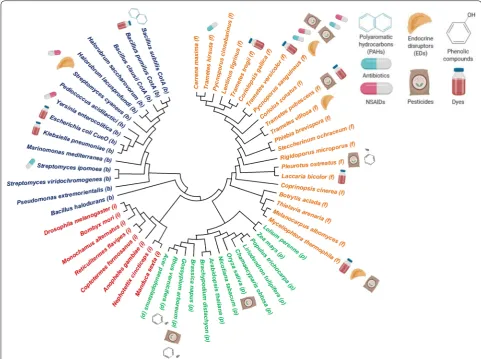
bacteria, lichen, and insects (Fig. 2), with laccases from
each species exhibiting particular catalytic
characteris-tics and sequences [20–22]. UniProtKB search results
for “laccase” with sequence sizes between 220 and 800 amino acids, revealed approximately 7300 cellular-organism sources, with 1026 bacteria, 6258 eukaryotes, and 16 halobacteria (archaea). Hence, it can be pre-dicted that this large number of enzymes produced by different organisms could have a wide range of
applica-tions in water bioremediation (Fig. 2). To date, many
of these enzymes have been applied in processes like electrocatalysis, delignification, and ethanol production
[23]. In this review, we aim to describe laccases from
different organisms, used in water bioremediation, their varying properties based on their origin, their biotech-nological prospectives for pollutant degradation (fab-ric discoloration, herbicide degradation, and emerging pollutants transformation), and the different strategies that have been explored to increase their activity and application.
Sources of laccases that are useful in water bioremediation
Fungal laccases
The first fungal laccase was reported by Bertrand [24],
who observed that this enzyme was responsible for the
color change in mushrooms of the Boletus genus when
in contact with air. A large number of fungi have been confirmed as laccase producers, with white rot fungi being the most recognized. Among fungal species, the
basidiomycetes, specifically Agaricus bisporus,
Pleu-rotus ostreatus, Trametes versicolor, Phanerochaete chrysosporium, and Coprinus cinereus, produce various
laccase isoforms (Table 1) [23, 25, 26].
Fungal laccases are involved in sporulation, pigment production, fruiting body formation, stress defense,
plant pathogenesis, and lignin degradation [27, 28].
Although most purified laccases are extracellular
enzymes, wood-rotting fungi also contain intracellu-lar laccases. It has been suggested that the localization of laccase is probably connected with its physiological function and determines the range of available
sub-strates [29]. Laccases exist in a variety of structures;
most of them are monomeric, but some are also present in homodimeric, heterodimeric, and multimeric forms. Their molecular mass ranges from 50 to 140 kDa, depending on the organism, although a typical fungal laccase will range from 60 to 70 kDa with an
isoelec-tric point around pH 4.0 [29, 30]. Fungal laccases are
usually glycosylated, with a 10–25% increase in mass, although some laccases present with a > 30% increase. The carbohydrate portion of laccases has been demon-strated to ensure their conformational stability and to protect the enzyme from proteolysis and inactivation
by radicals [31, 32].
Table
1
A
pplic
ation of some in
ter
esting fungal lac
cases tha t degr ade diff er en t c omp ounds and ma y b
e useful in w
at er tr ea temen t Lac case sour ce A
pplied enzyme form
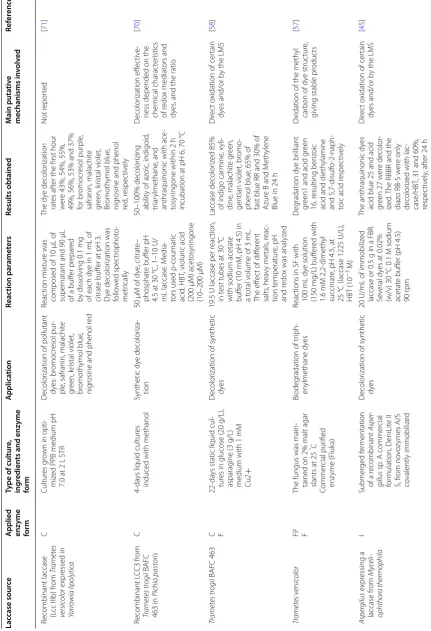
Type of cultur e, ing redien ts and enz yme form A pplica tion Reac tion par amet ers Results obtained M ain puta tiv e mechanisms in volv ed Ref er enc es Phar maceutical compounds Py
cnoporus sanguineus CCT‑
4518
C CI
The fungus was g
ro
wn in
PD
A solid medium f
or
7
da
ys at 28
°C. Laccase
ex
trac
t was pr
oduced in
50
mL of liquid media,
at 28 °C f or 72 h and suppor ted Laccase r emo val of 17 ‑alpha ‑eth ynilestradiol (EE2) Fr
ee and immobiliz
ed laccase ex trac t (100 U/L) w er e mix
ed with 10
mL
of EE2 at 10
mg/L, 10
mL
of acetat
e buff
er
, pH 4
or 5 or 10
mL of distilled
wat
er
, all of this at 28
°C
80% of r
emo
val of EE2
af
ter 24
h b
y the fr
ee
and immobiliz
ed laccase
ex
trac
t at pH 4 and 5.
The immobiliz ed f or m had thr ee c ycles of reusabilit
y with high
transf
or
mations
The laccase is able f
or
m
dimers of the EE2 b
y
polymer
ization of it
[ 49 ] Py cnoporus sanguineus C The Theobr oma gr andi -florum A
W was used as
Py
cnoporus sanguineus
laccase (Lac) inducer
, cultivat ed f or 7 da ys at 28 ± 2
°C and 150
rpm
D
eg
radation of estr
ogens
test
ed
100
U/L of laccases
, with 17 ‑α ‑ethin ylestradiol at 10
μg/mL, and 1% of
inducer b
y 24
h
Remo
val 96% of estr
ogens
af
ter 8
h of r
eac
tion
The
y suggest the
deg radation pr oduc t, with h ydr ox ylation of estr ogens [ 56 ] Tr ametes v ersic olor F Commer cial laccase po w der fr om T. v ersic olor (ac tivit y ≥ 0.5 U/mg) from Sig ma ‑Aldr ich D eg
radation of P
hA C: diclof enac , tr imethopr im, car bamaz epine , and sulfametho xaz ole Selec ted P hA C concentra ‑ tions w er
e added t
o
the enz
yme solution in
individual beak ers . T he beak ers w er e incubat ed
on a r
otar
y shak
er f
or
48
h at 80
rpm and 25
°C
The r
esults of this study
re
vealed that laccase
can eff ec tiv ely deg rade diclof enac (100%), tr imethopr im (95%), car bamaz epine (85%), and sulfametho xaz ole (56%) Not r epor ted [ 50 ] Tr ametes hirsuta C
It was g
ro
wn on PD
A medium f or 5 da ys at 28
°C and then on petr
i
plat
es
, pH 5 in static
condition f or 10 da ys , on K irk ’s medium. The super
natant was used
D
eg
radation of chloram
‑ phenicol ( CAP) Diff er ent mediat ors lik e syr ingaldeh yde , naph ‑ thol
, vanillin and ABT
S
w
er
e added at 0.25, 0.50,
1, 3, 5 and 10
mM, t
o
the r
eac
tion with 100
U
of laccase enz
yme and
10
mg/L of CAP in 0.1
M
acetat
e buff
er pH 5, b
y
48
h
The laccase enz
yme deg raded 0.5 mg/L CAP within 7 da ys without mediat
ors and was
efficiently deg
raded in
the pr
esence of laccase
mediat or syst em (syr in ‑ galdeh yde
, vanillin, ABT
S
and α
‑naphthol)
D
ehalogenation and o
xi
‑
dation of CAP b
y laccase
to f
or
m chloramphenicol
aldeh
yde which was
non
‑t
oxic t
o the micr
o‑ or ganisms studied [ 53 ] Tr ametes v ersic olor C SF (500
mL) with 20
g of
dr
ied apple pomace
,
Tw
een 80 (0.1%) and
moistur
e of 75% (w/w),
inoculat
ed with m
ycelia by 14 da ys , 30 °C with 200 mL. Enz yme fr om super natant ex trac t Chlor tetrac ycline ( C TC ) deg radation C
TC at 2
mg/L, laccase
dose at 0.5
IU
, pH 4.5 or
6.0, and ultrasonication
60% of C
TC, consider ed as a r ecalcitrant pollutant, was r emo
ved in 2
h
by ultrasonication and assist
ed laccase at pH
6.0.
While at pH 4.5, 80%
of C
TC was deg
raded
,
resulting non estr
ogenic
by pr
oduc
ts
Oxidation of C–
C and C–
O
bonds
[
51
Table 1 (c on tinued) Lac case sour ce A
pplied enzyme form
Type of cultur e, ing redien ts and enz yme form A pplica tion Reac tion par amet ers Results obtained M ain puta tiv e mechanisms in volv ed Ref er enc es Pleur otus ostr eatus FP PD
A medium at 25
°C, and added cipr oflo xacin ( CIP :
at 100, 200, 300, 400 and 500
ppm). The enz yme was secr et ed D eg
radation of cipr
oflo xa ‑ cin ( CIP) Fung i g ro wth b y 14 da ys
with 100, 200, 300, 400 and 500
ppm of CIP
Antibiotic deg
radation of
about 68.8, 94.25 and 91.34% was estimat
ed
af
ter 14
da
ys of incuba
‑
tion at 500
ppm CIP Not r epor ted [ 52 ] Py
cnoporus sanguineus CS43
f
F
STR of 10
L with 36.8%
tomat
o juice medium,
by 15
da
ys
, induced with
CuSO
4
and so
ybean oil at
48
h. LacI and LacII w
er e pur ified D eg
radation of endocr
ine
disrupting chemicals (EDCs): non
ylphenol and
tr
iclosan (a biocide)
EDC at 10
ppm final concentration w er e pr e‑ par
ed in pH 5 M
cI
lvaine
buff
er with 100
U/L
laccase
. Samples w
er e test ed e ver y 30 min f or 8
h at 25
°C
M
or
e than 95% r
emo
val
af
ter 8
h of tr
eatment
with 100
U/L at pH 5
Enz yme ‑dr iv en o xidation [ 41 ] Plastics
, personal car
e and her
bicide compounds
Py
cnoporus sanguineus (CS43)
CI
11
‑da
ys cultur
es in 10
‑L
STR in complex liquid medium at 28
°C. Crude ex trac t enz yme immo ‑ biliz ed D eg
radation of emer
ging endocr ine disrupt or (bisphenol A) 800 μL M cI lvaine buff er
(pH 3), 100
µL of ABT
S
(5
mM, 1.0% w/v) and
100
µL of laccase ex
trac t of P. sanguineus (CS43) 100% deg radation of
bisphenol A (20
mg/L)
was achie
ved in less
than 24
h
Pr
obably deg
radation
ends in the f
or mation of 4‑ isopr open ylphenol [ 42 ] Tr ametes v ersic olor BAFC 2234 MI 7‑ da ys cultur
es in 30
‑L
STR with complex liquid medium (50% t
omat o juice). P ur ified enz ymes In vitr o o
xidation of phenol
The r eac tion mix tur e in 1.5
‑mL contained dis
‑
solv
ed phenol (0.5
mM),
50
mM sodium citrat
e
pH 4.5 and 0.1
U/mL
laccase
84% phenol r
emo val in 4 h. Dar k color ed pr od ‑ uc ts par tly pr ecipitat ed w er e f ound Oxidativ
e coupling of phe
‑
no
xy radicals as major
path
wa
y of phenol
con version [ 43 ] R
ecombinant laccase from
Tr ametes san -guineus in Trichoderma atr oviride F Cultur es g ro
wn in 50
mL, incubat ed f or 4 da ys at 28 °C/150 rpm. P ur ified laccase D eg
radation of x
enobiotic
compounds (phenan
‑
thr
ene and benz
o[α] pyr ene) Phenanthr ene and benz o[α]p yr ene w er e added int o super na ‑
tants up t
o at 10
ppm,
incubat
ed at 28
°C and
shak
en at 150
rpm f
or
24
h
57.5
U/L of laccase in
super natant r emo ved phenanthr ene and benz o[α]p yr
ene (97 and
99% r
espec
tiv
ely) pr
e‑
sent in wast
ewat
er fr
om
a biofuel industr
y plant Not r epor ted [ 67 ] Nic
otiana tabacum expr
essing a laccase
from Pleur otus ostr eatus C Plants w er e g ro wn f or 16 da
ys in a g
ro
wing
chamber at 24
°C under
a phot
oper
iod of 16:8
h (light:dar kness). Enz yme secr et ed int o r hiz ospher e Ph yt or
emediation of phe
‑ nol cont ent fr om oliv e mill wast ewat ers Laccase ac tivit
y of trans
‑ genic r oot exudat es was evaluat ed b y o xidation of 2 mM ABT
S at 420
nm
in 0.1
M citrat
e buff
er pH
3.0 at 25
°C
Transgenic t
obacco plants
cultivat
ed in a h
ydr
o‑
ponic solution with oliv
e mill wast ewat ers w er e able t o r
educe the t
otal
phenol cont
ent up t
Table 1 (c on tinued) Lac case sour ce A
pplied enzyme form
Type of cultur e, ing redien ts and enz yme form A pplica tion Reac tion par amet ers Results obtained M ain puta tiv e mechanisms in volv ed Ref er enc es Anthr ac ophyllum disc olor MI
It was g
ro
wn in K
irk liquid
medium with
Tw
een 80
or soil supplement
ed
with
Tw
een 80 and
wheat grains . Whole cultur es D eg
radation of poly
cy clic ar omatic h ydr ocar bons (P AH) Cultur
es and 50
mg/L of
PAH at 30
°C b y 28 da ys . 10
g soil and 0.5
g wheat
grains in 30
mL tubes
contaminat
ed with a
50
mg/k
g of P
AHs at 30 °C b y 60 da ys
54 up t
o 75% r
emo val of phenanthr ene , anthra ‑ cene . fluoranthene , p yr ‑
ene and benz
o (a)p
yr
ene
in soil with
A. disc
olor
Pr
oduc
ts of deg
radations w er e anthraquinone , phthalic acid , 4 ‑h ydr ox y‑ 9‑ fluor enone , 9 ‑flu ‑ or
enone and 4,5
‑dih y‑ dr op yr ene [ 39 ] Tr ametes pubesc ens CBS 696.94 C
1L SF with synthetic liquid medium supplement
ed with dr y coff ee husk . 23 da
ys static incubation
at 30
°C. Crude ex
trac ts filt er ed Biodeg
radation of a mix
‑
tur
e of 2
‑chlor ophenol (CP), 2,4 ‑dichlor ophenol (DCP), 2,4,6 ‑tr ichlor ophe ‑ nol ( TCP), pentachlor o‑ phenol (PCP) D eg
radation of CP
s dur
ing
8
h at 40
°C, 200
rpm in
flasks containing 100
mL
of a CP mix
tur
e, with
15
mg/L of each CP
in 50
mM phosphat
e
buff
er
, pH 6.0. Enz
ymatic
ex
trac
t (5
mL) and 10
U/L
Biodeg
radation of 100%,
99%, 82.1% and 41.1% of CP
, DCP
, T
CP and PCP
, respec tiv ely , af ter 4 h. The r educ
tion in chlo
‑ rophenols , allo w ed 90% reduc tion t oxicit y Not r epor ted [ 44 ] Neosar tor ya fischeri C 50
mL SF with modified Czapek medium and 20 mg of asphalt
enes as
car
bon sour
ce
, at 37
°C 100 rpm, 4 w eeks . Whole cultur es M
etabolization and miner
‑
alization of asphalt
enes (recalcitrant petr oleum frac tion) A sphalt ene mineralization
was quantified b
y meas ‑ ur ing CO 2 pr oduc tion. Cell ‑fr ee ex tracellular
medium was solv
ent
ex
trac
ted and analyz
ed by GC–MS A ft er 11 w
eeks of g
ro
wth,
the fungus metaboliz
e
15.5% of the asphalt
enic
car
bon, including 13.2%
transf or med t o CO 2 G
eneration of o
xidiz
ed
metabolit
es such as
hy dr ox yp yr enedione and hy dr ox yphen ylacetic acid [ 40 ] Coriolopsis rigida LPSC 232 C 15 ‑da
ys liquid cultur
es in
modified Czapek D
ox
medium (0.5% pept
one and 0.15 mM Cu 2 +) D et
oxification of wat
er soluble frac tion fr om ‘‘alpeorujo ” ( W SF A) Reac tion mix tur es contain ‑ ing W SF
A 20% (v/v)
and 20
U laccase w
er
e
incubat
ed 24
h at 28
°C
and 150
rpm
Reduc
tion of fr
ee phenols
from
the W
SF
A
Oxidation of fr
ee phenols
,
resulting in radical formation, leading t
o
polymer
ization as w
ell as det oxification [ 208 ] Tr ametes villosa C F Ex trac
ted and pur
ified
enz
yme (No
vo
zymes)
Bisphenol A (BP
A) deg rada ‑ tion 2.2 mM BP A incubat ed for 1
h with 1.0
unit/mL of laccase . T he r eac tion mix tur e: 0.5 mM ABT S, 0.1
M sodium acetat
e,
pH 5.0, and an enz
yme
in a t
otal v
olume of
1.0
ml was incubat
ed
at 37
°C
BP
A was deg
raded b
y
a laccase
, which was
ex
trac
ted and pur
ified
from D
eniLit
e, a No
vo ‑ zymes ’ pr oduc t. T rans ‑ for
ming and impor
tant endocr ine ‑distur bing compound BP
A was metaboliz
ed t
o
tw
o compounds: one
with high molecular weight due t
o o
xidativ
e
condensation, and another identified as 4‑isopr
Table 1 (c on tinued) Lac case sour ce A
pplied enzyme form
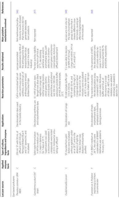
Type of cultur e, ing redien ts and enz yme form A pplica tion Reac tion par amet ers Results obtained M ain puta tiv e mechanisms in volv ed Ref er enc es D ye ‑based pollutants Pleur otus ostr eatus URM 4809 C 250
‑mL SF with 50
mL of
effluent with 0.05
g/L
Remaz
ol Br
illiant Blue R
and
10
7 spor
es/mL and incubat ed D ecolor ization dy es used
in the t
ex
tile industr
y
Cell in a micr
obial fuel cell
with continuous laccase synthesis; and 0.05
g/L of
anthraquinone r
emaz
ol
br
illiant blue R dy
e Laccase pr omot ed decolor ization b
y 86% of
the anthraquinone dy
e
remaz
ol br
illiant blue
R (used in the t
ex
tile
industr
y)
Not mentioned
, but ph
y‑ tot oxicit y r esults sho w ed
that the pr
ocess did not
generat e det ec table to xic pr oduc ts [ 46 ] G anoderma lucidum E47 strain C Solid ‑stat e f er mentation in MY
SA medium, pH
5.5, k
ept in dar
kness
for 7
da
ys at 25
°C.
Super
natants was used
as enz yme pr eparation. 5 compounds w er e t est ed in 0.5 L minir eac tor simu ‑
lating an effluent
D ecolor izing xanthene , az o and tr iar ylmethane dy es 0.1
mM of or
ganic dy
es:
Bengal r
ose; blue black
naphthol; congo r
ed;
meth
yl orange; br
omo ‑ cr esol g reen; br omo ‑ cr esol pur ple; br omo ‑
phenol blue; and phenol red
, 550 nm; 100 mM potassium acetat e buff er
pH 4.8, 5% but
yl acetat
e,
25
°C
The best ac
tivit
y‑
stabilit
y
reached in pH 4.8 at 37 °C, decolor
izing
xanthene
, az
o and tr
i‑ ar ylmethane dy es , with selec tivit
y on br
omo
‑
cr
esol g
reen and br
omo ‑ cr esol pur ple . A ctivit y on effluent biotr eatment Not r epor ted [ 47 ] O udemansiella c anarii C SF , m ycelial fr om petr i dishes w er e incubat ed without ag itation under
air at 28
°C and in the
absence of light b
y 14 da ys . Ex trac t was dialyz
ed and par
tially
pur
ified
D
ecolor
ization of congo
re d 50 mM acetat e buff er (pH
5.5) in 250
‑mL
140 SF with 50
mL and
containing 50
mg/L of
Congo r
ed and nativ
e
141 laccase (5
U). The mix tur es w er e incubat ed at 30
°C in the dar
k in a
rotar
y shak
er at 100
rpm
5 U w
er
e able t
o decolor
‑
iz
e 80% of 50
mg/L
Congo r
ed within 24
h at
30
°C and pH 5.5
Laccase ac
ts not only on
the dy e chr omophor e gr oup
, but also that it
clea ves diff er ent co va ‑ lent bonds
, causing an
eff
ec
tiv
e frag
mentation
of the molecule
[ 48 ] P. pastoris or A. thaliana expr essing L cc9 fr om Lac caria bic olor F
SF in BMGY medium at 28
°C, the cells w
er
e
suspended in of BMMY
.
M
ethanol was added t
o 1% e ver y 24 h D ecolor
ization of tr
iph ‑ en ylmethane dy es , emplo
yed in industr
ial dy eing pr ocesses The r eac tion mix tur e f or the decolor ization assa y contained 0.1 mM of cr
ystal violet, M
cI
lvaine
buff
er and 50
μL of
the enz
yme in a t
otal
of 200
μL. ABT
S, as the
mediat
or
, was added if
necessar y. I ncubat ed in dar k f or 24 h
In the pr
esence of ABT
S,
the decolor
ization rat
es
of Cr
ystal violet b
y lac ‑ cases in P. pastoris or A. thaliana reached 90.7%
and 83.6%, r
Table 1 (c on tinued) Lac case sour ce A
pplied enzyme form
Type of cultur e, ing redien ts and enz yme form A pplica tion Reac tion par amet ers Results obtained M ain puta tiv e mechanisms in volv ed Ref er enc es R
ecombinant laccase (Lcc III
b) fr om Tr ametes versic olor expr essed in Yarr owia lipolytic a C Cultur es g ro
wn in opti
‑
miz
ed PPB medium pH
7.0 at 2
L STR
D
ecolor
ization of pollutant
dy es: br omocr esol pur ‑ ple
, safranin, malachit
e gr een, k ristal violet, br omoth ymol blue , nig
rosine and phenol r
ed
Reac
tion mix
tur
e was
composed of 10
µL of
super
natant and 90
µL
of a buff
er pr
epar
ed
by dissolving 0.1
mg
of each dy
e in 1
mL of
citrat
e buff
er at pH 3.
D ye decolor ization was follo w ed spec tr ophot o‑ metr ically The dy e decolor ization rat es af
ter the first hour
w
er
e 43%, 54%, 55%,
49%, 56%, 53% and 37% for br
omocr esol pur ple , safranin, malachit e gr een, k ristal violet, Br omoth ymol blue , nig
rosine and phenol
red , r espec tiv ely Not r epor ted [ 71 ] R ecombinant L CC3 fr om Tr ametes tr ogii BAFC 463 in Pichia pastoris C 4‑ da
ys liquid cultur
es
induced with methanol
Synthetic dy e decolor iza ‑ tion 50
µM of dy
e, citrat
e–
phosphat
e buff
er pH
4.5 at 30
°C, 1–10 U/ mL laccase . M edia ‑
tors used ρ
‑coumar ic acid , HBT , violur ic acid (200 µM) acet osyr ingone (10–200 µM) 50–100% decolor izing abilit
y of az
oic , indigoid , tr iar ylmethane , and
anthraquinonic with ace
‑
tosyr
ingone within 2
h
incubation at pH 6, 70
°C D ecolor ization eff ec tiv e‑
ness depended on the chemical charac
ter istics of r edo x mediat ors and dy es
, and the ratio
[ 70 ] Tr ametes tr ogii BAFC 463 C F 22 ‑da
ys static liquid cul
‑
tur
es in glucose (20
g/L),
asparag
ine (3
g/L)
medium with 1
mM Cu 2 + D ecolor
ization of synthetic
dy
es
19.5 U laccase per r
eac
tion,
in t
est tubes at 30
°C
with sodium acetat
e
buff
er (10
mM, pH 4.5) in
a t
otal v
olume of 3
mL.
The eff
ec
t of diff
er ent salts , hea vy metals , r eac ‑ tion t emperatur e, pH and r edo
x was analyz
ed
Laccase decolor
iz
ed 85%
of indigo car
mine , x yli ‑ dine , malachit e g reen,
gentian violet, br
omo
‑
phenol blue
, 65% of
fast blue RR and 30% of Azur
e B and M
eth
ylene
Blue in 24
h
Dir
ec
t o
xidation of cer
tain
dy
es and/or b
y the LMS
[ 58 ] Tr ametes v ersic olor FP F
The fungus was main
‑
tained on 2% malt agar slants at 25 °C Commer cial pur ified enz yme (F luk a) Biodeg
radation of tr
iph ‑ en ylmethane dy es Reac
tions in SF with
100 mL dy e solution (150 mg/L) buff er ed with 1.6 mM 2,2 ‑dimeth yl succinat
e, pH 4.5, at
25
°C (laccase 1225
U/L),
HBT
(10
−
3 M)
D eg radation dy e br illiant gr
een1 and acid g
reen
16. r
esulting benz
oic
acid and dieth
ylamine and 5,7 ‑disulf o‑ 2‑ naph ‑
toic acid r
espec
tiv
ely
Oxidation of the meth
yl
car
bon of dy
e struc
tur
e,
giving stable pr
oduc ts [ 57 ] Asper gilus expr essing a laccase fr om M yc eli -ophthor a thermophila I Submer ged f er mentation
of a r
ecombinant
Asper
-gillus
sp
. A commer
cial for mulation, D eniLit e II S, fr om no vo zymes A/S co valently immobiliz ed D ecolor
ization of synthetic
dy
es
20
U/mL of immobiliz
ed
laccase or 0.5
g in a FBR.
Se
veral dy
es at 0.02%
(w/v) 30
°C 0.1
M sodium
acetat
e buff
er (pH 4.5)
90
rpm
The anthraquinonic dy
es
acid blue 25 and acid green 27 w
er e decolor ‑ iz ed . T
he RBBR and the
diaz o RB ‑5 w er e only decolor iz
ed with lac
‑
case/HBT
, 31 and 60%,
respec tiv ely , af ter 24 h Dir ec t o
xidation of cer
tain
dy
es and/or b
y the LMS
[
45
Table 1 (c on tinued) Lac case sour ce A
pplied enzyme form
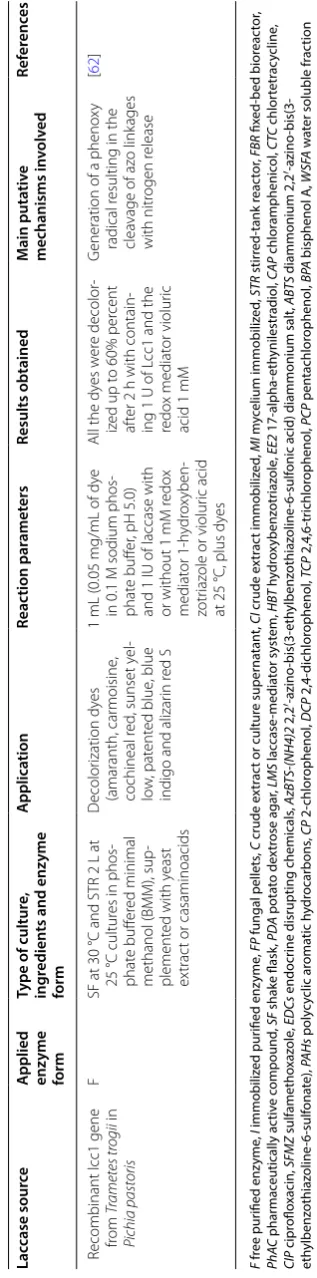
Type of cultur e, ing redien ts and enz yme form A pplica tion Reac tion par amet ers Results obtained M ain puta tiv e mechanisms in volv ed Ref er enc es R
ecombinant lcc1 gene from
Tr ametes tr ogii in Pichia pastoris F
SF at 30
°C and STR 2
L at
25
°C cultur
es in phos
‑
phat
e buff
er
ed minimal
methanol (BMM), sup
‑
plement
ed with y
east
ex
trac
t or casaminoacids
D ecolor ization dy es (amaranth, car moisine , cochineal r ed
, sunset y
el ‑ lo w , pat ent ed blue , blue
indigo and alizar
in r
ed S
1
mL (0.05
mg/mL of dy
e
in 0.1
M sodium phos
‑
phat
e buff
er
, pH 5.0)
and 1
IU of laccase with
or without 1
mM r edo x mediat or 1 ‑h ydr ox yben ‑ zotr iaz
ole or violur
ic acid
at 25
°C, plus dy
es
All the dy
es w
er
e decolor
‑
iz
ed up t
o 60% per
cent
af
ter 2
h with contain
‑
ing 1
U of L
cc1 and the
redo x mediat or violur ic acid 1 mM G
eneration of a pheno
xy
radical r
esulting in the
clea
vage of az
o link ages with nitr ogen r elease [ 62 ] F fr ee pur ified enz yme , I immobiliz ed pur ified enz yme , FP fungal pellets , C crude e xtr ac
t or cultur
e super na tan t, CI crude e xtr ac t immobiliz ed , MI m yc elium immobiliz ed , STR stir red-tank r eac tor , FBR fix ed-bed bior eac tor , Ph AC phar mac eutically ac tiv e c ompound , SF shake flask , PDA pota to de xtr ose agar , LMS lac case -media tor sy st em, H BT h ydr ox ybenz otr iaz ole , EE2 17-alpha-eth ynilestr adiol , CAP chlor amphenic ol , CT C chlor tetr ac ycline , CIP cipr oflo xacin, SF MZ sulfametho xaz ole , EDCs endocr
ine disrupting chemicals
, AzB TS -(NH4)2 2,2 ′ -azino -bis(3-eth ylbenz othiaz oline -6-sulf
onic acid) diammonium salt
, ABT S diammonium 2,2 ′ -azino -bis(3-eth ylbenz othiaz oline -6-sulf ona te), PAHs poly cy clic ar oma tic h ydr ocar bons , CP 2-chlor ophenol , DCP 2,4-dichlor ophenol , TC P 2,4,6-tr ichlor ophenol , PCP pen tachlor ophenol , BPA bisphenol A, W SFA w at
er soluble fr
The redox potential (E°) of laccases has a direct rela-tionship with the energy required to remove an electron from the reducing substrate, constituting one of the
fun-damental characteristics of these enzymes [33].
There-fore, laccases with a high E°, like fungal laccases, are of
special interest in biotechnology cause they are capable
of oxidizing substrates with high E° (E° > 400 mV) [33–
36]. For example, the E° of bisphenol A (BPA), p
-nonyl-phenol and azo dye BR114 are above 600 mV [37, 38].
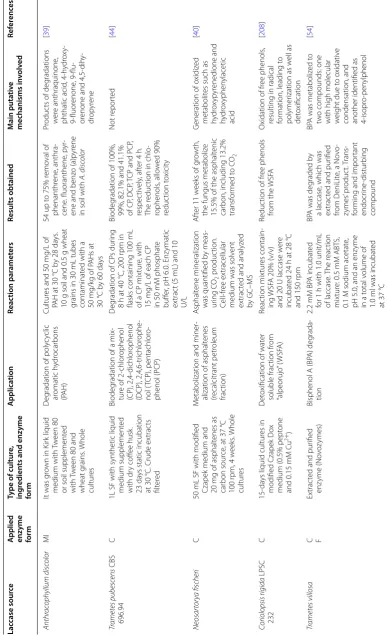
Fungal laccases aid bioremediation through the
oxida-tion of polycyclic aromatic hydrocarbons (PAHs) [39,
40], plastics and phenolic compounds [41–44], dyes
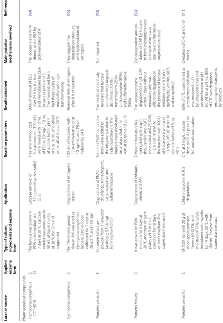
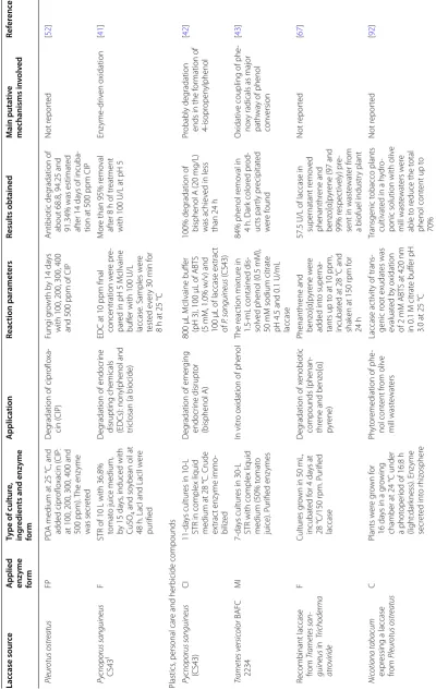
[44–48], and the degradation of pharmaceutically active
compounds [49–52], among others (Table 1). Given
that laccases from white-rot fungi have the potential for phenolic compound degradation, different studies have involved the immobilization of microorganisms, such as T. versicolor, into silica-alginate and loofa sponges as
supports for phenol removal [43]. While crude extract
from Trametes pubescens has been used for the
degrada-tion of chlorophenols (Table 1) [44]. Also, crude extract
from the white-rot fungus Trametes hirsuta, proved
capable of degrading chloramphenicol (one of the most persistent micro-pollutants in pharmaceutical wastes),
with or without mediators (Table 1) [44, 53]. Fukuda et al.
[54] used a free purified laccase from Trametes villosa to
degrade BPA, another hazardous pollutant discharged into rivers and seas, without the requirement of
media-tors. Meanwhile, Barrios-Estrada et al. [42] reported that
the degradation of BPA (20 mg/L) occurred within the
first 24 h when using Pycnoporus sanguineus (CS43) and
T. versicolor laccases immobilized onto ceramic
mem-branes (Table 1). Different steroidal estrogens can be
removed or degraded from aqueous systems by the free
laccases from P. sanguineus or laccases from T. versicolor
or Myceliophthora thermophila that have been
immobi-lized onto ceramic membranes (Table 1) [55, 56]. Other
problematic compounds in effluents of textile and paper industries include synthetic dyes, of which, many are toxic for mammals. Therefore, efforts have been made towards their elimination from industrial wastewaters
using laccases from T. versicolor and Trametes trogii
(Table 1) [49, 58].
The laccase yield from native fungal sources fails to meet the industrial need, as natural hosts often produce several laccase isozymes making it challenging to isolate the laccase of interest, more so when the enzyme is silent or not abundantly expressed. Therefore, heterologous laccase expression has become a promising alternative
[23, 59–71]. Heterologous expression of many fungal
lac-cases has been reported in bacteria such as E. coli [60],
yeasts like Pichia pastoris and Yarrowia lipolytica [43,
61–64], filamentous fungi such as Aspergillus oryzae, A.
niger, and Trichoderma atroviride [65–67], and plants
like Arabidopsis thaliana and Zea mays (Table 1) [68,
69]. Yeasts and filamentous fungi are usually more
attrac-tive hosts for heterologous protein production owing to their faster microbial growth, ease of gene manipulation, their ability to secrete large amounts of proteins into the growth medium, as well as the ability to perform
post-translational modifications [30, 49].
Recombinant fungal laccases have also been widely applied for bioremediation purposes. For instance, the
recombinant proteins Lcc1 and Lcc3 from T. trogii,
pro-duced in P. pastoris proved to be a useful biocatalyst for
the oxidative degradation of several polluting dyes, such as indigo carmine, the most important dye used for
man-ufacturing blue jeans [62, 70]. Moreover, Darvishi et al.
[71] expressed and produced a recombinant laccase (Lcc
IIIb) from T. versicolor in Y. lipolytica, proving its
capa-bility of decolorizing five phenolic azo dyes with > 40%
efficiency after 4 h (Table 1). Similarly, Wang et al. [68]
expressed a laccase from the ectomycorrhizal fungus Laccaria bicolor in P. pastoris and A. thaliana, which proved capable of decolorizing > 80% of the crystal violet dye, tested using laboratory-scale studies, providing an alternative to the decolorization of industrial wastes. In
another study, Balcázar-López et al. [67] expressed a
lac-case from P. sanguineus in the filamentous fungus T.
atro-viride; the heterologously expressed laccase maintained similar properties to those of the native enzyme, although the recombinant showed the potential to remove > 90% of the phenanthrene and benzo[α]pyrene present in waste-water from a biofuel industry plant using
laboratory-scale studies [67].
Plant and insect laccases
The first identified and reported laccase from plants was
from the Japanese lacquer tree Toxicodendron
vernicif-luum (Rhus vernicifera) [72]. However, studies on plant laccases are rare. Plant laccases share their molecular architecture and reaction mechanisms with fungal
lac-cases. In general, they have a lower E° like bacterial
lac-cases (0.41 V for R. vernicifera and a pI between 7.0 and
9.6) [22, 31, 73]. These proteins show a higher
glycosyla-tion pattern (22–45%) [74, 75], consist of 500–600 amino
acids, and weigh approximately 60–130 kDa [31]. Plant
laccases have been described and associated with
biosyn-thesis and polymerization of lignin [76, 77], elongation
[78–80], and the stress response [81–83].
Although plant laccases have not been largely involved in bioremediation, some applied cases have been
reported. Wang et al. [84] presented a system of
phy-toremediation ex planta based on the overproduction
in A. thaliana of a secretory laccase (LAC1), which was
LAC1 expression in A. thaliana conferred resistance to several toxic phenolic compounds, probably attributable to LAC1-induced transformation. Recombinant LAC1 plants were resistance to phenolic compounds under greenhouse conditions, helping to detoxify their growth
environment [84]. Watharkar et al. [85] showed that
lac-cases and other enzymes from Asparagus densiflorus
could be applied in the treatment of industrial textile effluents. For lab scale studies, they used a vertical sub-surface flow phytoreactor based on vertical percolation of wastewater through layers of soil, root zone and a netted
bottom. For large scale studies, they planted beds of A.
densiflorus on a high rate transpiration system (HRTS), which has been used successfully for some industries. In
both cases, A. densiflorus showed the ability to degrade
dyes and reduced levels of toxic heavy metals.
Laccases from other plants have been proposed and successfully tested for dye degradation using suspension
cells and purified laccases [86–90]. Huang et al. [91]
iden-tified laccases in rice (Oryza sativa), possibly involved
in atrazine and isoproturon (herbicides) catabolism or
detoxification. The two Oryza sativa laccases expressed
heterologously in P. pastoris, led to the increased
resist-ance of cells to atrazine and isoproturon, suggesting that some of the laccases could be involved in detoxification
or degradation of these herbicides [91].
Plants have been successfully used as recombinant expression systems of fungal and plants laccases.
Chi-aiese et al. [92] expressed a laccase from P. ostreatus in
Nicotiana tabacum, capable of reducing 70% of the total
phenol content from olive mill wastewaters (Table 1).
Other authors have expressed fungal laccases with
indus-trial applications in rice-based [93] and tobacco plants
[94, 95], as well as maize seeds [69]. Conversely, insect
laccases have been reported to play an important role in
cuticle sclerotization and pigmentation, as well as other processes such as wound healing and immune system
development and maintenance [96, 97]. To the best of
our knowledge, no insect laccase has been reported for bioremediation processes.
Bacterial laccases
Laccase activity in bacteria was detected for the first time in Azospirillum lipoferum, isolated from a rice
rhizos-phere in 1993 [98]. Several laccases were then gradually
discovered in bacteria from different genera, such as Bacillus, Streptomyces, Klebsiella, Pseudomonas, Yers-inia, Proteobacterium, and Marinomonas, among others
(Table 3) [99, 100]. Moreover, these enzymes have also
been found in microorganisms of the Archaea domain
such as Haloferax volcanii [101].
Under native conditions, bacterial laccases are involved in pigmentation processes, morphogenesis, toxin oxi-dation, and protection against oxidizing agents and UV
light [100, 102]. The molecular weight of these enzymes
is in the range of 50–70 kDa, with a majority being mon-omeric intracellular proteins, except those from
bacte-ria in the Streptomyces genera and some other examples
[103–106], such as the laccase produced by Bacillus
teq-uilensis SN4, an extracellular enzyme [104].
One of the most well-known bacterial laccases is the
outer endospore coat protein CotA from Bacillus subtilis,
which has three cupredoxin domains (Fig. 3) [107]. Other
similar bacterial MCOs include the copper homeostasis
protein CueO from E. coli [108]. Bacterial laccases with
three-dimensional structures of two-domain laccases
have been found in Streptomyces, Amycolatopsis, and
Nitrosomonas, belonging to the group denoted as SLACs (small laccases). The implication of the absence of this
Fig. 3 Cartoon structures of the three‑domain laccase from Bacillus subtilis (PDB 1GSK) and the homotrimeric two‑domain laccase from
domain is the need to form a homotrimer to be
catalyti-cally active (Fig. 3) [109–111].
The most significant biochemical properties of bacte-rial laccases are their stability under various conditions of pH, temperature, organic solvents, and salt
concentra-tions [105]. Usually, bacterial laccases are highly stable at
elevated temperatures, as seen in the B. subtilis laccase
at 70 °C, with a thermal half-life (t1/2) of 250 min, or the
t1/2 of 30 min at 80 °C of the Streptomyces
viridochro-meogenes laccase, compared with the 10 min t1/2 of
Cer-rena unicolor fungal laccase at the same temperature
[112–115]. With respect to media pH, bacterial laccases
usually work better in neutral to alkaline pH, similar to plant laccases, but unlike fungal laccases, which have optimum activities in acidic pH. Nevertheless, its optimal pH is dependent on the substrate. For instance, for
phe-nols, such as 2,3-dimethoxyphenol, the optimal pH for B.
subtilis, B. clausii, and Streptomyces coelicolor are pH 7, 8 and 9, respectively, while for ABTS, all three enzymes
require a pH of 4 [112, 116]. However, bacterial laccases
have shown greater tolerability to high concentrations of sodium chloride, being active in 1 M or higher
concentra-tions, as seen with the laccases of Marinomonas
mediter-ranea and Bacillus halodurans, among others [115, 117]. Some bacterial laccases have exhibited high tolerance to different solvents, including ethanol, methanol, dimethyl-formamide, acetonitrile, acetone, and dimethylsulfoxide,
as observed in the Bacillus pumilus W3 laccase, which
generally retains > 50% of its activity in solvent–water
mixtures [118].
Although bacterial laccases are generally more robust and stable enzymes in comparison to fungal laccases,
their application has been restricted by their low E° (E°
T1 < +460 mV) [22, 112]. Nevertheless, bacterial laccases
represent a good option for the treatment of contami-nated wastes such as textile effluents, which usually have high salt concentrations (40–100 g/L) and alkaline pH
[119].
Heterologous overexpression of bacterial laccases has
been reported in E. coli [99, 112, 115, 118, 120–125], P.
pastoris [126–128], and Streptomyces coelicolor [116].
Although E. coli is the most used expression system for
bacterial laccases, the production of MCOs in its cyto-plasm has a major drawback as its copper homeostasis systems maintain a cellular copper concentration around
10 µM under aerobic conditions [129–131], which is
insufficient to achieve fully loaded copper laccases [132].
Copper-depleted laccases are incapable of reaching their
maximum catalytic activity [122, 132]. This limitation
can be overcome by changing the oxygen concentration
when cultivating recombinant E. coli expressing laccases,
because under anaerobic (or microaerobic) conditions, the intracellular copper accumulation is 80-fold higher,
compared with that attained under aerobic conditions
[103, 122, 132, 133].
Bacterial laccases have been used in bioremediation, mainly for the degradation of synthetic dyes. Liu et al.
[99] reported a thermostable and pH-stable Klebsiella
pneumoniae laccase which degrades diverse dyes used in industrial processes (such as reactive brilliant blue X-BR, reactive dark blue M-2GE, congo red, bromophenol blue, and malachite green, among others) in short reaction
times (90 min) under diverse pH values at 70 °C (Table 3).
Another case is the B. pumilus CotA-laccase mutant
WLF, obtained by Luo et al. [125], which has an improved
expression in E. coli and has been tested for the
degra-dation of diverse dyes, obtaining higher decoloration yields with anthraquinonic and triphenylmethane dyes,
compared with aromatic heterocyclic dyes (Table 3).
Meanwhile, high decolorization of toluidine, malachite green, and reactive black 5 by the azide-resistant spore
laccase from halotolerant Bacillus safensis, has also been
reported [134]. Recombinant laccases from E. coli [116]
or Thermus thermophiles [126] expressed in P. pastoris, efficiently decolorized congo red and remazol brilliant
blue R (Table 3). Recently, the recombinant Streptomyces
ipomoea SilA laccase expressed in E. coli, in the presence of mediators such as acetosyringone and methyl syrin-gate, enhanced the decolorization and detoxification of a variety of textile dyes, like reactive black 5, orange II, and indigo carmine, also diminishing the toxicity of acid
orange 63, tartrazine and its products [135].
Outside these pollutants, other contaminant com-pounds have been degraded with laccases. Singh et al.
[136] used recombinant Yersinia enterocolitica laccase to
biotransform two nonsteroidal anti-inflammatory drugs (diclofenac (DF) and aspirin), obtaining complete
trans-formation of these molecules in 24 h (Table 3).
Further-more, DF and mefenamic acid were also transformed by
laccases produced by Streptomyces cyaneus [137], and
Streptomyces mutabilis laccases transformed
antibiot-ics like sulfadiazine and sulfathiazole [138]. Similarly,
the recombinant S. ipomoea SilA laccase expressed in E.
coli, has shown a high percent conversion of
ciprofloxa-cin and norfloxaciprofloxa-cin [139]. Interestingly, PAHs such as
anthracene, pyrene benzo[α]pyrene, phenanthrene, and fluoranthene, have been oxidized by the recombinant
laccase CotA from B. subtilis produced in E. coli [123].
Moreover, laccases from S. cyaneus have demonstrated
full BPA degradation after 2 days [137].
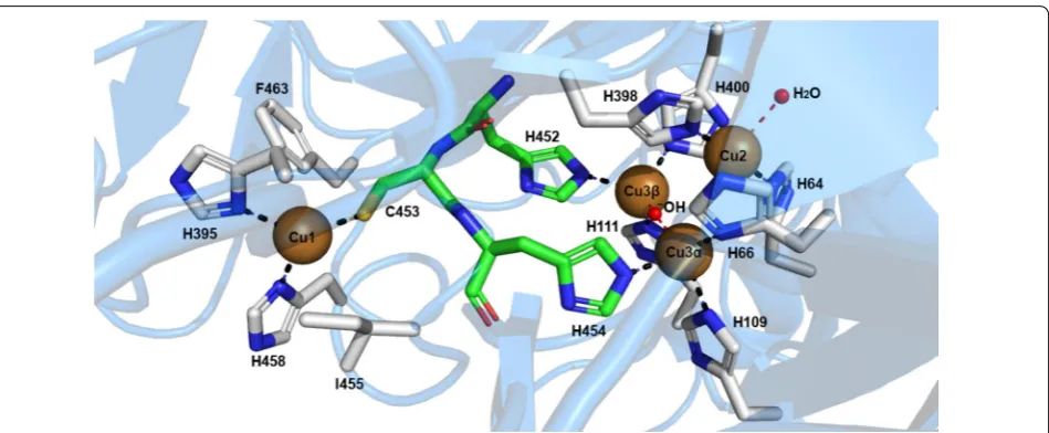
Structure of laccases and comparative structure analyses
Laccases as MCOs, have four copper atoms in remarkably special oxidation states: one type-1, one type-2, and two
are members of the cupredoxin superfamily, particularly the family of multi-domain cupredoxins. This family is characterized by the cupredoxin fold, which consists of two β-sheets arranged into a Greek-key barrel. The Greek-key motif has at least seven antiparallel β-strands twisted to form a closed barrel structure, in which some
β-strands are adjacent in space but not in sequence [109].
The classification of the copper atoms is based on the environment of the metal ion and its spectroscopic characteristics; T1: paramagnetic ‘blue’ copper, with an absorbance at 610 nm, T2: paramagnetic ‘non-blue’ cop-per, and T3: a diamagnetic spin-coupled copper–copper
pair, with an absorbance at 330 nm [140]. T1 copper has
the highest E° and is the substrate oxidation site. This ion
has a trigonal orientation, with two conserved histidines and one cysteine as equatorial ligands, and an axial ligand of variable nature, usually methionine in bacteria and leucine or phenylalanine in fungal laccases. Type-2 and the two type-3 coppers form a cluster, where molecular oxygen is reduced, and water is released. Types-2 and 3 copper atoms are coordinated by histidine side chains
(T2 by two of them and T3 by six) (Fig. 4). A hydroxyl
bridge maintains the antiferromagnetic coupling between
T3 copper atoms [141].
Common laccases contain three homologous cupre-doxin domains. Their mononuclear copper site exists in domain 3 and their trinuclear cluster is formed at the
interface between domain 1 and 3 [109]. In laccases with
this topology, the function of domain 2 is to join and position domains 1 and 3, enabling the formation of the
trinuclear cluster [106]. In contrast, in two-domain
lac-cases, which are from bacteria and are so called small
laccases, their mononuclear copper site exists in domain 1 or 2, but for the formation of their trinuclear cluster they need to oligomerize as homotrimers, generating this catalytic site at the interface between the domain 1 of one
monomer and the domain 2 of the other monomer [106,
142].
In both cases, the distance and relative position between the copper sites are conserved (the distance between T1 copper and the cluster); about 12 Å in all
lac-cases [109].
There are several hypotheses on the evolution of lac-cases; all of them consider that the cupredoxin domain, with one copper atom in its structure, developed in dif-ferent forms of MCOs, including dicyanin, ascorbate oxidase, nitrite reductase, ceruloplasmin, SLACs, and
three-domain laccases [140, 143]. These hypotheses
pos-tulate different pathways and intermediate species that led to the development of the trinuclear cluster and the origin of the different MCOs. These structures maintain the original cupredoxin domain but are associated in dimers of independent chains or form longer chains by
gene fusion [111]. Some of these domains maintain the
copper-binding site, and different forms of interdomain association were evolutionarily explored by independent divergence to develop at least two cluster types of three
copper atoms [142]. Other interesting schemes of this
hypothesis have also been reported [144, 145].
Three-domain laccases are mainly studied in fungi, but have also been observed in some bacteria, archaea,
plants, and insects [22, 146]. The database
contain-ing information on different laccases and MCOs is
Bio-CatNet [147]. Laccases are considered “moonlighting”
proteins, owing to their multiple biological activities
[108]. PDB structures of > 70 fungal and a few bacterial
laccases have been reported, crystallized in their wild-type, mutant, and derivative forms, as well as complexed to a variety of substrate-like ligands and oxygen
reac-tive species [109, 148]. This set of structures has shed
light on their stabilities and functional characteristics, as described above.
Nevertheless, no structures from other species have been reported, except a plant (zucchini)
ascorbate-oxi-dase closely homologous to laccases [149, 150]. The
gen-eral three-domain structure of laccases is maintained in different species, with the loops protruding the cupre-doxin domains being the most conspicuous difference
[111, 151], as well as the form and by consequence, the
selectivity at the substrate binding site [152]. More
sub-tle differences are situated in the axial position of the
T1 copper atom, causing the span of E°s from 400 mV in
plant and bacterial laccases to approximately 800 mV in
the majority of fungal types [22, 139, 153]. In Fig. 5, we
compare the structures of representative fungal, bacte-rial, plant, and insect laccases. This latter structure was homology modeled from its amino acid sequence. The
conserved orientation of the coppers and the cavities for substrates and products are also presented.
Several review papers have been published discuss-ing the structure of laccase and its implications on function. For instance, the description of the molecu-lar mechanism of substrate oxidation in the T1 site, the intramolecular electron transfer to the trinu-clear cluster located about 12 Å away, and the oxygen reduction to water, can be understood in the scheme
of Hakulinen and Rouvinen [109], and the detailed
descriptions of Mot and Silaghi-Dumitrescu [140],
Pardo and Camareno [154], and Sitarz et al. [155].
These references show the complexity and subtlety of the reaction pathway through enzyme structure. This
mechanism involves a substrate binding pocket [154,
156], which confers selectivity by proper docking, and
also affects the E° by induced fitting to the active site.
There, the T1 copper atom extracts an electron from the substrate, followed by a relay of protein functional groups, namely thiol, carbonyl, and imidazole groups, which transfer that electron through the trinuclear cluster, where they are gathered until four electrons are collected. T3 copper atoms transfer such electrons
Fig. 5 Laccase structure conservation and function. a Structure of Trametes versicolor (PDB ID 1GYC), and Bacillus subtilis (PDB ID 1GSK) laccases compared to Cucurbita pepo (zucchini) ascorbate oxidase (PDB ID 1AOZ) from left to right. Domain 1 (D1) is at the front and right of the structure, domain 2 (D2) is behind and in the upper portion, domain 3 (D3) is at the left. Brown spheres symbolize the position of copper atoms, T1 above the trinuclear cluster. b The molecular surface shows protruding chemical groups, in red, and concave or cavity regions, in green. Some of these latter regions correspond to the ligand‑binding site (LB) along with the dioxygen molecule entrance (O2) and the water exit (H2O) channels. Central and
to the T2 copper, and an oxygen channel allows an oxygen molecule to reach this buried metal ion and be
reduced [140].
The reduced oxygen atoms are then converted to water following assistance from two carboxylate groups from aspartic and glutamic acids, which transfer the required hydrogen atoms. At least two structural water molecules also contribute to the electron transfer pro-cess. The generated water molecules finally go out a second channel formed by polar residues of the protein. Copper atoms undergo a series of at least five stages
during this process [109, 154, 157]. This depiction of
the process exposes the number of chemical species involved during oxidative catalysis, and the essential
participation of the molecular structure [156].
Characteristics of the biological activity of laccases enable the name “green catalysts,” as they oxidize differ-ent substrates, only require oxygen molecules as reac-tants, and only produce water molecules as byproducts
[22]. The structure of the molecular system is
com-plex, involving its protein structure as well as its
car-bohydrate moiety as a stabilizing fastener [140, 156]
and functional coadjuvant, along with structural water molecules, a C-terminus rearrangement, the coordina-tion state of copper atoms, electron transfer through
main and side-chains, and mediators [158].
Moreo-ver, solvent composition is also a determinant in lac-case stability, for example the presence of polyhydroxyl
compounds [159].
Different approaches have been employed to handle such complexity for the development of laccases tai-lored to specific industrial and bioremediation
pro-cesses [22, 152, 156]. These approaches can be classified
as rational (computer prediction based on molecular modeling, quantum mechanics, and molecular
dynam-ics simulations) [152, 160], semi-rational
(experimen-tal assays of trial and error mutants on a structural position identified by knowledge-based analyses or calculation), directed evolution screenings, assays of
chimeric structures and laccase immobilization [152,
154, 161], and recent synthetic biology schemes [162].
These approaches have successfully produced laccase mutants or derivatives with enhanced temperature or organic solvent stability; activities tailored to develop
specificity to certain substrates; higher E° in the T1
site, enhanced heterologous expression, the shift of pH-activity profiles, and tolerance to chemical inhibitors. In structural terms, these improvements were achieved by modifying the functional groups in the substrate
binding site and T1 copper coordination [22], as well as
introducing stabilizing mutants in the domain interface
[163]. Nevertheless, the precise prediction of the effect
of a specific mutation remains elusive [152, 155].
Mechanism of action of laccases
The potential application of laccases in numerous and different biocatalytic processes for industry and envi-ronmental solutions has increased the interest in under-standing their mechanism of action. In general, laccases oxidize a wide range of substrates; typically substituted phenols and aromatic amines, which are transformed
into free radicals (Fig. 6a) [164, 165]. Unstable chemical
products and primarily generated free radicals commonly
start domino reactions (Fig. 6b), leading to complex
chemical transformations of biological relevance such as
lignin synthesis and degradation [166].
The overall laccase reaction involves one electron (1e−),
sequential oxidations of four molecules of reducing
sub-strates, concurrently with two double electron (2 × 2e−)
reductions of oxygen atoms into their respective H2O
molecules. This process is accompanied by a catalytic
exchange of 4 H+ equivalents [167]. From the structural,
mechanistic, and kinetical points of view, a laccase reac-tion is approached as two half-reacreac-tions connected by an internal electron transfer (IET) step, assisted by the catalytic copper ions located at the T1 Cu and T2 Cu/
T3 Cuα/T3 Cuβ trinuclear cluster (TNC) sites [157, 167,
168].
The fully conserved nature of the eleven (one Cys and ten His) residues forming the T1 copper and TNC laccase sites, and in general all MCOs, explain their essential role in the catalytic action. This relationship has been experi-mentally demonstrated by the comparison of sequences
and mutagenic approaches in many studies [168–170].
Similarly, other fully or highly conserved residues achiev-ing important roles in different catalytic steps involved in laccase action have been identified and include the recog-nition and docking of reducing substrates, IET from the T1 copper ion into the TNC site, and reduction of oxy-gen atoms at the TNC site. As a rule, these residues are located in the vicinity of their respective sites of action,
where they appear as second sphere residues [171].
Despite these advances in the understanding of the action of laccases in terms of structure–function, a com-plete picture relating their molecular properties and mechanisms with their kinetic performance remains unclear. This condition could be understood based on the evolutionarily adjusted broad range of organic molecules capable of being oxidized by a laccase, and the relative ability of diverse laccases to drag substrates into recog-nition sites and favorably orientate them to be oxidized,
limiting an integrated scheme [172–175]. A brief review
In the first half semi-reaction, 1e− substrate oxidation takes place at the T1 copper site located at the bottom
of the substrate binding pocket. In T. versicolor laccase
2 (TvL), this substrate interaction region is delimited by several highly conserved hydrophobic residues; Phe 162, Leu 164, Phe 265, Phe 332, and Pro 391, that form a favorable environment for the docking of hydrophobic molecules, such as the typical aromatic phenol/amine substrates to be oxidized by laccase. In addition, the fully conserved residue Asp 206 (near His 458 of the T1 cop-per site), located at the bottom of the substrate bind-ing pocket, contributes to substrate stabilization and
orientation (through O–H interactions) at the catalytic T1 copper site, through the participation of a fully
con-served His 458 [174, 176]. This last residue is exposed to
the solvent at the interface of the substrate binding cavity. In this manner, Asp 206 acts as an essential mechanistic element by promoting electron subtraction and transfer from substrate donor molecules into the T1 copper ion
(Cu2+→ Cu1+) through a direct interaction with His 458,
in the T1 copper site. Moreover, the high E°′ observed
on this TvL has been directly related to the presence of the non-ligating semi-conserved hydrophobic residue
Phe 463 at the axial position in this center [177]. Based