*Corresponding author:Vijay Sarukte
Department of Orthopaedics, K.B .Bhabha Hospital, Bandra (w) ISSN: 0976-3031
Research Article
THE STUDY OF THE FUNCTIONAL OUTCOME OF OPEN REDUCTION AND INTERNAL FIXATION OF
THREE AND FOUR PART PROXIMAL HUMERUS FRACTURE (ACCORDING TO NEER’S
CLASSIFICATION) WITH PHILOS (PROXIMAL HUMERUS INTERNAL LOCKING SYSTEM)
*Vijay Sarukte
1., Jay Prakash Varma
2., Ravi Bhanushali
3and Umesh Nagrale
41,2,3,4
Department of Orthopaedics, K.B .Bhabha Hospital, Bandra (w)
ARTICLE INFO ABSTRACT
Proximal humerus fractures one of the most common fracture in elderly osteoporotic bones remains an unsolved mystery due to higher rate of complications affecting activities of daily living. Hence we aim to assess the functional outcome of proximal humeral fractures (3-part and 4-part) treated with the PHILOS plate. Method-In our study we prospectively studied 25 cases of which fifteen were type 3 and ten were type 4 fracture proximal humerus fracture (neers classification) and fixation with PHILOS plate was done in our institution. Clinical outcome was measured using the patient-based UCLA and DASH scoring systems. Two patients had complications, one with screw perforation and delayed union while other with stiffness and functional limitation. Radiological union was achieved within 11.2 weeks. Functional ROM forward flexion 153.6, abduction 129.2, external rotation 54,internal rotation 48.2degree. Mean UCLA Scores at final follow up was 27.8 with good to excellent results in 72% cases and fair to poor result in 28% cases. Average DASH Score at final follow up was 22.53.From this clinical study, we consider PHILOS plating is safe and effective for unstable 3 and 4 part proximal humeral fractures. This technique offers a stable fixation and early recovery. During this study complications were minimal and excellent functional results were obtained.
INTRODUCTION
The incidence of proximal fractures of the humerus is between 4% and 5% of all fractures occuring most commonly in the elderly [Habermeyer P et al]. Most of these fractures, in young as well as in the elderly, are stable and minimally displaced and can be treated conservatively [flatow et al]. In younger patients, high-energy trauma is the cause and displacement is often more severe and associated with fracture dislocation [Young TB et al].Unstable displaced fractures have high morbidity if not treated properly. Secure fixation of these displaced three and four part fractures of the proximal humerus remains a problem[Zyto et al & Rees j et al].Various methods have been described, including Kirschner (K)-wires, external fixation, tension band fixation, Rush pins, intramedullary nails and plating. Complications of these techniques include cutout or backout of the screws and plates, avascular necrosis, nonunion, malunion, nail migration, rotator cuff impairment, and impingement syndromes. Insufficient anchorage from conventional implants may lead to early loosening and failure, especially in osteoporotic bones.
The proximal humeral internal locking system (PHILOS) plate (Synthes, Stratec Medical Ltd, Mezzovico, Switzerland) has
been developed to improve screw fixation in osteoporotic bone and to minimize soft-tissue dissection. It combines the principles of fixation with a conventional plate with those of locking screws. The screw holes in the shaft can take either standard or locking screws. The plate is preshaped and contoured for the proximal humerus. No compression of the plate is required, which reduces the risk of loss of reduction and preserves the blood supply of the bone. Locking the screws into the plate ensures angular as well as axial stability and reduces the risk of loss of reduction. The locked interface also provides fixed stability, which helps to prevent subsidence in the metaphyseal areas.
This prospective study was undertaken to assess the outcome of 25 Cases of 3 part and 4 part proximal humerus fracturesand also to evaluate the efficacy of the PHILOS plate in its treatment.
MATERIALS AND METHODS
Our study was performed at secondary care municipal hospital in mumbai from 12/03/2014 to 11/06/2015.Inclusion criteria was 25 fresh close Proximal Humeral Fractures either 3 or 4 part, presenting to our OPD/ Emergency Room. Exclusion
Recent Scientific
Research
International Journal of Recent Scientific Research
Vol. 7, Issue, 12, pp. 14670-14675, December, 2016
Copyright © Vijay Sarukte et al., 2016, this is an open-access article distributed under the terms of the Creative Commons
Attribution License, which permits unrestricted use, distribution and reproduction in any medium, provided the original work is properly cited.
Article History:
Received 15thSeptember, 2016
Received in revised form 25th October, 2016
Accepted 23rdNovember, 2016 Published online 28thDecember, 2016
Key Words:
Vijay Sarukte et al., The Study of The Functional Outcome of Open Reduction And Internal Fixation of Three And Four Part Proximal Humerus Fracture (According To Neer’s Classification) With Philos (Proximal Humerus Internal Locking System)
14671 |
P a g e
criteria was pathologic fractures from primary or metastatic tumours and age under 18 years.
All patients were subjected to radiographs of the involved shoulder AP and Axial views (in 19 cases), and CT Scan (9 cases) if needed. The fractures were classified as per Neer’s
Classification. Three and four part fractures were enrolled for the study after written informed consent, & were treated by Open Reduction & internal fixation with PHILOS (Proximal Humerus Internal Locking System) plate. Basic patient
demographics, mechanism of injury and Neer’s fracture
classification were recorded. The functional outcome of patients was assessed using UCLA and DASH (Disability of arm, shoulder and hand) scoring system. Postoperative radiographs were reviewed for evidence of bony union or complications (non-union, avascular necrosis, implant failure, etc.).This information was entered into a Microsoft Excel database for statistical analysis. After medical evaluation and pre-anesthetic check up, informed written consent was taken from patient and were taken up for surgery. Patients were operated under general anesthesia in beach chair position One of the two approaches were routinely used for exposure of the fragments– Deltopectoral & Direct lateral approach. This was decided preoperatively based on fracture configuration on X-rays & CT scan. Deltopectoral approach was prefered in cases of fracture dislocation or minimally displaced greater tuberosity fragments. Lateral approach was preferred in cases of large greater tuberosity fragment with displacement. Various techniques for reduction of fracture fragment were used to reduce anatomically like Flexing and abducting the arm to reduce extension at the fracture site. If required, traction sutures were placed around the tendon-bone interfaces of the rotator cuff and the tuberosity fragments. The PHILOS plate was then applied lateral to the bicipital groove,5-8 mm distal to the upper end of the greater tuberosity and 2-4 mm lateral to the bicipital groove. A conventional non-locking screw was first inserted into the slotted gliding hole on the plate, which will bring the plate to the bone and allow for minor adjustments in the plate height and position when checked on fluoroscopy. The proximal targeting device was then used to insert atleast 4 polyaxial locking screws into the head; locking screws were also be inserted into the shaft.
In lateral approachskin incision was made beginning at the anterolateral tip of acromion extending approximately 5 cm distally. Axillary nerve was identified and taped meticulously, then pulled aside. A subdeltoid extraperiosteal tunnel was created along the humerus with a periosteal elevator. PHILOS plate was then advanced through the created tunnel antegradely, along the lateral surface of the humerus, beneath the axillary nerve and the deltoid muscle maintaining the distal plate tip on the bone.
In the post operative period patients were given i.v antibiotics, Drains were removed on 2nd post operative day, followed by suture removal on 14th day. Postoperative rehabilitation programme and immobilization period altered depending on the type of fracture and was decided by the operating surgeon depending on the internal fixation.
All the patients was seen at 2 weeks for suture removal,6 weeks and 3 month intervals until union is achieved on radiographs. The patients were assessed with respect to healing of fracture, time to union, nerve palsy, delayed or non-union (> 6 months),plate pull-out, implant breakage, infection, additional procedures required, and shoulder functions. Functional results were analyzed at 3 months, 6 months and at 1 year using the DASH Score and UCLA Score.
Patients reporting with 3 & 4 part fractures is small so we used purposive sampling procedure to select the sample. The data were summarized through frequency tables & were expressed as mean + S,D. wherever appropriate and compared using paired t test /Wilcoxon test using SPSS version 15.0 software.
RESULTS
Of the 25 patients in our study 12 were males and 13 were females with Mean age of 57.7 years.52 % of the cases were in the age group of 30-60 years&44% were >60 years. In our study we found that in younger age group i.e (30-60 years) males dominated (54% vs. 46%). In elderly age group, fracture incidence in females rises sharply to 64 % vs. 36% males. Domestic falls were responsible for fracture in 72% (n=18) of cases while Road Traffic Accidents were responsible for 28% (n=7) cases.
(40%).We used deltopectoral approach in 7(32%) cases and lateral approach in 18(68%) cases All surgical incisions healed by primary intention. with no cases of wound dehiscence or infectionor nerve injury. Operative time <60 min was in 40% &> 60 min in 60% cases. In type 3fractures 60 % (n=9/15) of cases have taken < 60 min time whereas 40% (n 6/15) has taken > 60 min time. In type 4 fractures maximum i.e.90% took > 60 min. All fractures united with an average time of union 11.2 weeks. Mean Flexion at shoulder was 75.800 + 15.520 at 6 weeks which progressed to 15.50 + 24.010 at 1 year. Forward flexion shows significant improvement (p value <0.001) across all the follow up. Similarly Mean abduction, Mean external rotation and Mean internal rotation were calculated at 6 weeks and 1 year as shown in Table no 1.All four movements shows significant improvement (p value <0.001) across all the follow ups.
As analyzed from the graphs 1&2, mean DASH score and mean UCLA Score was also calculated on follow ups as shown in table no 1.Mean UCLA Scores at final follow up was 27.8 with good to excellent results in 72% cases and fair to poor result in 28%cases. Average DASH Score at final follow up was 22.53.
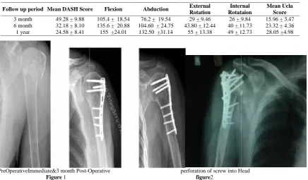
We had failure in two cases. One patient had screw perforation in head at 6 weeks and had delayed union, with final UCLA Score of 14 & decreased strength of forward flexion and difficulty in routine functions by affected limb. Another patient had malunion and developed stiffness of shoulder and undergone manipulation under anesthesia & got sufficient shoulder movement to allow her to do her routine activities.
DISCUSSION
Proximal humeral fractures are the second most common upper-extremity fracture and third most common fracture, after hip and distal radial fractures, in patients who areolder than sixty-five years of age6[Shene et al 2007]. Majority of them
treated with sling immobilization and physical therapy7[Ianotti
et al 2003]. Approximately 20% of proximal humeral fractures are displaced & maybenefit from operative treatment6[shene et
al]. Many surgical fixation techniques have been described inthe literature, but no single surgical fixation technique is considered to be the gold standard of care8[Robert j et al 2009]. There are various treatment options available like conservative treatment with immobilisation and gradualphysiotherapy, operative treatment including transosseous suture fixation, percutaneous k wire fixation, open reduction and internal fixation with conventional or locked-plate fixation, and hemiarthroplasty6,9[Shene et al and Koval et al].There is a uniform agreement that when the tuberosities and medial calcar are anatomically reduced the successful outcome is most likely and the range of motion occurs early in the rehabilitation process10[Gallo et al 2005]. Open reduction internal fixation offers best chance at accurate reduction and union of all fracture fragments, including the greater tuberosity and therefore, good and excellent functional results can be achieved10. However, this method has been limited by difficulty in obtaining adequate exposure especially if greater tuberosity is diplaced and rigid fixation without compromising soft tissue structures. There are several fixation options which have different methods & principles of maintaining reduction, however they also have specific implant related problems as well. [Gallo et al]
Open Reduction and Internal Fixation with Conventional Plate was used extensively earlier for management of proximal humeral fractures.
Wanner et al11used two one-third tubular plates with astandard deltopectoral approach. Traditional plate constructs are usually reserved for young patients with an intact medial hinge, an adequate diaphyseal cortex (>4 mm), and no
Table No 1-DASH, UCLA Score and range of movements at follow up
Follow up period Mean DASH Score Flexion Abduction External Rotation
Internal Rotataion
Mean Ucla Score
3 month 49.28 + 9.88 105.4 + 18.54 76.2 + 19.54 29 + 9.46 26 + 9.84 15.96 + 3.47 6 month 32.18 + 8.10 135.6 + 20.88 104.60 + 24.75 43.80 + 12.44 40 + 11.73 23.32 + 4.36 1 year 24.58 + 8.41 155 +24.01 132.50 +31.14 55 + 13.38 49 + 12.73 28.05 +4.98
PreOperativeImmediate&3 month Post-Operative perforation of screw into Head
Figure 1 figure2
(40%).We used deltopectoral approach in 7(32%) cases and lateral approach in 18(68%) cases All surgical incisions healed by primary intention. with no cases of wound dehiscence or infectionor nerve injury. Operative time <60 min was in 40% &> 60 min in 60% cases. In type 3fractures 60 % (n=9/15) of cases have taken < 60 min time whereas 40% (n 6/15) has taken > 60 min time. In type 4 fractures maximum i.e.90% took > 60 min. All fractures united with an average time of union 11.2 weeks. Mean Flexion at shoulder was 75.800 + 15.520 at 6 weeks which progressed to 15.50 + 24.010 at 1 year. Forward flexion shows significant improvement (p value <0.001) across all the follow up. Similarly Mean abduction, Mean external rotation and Mean internal rotation were calculated at 6 weeks and 1 year as shown in Table no 1.All four movements shows significant improvement (p value <0.001) across all the follow ups.
As analyzed from the graphs 1&2, mean DASH score and mean UCLA Score was also calculated on follow ups as shown in table no 1.Mean UCLA Scores at final follow up was 27.8 with good to excellent results in 72% cases and fair to poor result in 28%cases. Average DASH Score at final follow up was 22.53.
We had failure in two cases. One patient had screw perforation in head at 6 weeks and had delayed union, with final UCLA Score of 14 & decreased strength of forward flexion and difficulty in routine functions by affected limb. Another patient had malunion and developed stiffness of shoulder and undergone manipulation under anesthesia & got sufficient shoulder movement to allow her to do her routine activities.
DISCUSSION
Proximal humeral fractures are the second most common upper-extremity fracture and third most common fracture, after hip and distal radial fractures, in patients who areolder than sixty-five years of age6[Shene et al 2007]. Majority of them
treated with sling immobilization and physical therapy7[Ianotti
et al 2003]. Approximately 20% of proximal humeral fractures are displaced & maybenefit from operative treatment6[shene et
al]. Many surgical fixation techniques have been described inthe literature, but no single surgical fixation technique is considered to be the gold standard of care8[Robert j et al 2009]. There are various treatment options available like conservative treatment with immobilisation and gradualphysiotherapy, operative treatment including transosseous suture fixation, percutaneous k wire fixation, open reduction and internal fixation with conventional or locked-plate fixation, and hemiarthroplasty6,9[Shene et al and Koval et al].There is a uniform agreement that when the tuberosities and medial calcar are anatomically reduced the successful outcome is most likely and the range of motion occurs early in the rehabilitation process10[Gallo et al 2005]. Open reduction internal fixation offers best chance at accurate reduction and union of all fracture fragments, including the greater tuberosity and therefore, good and excellent functional results can be achieved10. However, this method has been limited by difficulty in obtaining adequate exposure especially if greater tuberosity is diplaced and rigid fixation without compromising soft tissue structures. There are several fixation options which have different methods & principles of maintaining reduction, however they also have specific implant related problems as well. [Gallo et al]
Open Reduction and Internal Fixation with Conventional Plate was used extensively earlier for management of proximal humeral fractures.
Wanner et al11used two one-third tubular plates with astandard deltopectoral approach. Traditional plate constructs are usually reserved for young patients with an intact medial hinge, an adequate diaphyseal cortex (>4 mm), and no
Table No 1-DASH, UCLA Score and range of movements at follow up
Follow up period Mean DASH Score Flexion Abduction External Rotation
Internal Rotataion
Mean Ucla Score
3 month 49.28 + 9.88 105.4 + 18.54 76.2 + 19.54 29 + 9.46 26 + 9.84 15.96 + 3.47 6 month 32.18 + 8.10 135.6 + 20.88 104.60 + 24.75 43.80 + 12.44 40 + 11.73 23.32 + 4.36 1 year 24.58 + 8.41 155 +24.01 132.50 +31.14 55 + 13.38 49 + 12.73 28.05 +4.98
PreOperativeImmediate&3 month Post-Operative perforation of screw into Head
Vijay Sarukte et al., The Study of The Functional Outcome of Open Reduction And Internal Fixation of Three And Four Part Proximal Humerus Fracture (According To Neer’s Classification) With Philos (Proximal Humerus Internal Locking System)
14673 |
P a g e
metaphyseal comminution. Conventional plates can fail in osteoporotic bones due to poor screw purchase & high complication rates. With the advent of fixed-angle locked proximal humerus plates has marked a promising advance in the treatment of these fractures [Gallo et al].This device is positioned on the greater tuberosity and lateral proximal humerus and can provide rigid fixation despite purchase in only one cortex [Shene et al 2007 and Lee et al 2009]. This is a major advantage in dealing with fractures involving osteoporotic bone [Koval et al]. Because the plate-screw construct achieves stability through the plate-screw interface as opposed to the plate-bone interface that conventional plating requires, and preserves the remaining periosteal blood supply [Gallo et al]. Several approaches have been offered during the past 30 years tovisualize and fix these fractures.
Ranging from a radical deltoid detaching approach of Martini to more traditional deltopectoral and lateral deltoid splitting exposures.
PHILOS plate was developed to provide angular stability and achieve a favorable screw– bone interface, especially in osteoporotic bone. The plate incorporates multiple locking screws in convergent and divergent directions to improve pullout strength and fixation strength. [Thanasas et al 2009].This creates a fixed angled device that acts as a single unit that captures a volume of bone as shown bywanner et al. In our study we observed, falls resulting in fracture of proximal humerus increasing with age. With Age incidence was almost equal in 30-60 years age group, whereas females were affected largely in elderly age group >60 years age group. This observation is attributable to the rising incidence of osteoporosis in the elderly & specially women. From the observations, we also concluded that incidence of humeral fractures increases as age progresses. Similar results were shown in other studies by Zeng L et al, Seluk Keser et al, Kayalar M et al. In our series it was seen that Neer’s 3-part fractures had comparatively better functional outcome as compared to the 4-part fractures. Greater tuberosity displacement and communition of fracture fragments was responsible for this delay as it is difficult to hold the reduction while the plate was being applied. The union time in our study was average of 11.2 weeks. One case who had screw perforation in joint, which took 24 weeks for union.Atilla et al and Moonot et alin their series of 32 patients and 31 showed radiological union at around 12 weeks and 10 weeks. Which was similar to our study As per our study, functional outcome improved withthe time, and were better in younger population, and patients undergoing proper physiotherapy. Olerued et al, Konard Get al and Plecko et al in their study showed mean DASH score of 26, 15.2 +/- 16.8 and18 at final follow up.
The average UCLA score in our group was 27.8 at final follow up in our study. Handschin et al in his study of 31 patients showed that the UCLA scores were excellent in 10%, good in 67%, and fair in 23% of the patients treated with PHILOS plate fixation at the end of 19 months of follow up. similar result were shown byHessmann et al and Helwig et al.
Numerous complications have been seen in proximal humerus fracture resulting in poor functional outcomes requiring revision surgery. These complications are due to inappropriate
indications, inadequate preoperative evaluation of the fracture advanced osteoporosis and inadequate follow up and rehabilitation. This fracture behaves diversely from good union and early functional result to complete avascular collapse and arthritis of the shoulder joint. In our study we had 2 complications. One patient had screw perforation and other with shoulder stiffness.
Conservative management for displaced or unstable fracture patterns of proximalhumerus has not been favorable, resulting in persistent pain, stiffness,. Nonunion, malunion, and avascular necrosis resulting in a painful dysfunction of joint [Gerber et al 2004]
Percutaneous fixation with pins is a minimally invasive tecnique with lower rate of osteonecrosis, but is less stable than other forms. Done mainly for unstable two-part surgical neckfractures, with limitedind ication in three-part and valgus impacted four-part fractures. This form of fixation is generally reserved for patients with good bone quality, minimal comminution, particularly involving the tuberosity and an intact medial calcar. It is also essential that patients are compliant with postoperative follow-up and immobilization. However it is associated with malunion (28%) mainly varus, migration of pins (upto 1/3rdpatients) rarely into chest, Pin-Track Infection & neurovascular Injury. Hence weekly radiographic evaluation is required [Magovern et al].
Intramedullary nails is alsoan effective method for two-part surgical Neck fractures, although their use in more complex proximal humeral fractures has varied [Lanting et al 2008]. Locked intramedullary nails are axially and rotationally stable, whereas flexible intramedullary nails are not. Shoulder impairment and iatrogenic fractures are risks with locked intramedullary nails. Verbruggen and Stapert stated that rates of flexible nail migration as high as 29% and rates of fracture distraction of up to 41%.Other complication like Malunion, postoperative varus deformity ,radial nerve injury in closed reduction and rotator cuff impingement leading to shoulder pain.
In a comparison of a locked plate and locked nail, plates were found to be stronger in torsion, equivalent in axial stiffness, and superior in varus bending. In a cadaveric study byEdwards et
al the locking plate was biomechanically superior to intramedullary nailing of the proximal humerus when bending and torsion forces were applied.
In comparison with proximal humeral blade plates and conventional plates, locking plates provided better torsional fatigue resistance and stiffness with good results in weak osteopenic bone.
Blade plates are more rigid than conventional plates but have limited proximal screw options [Weinstein et al].
Various studies have been done for PHILOS Plate,
length positioning up to subchondral bone for better fixation on porotic bones. Joint penetration by screws can be reduced by accurate length measurement and shorter screw selection, for obtaining anatomical reduction of the tubercles and restoration of the medial support. Our preference is for the use of shorter screws, thus avoiding the possibility of collapse and accommodation.
In our study, Philos plate fixation provided stable fixation with minimal metal work problems and enabled early range-of-motion exercises to achieve acceptable functional results and excellent rate of union. However, there is no control group included in this study with which to compare these results & the number of patients were relatively small.
CONCLUSION
The fixation of fractures of proximal humerus by PHILOS plate markedly reduces the morbidity in patients, helps the patient to go back to his occupation as early as possible. From this clinical study, we consider PHILOS plating is safe and effective for unstable 3 and 4 part proximal humeral fractures. This technique offers a stable fixation and early recovery. During this study complications were minimal and excellent functional results were obtained. Though sample size in this study was small, but with the trend of results shown by present study, we can say that treatment of proximal humeral fractures by PHILOS plating is safe and effective. Further larger sample size studies are required to conclusively say whether PHILOS plating for proximal humerus fractures be considered as a standard technique of fixation.
Bibliography
1. Habermeyer P, Schweiberer L. Fractures of the proximal humerus. Orthopade 1989; 18:200-7.
2. Young TB, Wallace WA. Conservative treatment of fractures and fracture-dislocations of the upper end of the humerus. J Bone Joint Surg [Br] 1985; 67-B: 373-7. 3. Flatow EL. Fractures of the proximal humerus. In:
Bucholz RW, Heckman JD, eds. Rockwood and Greens fractures in adults. Vol. 1. Philadelphia: Lippincott, Williams and Wilkins, 2001:997-1035.
4. Zyto K, Ahrengart L, Sperber A, Tornkvist H. Treatment of displaced proximal humeral fractures in elderly patients. J Bone Joint Surg [Br] 1997; 79-B: 412-17. 5. Rees J, Hicks J, Ribbans W. Assessment and
management of three- and four-part proximal humeral fractures. ClinOrthop1998; 353:18-29.
6. Shane J. Nho, Robert H. Brophy, Joseph U. Barker, Charles N. Cornell and John D. MacGillivray. Managament of Proximal Humerus Fracture Based on Current Literature. J Bone Joint Surg Am. 2007; 89:44-58
7. Iannotti JP, Ramsey ML, Williams GR, Warner JJP. Nonprosthetic management of proximal humeral fractures. J Bone Joint Surg [Am] 2003; 85:1578-93. 8. Robert J. Gillespie, Vimala Ramachandran, Ethan S.
Lea, Heather A. Vallier. Biomechanical Evaluation of 3-Part Proximal Humerus Fractures: A Cadaveric Study. Orthopedics November 2009; 32(11):816.
9. Koval Kenneth, Blair Benjamin, Takei Robert, Kummer Frederick, Zuckerman Joseph. Surgical Neck Fractures
Ten Fixation Techniques. Journal of Trauma, 1996 -Volume 40 - Issue 5 - pp 778-783.
10. Gallo Robert, Zeiders Gregory, Altman Gregory. Two-Incision Technique for Treatment of Complex Proximal Humerus Fractures. Journal of Orthopaedic Trauma: 2005 - Volume 19 - Issue 10 - pp 734-740.
11. Wanner GA, Wanner-Schmid E, Romero J, Hersche O, von Smekal A, Trentz O, Ertel W. Internal fixation of displaced proximal humeral fractures with two one third tubular plates. J Trauma. 2003; 54:536-44.
12. Lee CW, Shin SJ. Prognostic factors for unstable proximal humeral fractures treated with locking-plate fixation. J Shoulder Elbow Surg. 2009; 18(1):83-8 13. Thanasas C, Kontakis G, Angoules A, Limb D,
Giannoudis P. Treatment of proximal humerus fractures with locking plates: a systematic review. J Shoulder
elbow surg. 2009;18(6):837-844
14. Zeng L, Chen Y, Wang L, Lu Y, Zhang W, Chen Q, Liu Y, Zhang W. Effectiveness of locking plates for Neer three- and four-part proximal humerus fractures. ZhongguoXiu Fu Chong Jian Wai Ke Za Zhi. 2012;26(12):1469-72
15. Seluk Keser · Seluk B_l_kbas¸ · Ahmet Bayar ·Ulunay Kanatlı · Jale Meray · HakanZdemir. Proximal humeral fractures with minimal displacement treated conservatively International Orthopaedics (SICOT) (2004) 28:231–234
16. Atilla Sancar P, Sami S, Ufuk O, Yavuz K. Locking plate fixation of three- and four-part proximal humeral fractures. Acta Orthop Traumatol Turc 2010; 44(2):97-104
17. Moonot p, N. Ashwood, M. Hamlet. Early results for treatment of three- and four part fractures of the proximal humerus using the PHILOS plate system. J
Bone Joint Surg [Br] 2007; 89-B: 1206-9.
18. Olerud P, Ahrengart L, Ponzer S, Saving J, Tidermark J .Internal fixation versus nonoperative treatment of displaced 3-part proximal humeral fractures in elderly patients: a randomized controlled trial. J Shoulder Elbow
Surg. 2011;20(5):747-55
19. Handschin AE, Cardell M, Contaldo C, Trentz O, Wanner GA. Functional results of angular-stable plate fixation in displaced proximal humeral fractures. Injury. 2008;39(3):306-13
20. Gerber, C. M. L. Werner, P. Vienne. Internal fixation of complex fractures of the proximal humerus. J Bone Joint
Surg [Br] 2004; 86-B: 848-55.
21. Magovern B, Ramsey ML. Percutaneous fixation of proximal humerus fractures. Orthop Clin North Am. 2008;39(4):405-16, v
22. Lanting B, MacDermid J, Drosdowech D, Faber KJ. Proximal humeral fractures:a systematic review of treatment modalities. J Shoulder Elbow Surg. 2008 ;17(1):42-54
23. Edwards SL,Wilson NA,Zhang LQ,Flores S,Merk BR. Two part surgical neck fractures of the proximal part of the humerus. A Biomechnical evaluation of two fixation techniques. J Bone Joint Surg Am.2006: 88(10):2258-64 24. Weinstein DM, Bratton DR, Ciccone WJ 2nd, Elias JJ.
Vijay Sarukte et al., The Study of The Functional Outcome of Open Reduction And Internal Fixation of Three And Four Part Proximal Humerus Fracture (According To Neer’s Classification) With Philos (Proximal Humerus Internal Locking System)
14675 |
P a g e
stabilization of three-part proximal humeral fractures. J
Shoulder Elbow Surg. 2006;15(2):239-43
25. Lungershausen W, Bach O, Lorenz CO. Locking plate osteosynthesis for fracture of the proximal humerus. Zentralbl Chir 2003; 128(1):28-33
26. Bjorkenheim JM, Pajarinen J, Savolainen V. Internal fixation of proximal humeral fractures with a locking compression plate: a retrospective evaluation of 72 patients followed for a minimum of 1 year. ActaOrthop Scand. 2004;75(6):741-5
*******
How to cite this article:Vijay Sarukte et al.2016, The Study of The Functional Outcome of open Reduction And Internal Fixation of Three And Four
Part Proximal Humerus Fracture (According To Neer’s Classification) With Philos (Proximal Humerus Internal Locking