*Corresponding author:Mohit N. ISSN: 0976-3031
Research Article
EFFICACY OF SONOELASTOGRAPHY IN DIFFERENTIATING BENIGN AND MALIGNANT
CERVICAL LYMPH NODES: CORRELATION WITH HISTOLOGIC NODAL FINDING
Mohit N., Lokesh Kumar T. and Vijayalakshmi K.
Department of Radiodiagnosis, Mahatma Gandhi Medical College and Research Institute,
Sri Balaji Vidyapeeth, Pillaiyarkuppam, Pondicherry - 607402.
DOI: http://dx.doi.org/10.24327/ijrsr.2019.1001.3084
ARTICLE INFO ABSTRACT
This study was undertaken to evaluate the diagnostic efficacy of sonoelastography in the differentiation of benign and metastatic cervical lymph nodes by correlating with histologic nodal findings.
This was a prospective single centre study wherein sonoelastography was performed for a period of fourteen months on fourty patients with palpable cervical lymph nodes suspected to have metastasis. These cervical lymph nodes were evaluated based on gray scale ultrasound (short axis diameter, short:long axis ratio, shape, echogenicity, border, presence or absence of central fatty hilum and calcification), colour doppler ultrasound (vascularity) and ultrasound elastography (elastography score and strain index) features and further compared with fine needle aspiration cytology and surgical histopathological findings.
Of the 40 patients included in this study and comparing the gray scale ultrasound, colour doppler ultrasound, ultrasound elastography features and fine needle aspiration cytology findings of cervical lymph nodes with gold standard reference histopathology finding, strain index, an ultrasound elastography feature was found to show good agreement ( K-value : 0.80 ) and the most promising variable to differentiate malignant and benign cervical lymph nodes with 97.06% sensitivity, 83.33% specificity, PPV of 97.06%, NPV of 83.33% and 95% accuracy, whereas ultrasound elastography scoring scale showed moderate agreement (K-value : 0. 61) with 97.06% sensitivity, 50% specificity, PPV of 91.67%, NPV of 75% and 90% accuracy.
In this study it was found that sonoelastography was a promising non invasive and rapidly developing imaging technique with easy availability, less expensive, short examination time, real-time display and immediate interpretation that can provide assistance in the differentiation of benign and metastatic cervical lymph nodes thus precluding unnecessary fine needle aspiration cytology in a significant number of patients.
INTRODUCTION
Lymph nodes (LNs) are secondary lymphoid organ necessary for efficient functioning of the immune system and acts by filtering non self particles and cancer cells. There are many LNs located at the neck which are classified at anatomical basis or more recently at level system. Minor infections to fatal cancers cause abnormal size or characteristics of cervical LNs (cervical lymphadenopathy). Thus, differentiation of malignant from benign cervical LNs is necessary to detect present status of the patients and provide correct treatment (1).
Sometimes metastatic cervical lymphadenopathy appears as the first symptom in patients having malignancy in the head and neck, lung, breast and the like. Five year survival rate is
reduced by 50% and further reduced to 25% by presence of a metastatic cervical LN and another metastatic node on opposite side of the neck respectively. Histopathogical examination (HPE) is considered gold standard for cervical lymphadenopathy evaluation (2). Fine needle aspiration cytology (FNAC) being most efficient, is an invasive method liable to sampling fallacy and analytical unreliability (3) with 12.5% to 25% false negative rate (4-6). Computed Tomography (CT) and Magnetic Resonance Imaging (MRI) have restricted ability in differentiating benign from malignant LNs (7).
Malignant tissues being harder in comparison to normal surrounding tissues, tissue elasticity measurement aids in differentiating malignant from benign cervical LNs. Ultrasound Elastography (USE) or sonoelastography, is a non-invasive
Available Online at http://www.recentscientific.com
International Journal of
Recent Scientific
Research
International Journal of Recent Scientific Research
Vol. 10, Issue, 01(F), pp. 30566-30573, January, 2019
Copyright © Mohit N., Lokesh Kumar T. and Vijayalakshmi K., 2019, this is an open-access article distributed under the terms of the Creative Commons Attribution License, which permits unrestricted use, distribution and reproduction in any medium, provided the original work is properly cited.
DOI: 10.24327/IJRSR CODEN: IJRSFP (USA)
Article History:
Received 06th October, 2018 Received in revised form 14th
November, 2018
Accepted 23rd December, 2018 Published online 28th January, 2019
Key Words:
Sonoelastography, Cervical lymph nodes, Fine needle aspiration cytology,
Correlation with Histologic Nodal Finding
imaging technique that helps detect the elasticity of the tissues and describes stiffness or displacement (i.e., strain) of tissues to an applied force (8,9). Hard tissues show less deformation and strain compared to tissues that are compliant when same amount of force is applied. This functional imaging mode being highly specific (10), easily available, economical and less time consuming may be performed along with routine US instead of biopsy/FNAC to confirm suspected findings when there is a low suspicion of malignancy, saving the patient an invasive procedure.
Aim and Objectives
1. To estimate the efficacy of sonoelastography in the differentiation of benign and metastatic cervical LNs. 2. To correlate the sonoelastography findings with
histologic nodal findings.
3. To estimate the accuracy of B-mode Ultrasonography (USG) in the differentiation of benign and metastatic cervical LNs.
METHODOLOGY
This study was carried out as a cross sectional comparative design with prospective recruitment of 40 patients with palpable cervical LNs suspected to have metastasis who also underwent FNAC and excision biopsy of the same to confirm the diagnosis in Mahatma Gandhi Medical College and Research Institute, Pondicherry, India, from February 2017 until June 2018.
All the patients included in the study were examined in MINDRAY DC-8 US equipment with hyper extended neck in supine position using high frequency probe (3 - 12 MHz). The high frequency probe provides the necessary penetration, an image with high resolution and elastography evaluation of the cervical LNs. The cervical LN characteristics were evaluated using gray scale, colour doppler and USE following which these cervical LNs were subjected to cytological evaluation and then compared with HPE results which was considered as gold standard to differentiate metastatic from benign cervical LNs.
Inclusion criteria
Patients who were referred to radio diagnosis department for US neck with palpable cervical LNs suspected to have metastasis.
Patients who underwent FNAC and surgical excision biopsy of palpable cervical LNs which were characterized on US.
Exclusion criteria
Patients not suspected to have metastatic cervical LNs. Patients who did not undergo FNAC or surgical
excision biopsy of palpable cervical LNs.
Patients who were not willing to participate in the study.
Statistical Analysis
All data of 40 patients including patients name, hospital number, age, sex, US findings, FNAC and HPE findings were collected and entered in Microsoft excel 2013 and analysed using SPSS version 21.0.
Categorical variables like age, gender, short axis diameter, S:L axis ratio, shape, central fatty hilum, echogenicity, border, calcification, vascularity, ESS, SI, FNAC and HPE were presented as percentages. Association of tumour characteristics with HPE findings were assessed using Chi square test and a p value of less than 0.05 was considered as statistically significant. Diagnostic validity of categorical US variables including FNAC for diagnosing malignancy was assessed using sensitivity, specificity, PPV (positive predictive value), NPV (negative predictive value) and accuracy against HPE findings, which was considered as gold standard.
RESULTS
Among the study participants suspected to have metastasis, cervical lymphadenopathy was prevalent in the age group of 41-60 years and included 23 patients (57.5%).
Table 1 Age distribution of study participants (n=40)
Age
(years) Frequency
Percent (%)
18-40 12 30
41-60 23 57.5
>60 5 12.5
Total 40 100
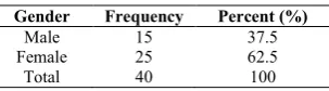
Cervical lymphadenopathy was found to be more common in females (62.5%) than in males (37.5%).
Table 2 Gender distribution of study participants (n=40)
Gender Frequency Percent (%)
Male 15 37.5
Female 25 62.5
Total 40 100
Among the 40 study participants, 34 (85%) cervical LNs were confirmed to be malignant on HPE and the other 6 (15%) benign. HPE was used as a reference gold standard.
Table 3 Distribution of study participants based on HPE (n=40)
Histopathology Frequency Percent (%)
Benign 6 15
Malignant 34 85
Total 40 100
Cervical Lymph Node Characteristics with Histopathological Correlation
International Journal of Recent Scientific Research Vol. 10, Issue, 01(F), pp.
Gray Scale Ultrasound
Short Axis
Figure 1 Correlation of short axis diameter and HPE 34 (85%) cervical LNs had short axis diameter
which 29 (85.3%) were proved to be malignant and 5 (14.7%) benign. Of the other 6 (15%) nodes with short axis diameter < 8mm, 5 (83.3%) were malignant and 1 (16.7%) benign. Thus, short axis diameter cut off 8mm was not significant to differentiate malignant and benign nodes (p value = 0.91)
Short: Long Axis
33 (82.5%) cervical LNs had S:L axis ratio ≥ 0.5, out of which 31 (93.9%) were proved to be malignant and 2 (6.1%) benign. Of the other 7 (7.5%) nodes with S:L axis ratio < 0.5, 3 (42.8%) were malignant and 4 (57.2%) benign. Thus, S:L ratio cut off 0.5 was proved to be a significant gray scale US characteristic to differentiate malignant and benign nodes (p value = 0.01).
Figure 2 Correlation of S:L axis ratio and HPE
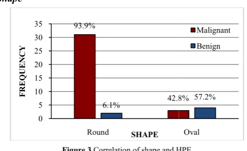
Shape
Figure 3 Correlation of shape and HPE
33 (82.5%) cervical LNs were round, out of which 31 (93.9%) were proved to be malignant and 2 (6.1%) benign. Of the other 7 (17.5%) oval nodes, 3 (42.8%) were malignant and 4 (57.2%) benign. Thus, round shape of LNs is also a significant
34 85%
SHORT AXIS
93.9%
42.8% 6.1%
0 5 10 15 20 25 30 35
≥0.5
FREQU
ENCY
S:L AXIS RATIO
93.9%
42.8% 6.1%
0 5 10 15 20 25 30 35
Round
FREQUEN
CY
SHAPE
International Journal of Recent Scientific Research Vol. 10, Issue, 01(F), pp. 30566-30573, January,
Correlation of short axis diameter and HPE
34 (85%) cervical LNs had short axis diameter ≥ 8mm out of malignant and 5 (14.7%) benign. Of the other 6 (15%) nodes with short axis diameter < 8mm, 5 (83.3%) were malignant and 1 (16.7%) benign. Thus, short axis diameter cut off 8mm was not significant to differentiate malignant and benign nodes (p value = 0.91).
≥ 0.5, out of which 31 (93.9%) were proved to be malignant and 2 (6.1%) benign. Of the other 7 (7.5%) nodes with S:L axis ratio < 0.5, 3 (42.8%) were malignant and 4 (57.2%) benign. Thus, S:L axis ratio cut off 0.5 was proved to be a significant gray scale US characteristic to differentiate malignant and benign nodes (p
Correlation of S:L axis ratio and HPE
of shape and HPE
33 (82.5%) cervical LNs were round, out of which 31 (93.9%) were proved to be malignant and 2 (6.1%) benign. Of the other 7 (17.5%) oval nodes, 3 (42.8%) were malignant and 4 (57.2%) benign. Thus, round shape of LNs is also a significant grey
scale US characteristic to differentiat nodes (p value = 0.01).
Central Fatty Hilum
Figure 4 Correlation of central fatty hilum and HPE
39 (97.5%) cervical LNs did not show central fatty hilum, out of which 34 (87.1%) were
(12.9%) benign. The only node (2.5%) with central fatty hilum was proved to be benign (100%). Thus, absence of central fatty hilum is significant enough to differentiat
benign nodes (p value = 0.016
Echogenicity
8 (20%) cervical LNs were homogenously hyperechoic, of which 6 (75%) were proved to be malignant and 2 (25%) benign. 24 (60%) LNs were homogenously hypoechoic, of which 21 (87.5%) were malignant and 3 (12.5%) benign. 5 (12.5%) nodes which appeared ho
malignant (100%). 2 (5%) LNs with central necrosis were proved to be malignant (100%). One node (2.5%) which appeared normal with central hyperechogenicity was concluded benign (100%). Thus abnormal echogenicity was proved to be statistically significant (p value = 0.016) in differentiating malignant from benign nodes.
Figure 5 Correlation of echogenicity and HPE
Border
6 15%
<8 mm
≥8 mm
42.8% 57.2%
<0.5 Malignant
Benign
42.8% 57.2%
Oval Malignant
Benign
87.1%
12.9%
0 5 10 15 20 25 30 35 40
Absent
F
REQUE
NCY
CENTRAL FATTY HILUM
100% 100% 0%
25% 12.5% 0% 0%
100%
0 5
Hyperechoic Hypoechoic Isoechoic Heterogenous with
anechoic area Hypoechoic with central hypergenicity
ECHOGENICITY
, January, 2019
scale US characteristic to differentiate malignant from benign
Correlation of central fatty hilum and HPE
39 (97.5%) cervical LNs did not show central fatty hilum, out proved to be malignant and 5 (12.9%) benign. The only node (2.5%) with central fatty hilum was proved to be benign (100%). Thus, absence of central fatty hilum is significant enough to differentiate malignant from benign nodes (p value = 0.016).
8 (20%) cervical LNs were homogenously hyperechoic, of which 6 (75%) were proved to be malignant and 2 (25%) benign. 24 (60%) LNs were homogenously hypoechoic, of which 21 (87.5%) were malignant and 3 (12.5%) benign. 5 (12.5%) nodes which appeared homogenously isoechoic were malignant (100%). 2 (5%) LNs with central necrosis were proved to be malignant (100%). One node (2.5%) which appeared normal with central hyperechogenicity was concluded benign (100%). Thus abnormal echogenicity was proved to be tatistically significant (p value = 0.016) in differentiating malignant from benign nodes.
Correlation of echogenicity and HPE
0% 100%
Present
CENTRAL FATTY HILUM
Malignant
Benign
75%
87.5% 100%
12.5%
10 15 20 25
FREQUENCY
ECHOGENICITY Column1
Benign
Figure 6 Correlation of border and HPE
25 (62.5%) cervical LNs showed lobulated borders, out of which 24 (96%) were proved to be malignant and 1 (4%) benign. Other 15 (37.5%) which showed regular border, 10 (66.7%) were malignant and 5 (33.3%) benign. Thus, there was statistically significant association between lobulated border and malignancy (p value = 0.012).
Calcification
Figure 7 Correlation of calcification and HPE
All 5 (12.5%) cervical LNs which showed presence of punctuate calcification were proved to be malignant (100%). Of the other 35 (87.5%) with no calcification, 29 (82.8%) were malignant and 6 (17.2%) benign.
Colour Doppler Ultrasound
Vascularity
28 (70%) cervical LNs showed peripheral vascularity, 26 (92.8%) of which were proved to be malignant and 2 (7.2%) benign. Further, 11 (27.5%) nodes showed both central and peripheral vascularity, of which 8 (72.7%) were malignant and 3 (27.3%) benign. One node (2.5%) showed normal central vascularity which was benign (100%). Thus, abnormal vascularity was proved to be statistically significant (p value = 0.01 ) to differentiate malignant from benign nodes.
96%
66.7%
4% 0
5 10 15 20 25 30
Lobulated Regular
FREQ
UENCY
BORDER
100%
82.8%
0% 0
5 10 15 20 25 30 35
Present
FREQUEN
CY
CALCIFICATION
Correlation with Histologic Nodal Finding
Correlation of border and HPE
25 (62.5%) cervical LNs showed lobulated borders, out of which 24 (96%) were proved to be malignant and 1 (4%) benign. Other 15 (37.5%) which showed regular border, 10 (66.7%) were malignant and 5 (33.3%) benign. Thus, there was statistically significant association between lobulated border
Correlation of calcification and HPE
All 5 (12.5%) cervical LNs which showed presence of punctuate calcification were proved to be malignant (100%). Of the other 35 (87.5%) with no calcification, 29 (82.8%) were
%) cervical LNs showed peripheral vascularity, 26 (92.8%) of which were proved to be malignant and 2 (7.2%) benign. Further, 11 (27.5%) nodes showed both central and peripheral vascularity, of which 8 (72.7%) were malignant and 2.5%) showed normal central vascularity which was benign (100%). Thus, abnormal vascularity was proved to be statistically significant (p value = 0.01 ) to differentiate malignant from benign nodes.
Figure 8 Correlation of vascularity and HPE
Sonoelastography
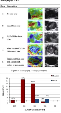
Elastography score
Figure 9 Elastography scoring system (1
Figure 10 Correlation of ES and HPE
2 (5%) cervical LNs showed central necrosis (ES5) and both (100%) were proved to be malignant. 17 (42.5%) nodes showed ES4 pattern and were proved to be malignant (100%).
66.7%
33.3%
Regular Malignant Benign
82.8%
17.2%
Absent
Malignant
Benign
92.8%
7.2%
0 5 10 15 20 25 30
Peripheral
FREQU
ENCY
VASCULARITY
100%
100%
0 0
0 2 4 6 8 10 12 14 16 18
ES5 ES4
F
REQUE
NCY
ELASTOGRAPHY SCORE
Correlation of vascularity and HPE
Elastography scoring system (11)
Correlation of ES and HPE
2 (5%) cervical LNs showed central necrosis (ES5) and both (100%) were proved to be malignant. 17 (42.5%) nodes showed ES4 pattern and were proved to be malignant (100%).
72.7%
0 27.3%
100%
Mixed Central
VASCULARITY
Malignant
Benign
82.4%
25%
0
17.6% 75%
0
ES3 ES2 ES1
ELASTOGRAPHY SCORE
Malignant
International Journal of Recent Scientific Research Vol. 10, Issue, 01(F), pp.
17 (42.5%) nodes showed ES3 pattern, of which 14 (82.4%) were malignant and 3 (17.6%) benign. 4 (10%) LNs showed ES2 pattern, of which 1 (25%) was malignant and 3 (75%) benign. No LN showed ES1 pattern. Thus, ESS was proved to be statistically significant to differentiate malignant and benign nodes (p value = 0.002).
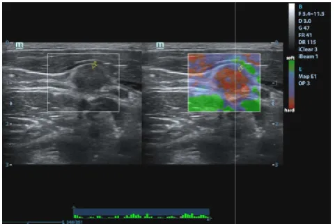
Figure 11 Level II oval homogenously hypoechoic cervical LN with ES 2 which was confirmed as well differentiated SCC on HPE
Figure 12 Level IV cervical LN with ES 3 which was confirmed as moderately differentiated SCC on HPE
Figure 13 Level II cervical LN with ES 4 which was confirmed as high grade mucoepidermoid CA on HPE
International Journal of Recent Scientific Research Vol. 10, Issue, 01(F), pp. 30566-30573, January,
17 (42.5%) nodes showed ES3 pattern, of which 14 (82.4%) benign. 4 (10%) LNs showed ES2 pattern, of which 1 (25%) was malignant and 3 (75%) benign. No LN showed ES1 pattern. Thus, ESS was proved to be statistically significant to differentiate malignant and benign
II oval homogenously hypoechoic cervical LN with ES 2 which was confirmed as well differentiated SCC on HPE
Level IV cervical LN with ES 3 which was confirmed as moderately differentiated SCC on HPE
ES 4 which was confirmed as high grade
Figure 14 HPE proved micropapillary CA showing necrotic (central anechoic areas) level III cervical LN with ES 5 (peripheral hard areas with central soft
areas suggestive of central
Strain Index
Figure 15 Correlation of SI and HPE 34 (85%) cervical LNs had a SI
were proved to be malignant and 1 (2.9%) benign. Of the other 6 (15%) with SI < 1.5, 1 (16.7%) was malignant and 5
benign. Thus, the SI cut off
from benign nodes was proved to be statistically significant ( p value = 0.01 ).
Figure 16 HPE confirmed papillary CA showing round, homogenously hyperechoic level III cervical LN wit
97.1%
2.9% 0
5 10 15 20 25 30 35
≥1.5
F
REQU
ENCY
STRAIN INDEX , January, 2019
HPE proved micropapillary CA showing necrotic (central anechoic areas) level III cervical LN with ES 5 (peripheral hard areas with central soft
areas suggestive of central necrosis) and SI 1.79
Correlation of SI and HPE
34 (85%) cervical LNs had a SI ≥ 1.5, 33 (97.1%) of which were proved to be malignant and 1 (2.9%) benign. Of the other 6 (15%) with SI < 1.5, 1 (16.7%) was malignant and 5 (83.3%) benign. Thus, the SI cut off ≥1.5 to differentiate malignant from benign nodes was proved to be statistically significant ( p
HPE confirmed papillary CA showing round, homogenously hyperechoic level III cervical LN with ES 4 and SI 3.51
16.7% 2.9%
83.3%
<1.5
STRAIN INDEX
Malignant
Figure 17 Oval, homogenously hypoechoic level III cervical LN with ES 2 and SI 1.05 which showed no evidence of malignancy on HPE in a known case of
oesophageal CA
Figure 18 HPE confirmed adenocarcinoma showing oval, hypoechoic level III cervical LN with ES 3 and SI 1.33
Figure 19 Oval, homogenously hypoechoic level V cervical LN with ES 3 and SI 1.82 which showed no evidence of malignancy on HPE in a known case of
nasopharyngeal CA
Correlation with Histologic Nodal Finding
Oval, homogenously hypoechoic level III cervical LN with ES 2 and SI 1.05 which showed no evidence of malignancy on HPE in a known case of
HPE confirmed adenocarcinoma showing oval, homogenously hypoechoic level III cervical LN with ES 3 and SI 1.33
Oval, homogenously hypoechoic level V cervical LN with ES 3 and SI 1.82 which showed no evidence of malignancy on HPE in a known case of
Cytology
Figure 20 Correlation of FNAC and HPE
29 (72.5%) cervical LNs turned out to be cytologically malignant of which 27 (93.1%) were proved to be malignant and 2 (6.9%) benign on HPE. Of the other 11 (27.5%) which were concluded as benign on cytology, 7
malignant and 4 (36.4%) benign on HPE. Thus, cytological evaluation of cervical LNs to differentiate malignant from benign was also statistically significant ( p value = 0.02 ).
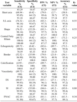
Table 4Diagnostic value of the following variables to differentiate malignant from benign cervical LNs
Test variable Sensitivity % (CI) Specificity % (CI) Short axis 85.29 (68.94 - 95.05) 16.67 (0.42 - 64.12) S:L axis 91.18 (76.32 - 98.14) 66.67 (22.28 - 95.67) Shape 91.18 (76.32 - 98.14) 66.67 (22.28 - 95.67) Central Fatty Hilum 100.00 (89.72 - 100.0) 16.67 (0.42 - 64.12) Echogenicity 100.00 (89.72 - 100.0) 16.67 (0.42 - 64.12) Border 70.59 (52.52 - 84.90) 83.33 (35.88 - 99.58) Calcification 14.7 (4.95 - 31.06) 100.0 (54.07 - 100) Vascularity 100.0 (89.72 - 100) 16.67 (0.42 - 64.12) ES 97.06 (84.67 - 99.93) 50.00 (11.81 - 88.19) SI 97.06 (84.67 - 99.93) 83.33 (35.88 - 99.58) FNAC 79.41 (62.10 - 91.30) 66.67 (22.28 - 95.67)
K-value (kappa value) Poor agreement : <0.20 Fair agreement : 0.20 - Moderate agreement : 0.40
93.1% 6.9% 0 5 10 15 20 25 30 Malignant FREQU ENCY CYTOLOGY
Correlation of FNAC and HPE
29 (72.5%) cervical LNs turned out to be cytologically malignant of which 27 (93.1%) were proved to be malignant and 2 (6.9%) benign on HPE. Of the other 11 (27.5%) which were concluded as benign on cytology, 7 (63.6%) were malignant and 4 (36.4%) benign on HPE. Thus, cytological evaluation of cervical LNs to differentiate malignant from benign was also statistically significant ( p value = 0.02 ).
Diagnostic value of the following variables to differentiate malignant from benign cervical LNs
Specificity PPV % (CI) NPV % (CI) Accuracy % (CI) K- value 85.29 (79.8 - 89.4) 16.67 (2.7 - 58.7) 75.0 (58.8 - 87.3) 0.02 93.34 (83.3 - 97.7) 57.14 (28.3 - 81.9) 87.5 (73.2 - 95.8) 0.55 93.34 (83.3 - 97.7) 57.14 (28.3 - 81.9) 87.5 (73.2 - 95.8) 0.55 87.17 (82.6 - 90.7) 100.0 (89.7 - 100) 87.5 (73.2 - 95.8) 0.25 87.17 (82.6 - 90.7) 100.0 (89.7 - 100) 87.5 (73.2 - 95.8) 0.25 85.0 (79.8 - 99.3) 33.33 (21.0 - 48.9) 72.5 (56.1 - 85.4) 0.33 100.0 (89.7 - 100) 17.14 (15.3 - 19.2) 27.5 (14.6 - 43.8) 0.05 87.17 (82.6 - 90.7) 100.0 (89.7 - 100) 87.5 (73.2 - 95.8) 0.25 91.67 (83.1 - 96.1) 75.00 (27.1 - 96.0) 90.0 (76.3 - 97.2) 0.61 97.06 (84.6 - 99.5) 83.33 (41.2 - 97.3) 95.0 (83.0 - 99.4) 0.80 93.10 (81.1 - 97.1) 36.36 (19.3 - 57.7) 77.5 (61.5 - 89.1) 0.34
Poor agreement : <0.20 0.40 Moderate agreement : 0.40 - 0.60
International Journal of Recent Scientific Research Vol. 10, Issue, 01(F), pp. 30566-30573, January, 2019
Good agreement : 0.60 - 0.80 Very good agreement : 0.80 - 1.00
HPE was considered Gold Standard. With reference to the above variables of cervical LN characterisation on gray scale US, colour doppler US, USE and FNAC findings, USE SI showed good agreement (K-value: 0.80) and the most promising variable to differentiate malignant from benign cervical LNs with 97.06% sensitivity, 83.33% specificity, 97.06% PPV, 83.33% NPV and 95% accuracy whereas USE score showed moderate agreement (K-value : 0. 61) with 97.06% sensitivity, 50% specificity, 91.67% PPV, 75% NPV and 90% accuracy. Thus, USE was superior to FNAC in differentiating malignant from benign cervical LNs which only showed fair agreement (K-value: 0.34) with 79.41% sensitivity, 66.67% specificity, 93.10% PPV, 36.36% NPV and 77.5% accuracy.
Grey scale US characteristics, S:L axis ratio and shape also showed moderate agreement ( K-value : 0. 55 ) with 91.18% sensitivity, 66.67 specificity, 93.34% PPV, 57.14% NPV and 87.5% accuracy. Abnormal echogenicity and absent central fatty hilum showed 100% sensitivity and NPV while presence of calcification showed 100% specificity and PPV.
Vascularity, a colour doppler US characteristic, also showed 100% sensitivity and NPV.
DISCUSSION
The gray scale and colour doppler features of cervical LN which helped predict malignancy were short axis ≥ 8mm, S:L axis ratio ≥ 0.5 (round shape), absence of normal central fatty hilum, abnormal echogenicity, lobulated border, presence of calcification and peripheral/mixed vascularity. USE score of 3-5 and SI ≥ 1.3-5 were promising predictors of metastatic cervical LNs.
These features have already been described in literature and current study also found similar results- namely Lyshchik et al. (12), Ishibashi et al. (11), Alam et al. (10) where they have addressed similar features to differentiate malignant from benign cervical LNs.
On sonoelastography, qualitative analysis and quantitative measurements were evaluated. Qualitative analysis was initially done using a 4-point elastography scoring scale (ESS) where scores of 1-2 indicated benign and scores of 3-4 indicated malignant cervical LNs with sensitivity, specificity, PPV and NPV of 66.7%, 57.1%, 52.2% and 71.0 % respectively (13). Alam et al. (10) used a 5-point elastography scale and reported sensitivity, specificity and accuracy of 83%, 100% and 89% respectively. Ishibashi et al. (11) further modified the 5-point scale and found 83.8% sensitivity, 82.5% specificity, 78.8% PPV, 86.8% NPV and 83.1% accuracy. Similar 5-point ESS was used in this current study which revealed sensitivity, specificity, PPV, NPV and accuracy of 97.06%, 50%, 91.67%, 75% and 90% respectively. Quantitative sonoelastography criterion, SI ≥ 1.5 showed best diagnostic efficacy of all the gray scale, colour doppler and sonoelastography criteria evaluated. This current study also revealed similar results with 97% sensitivity, 83% specificity, 97% PPV, 83% NPV and 95% accuracy. It thus showed good agreement (K-value = 0.80) and clinically significant
association (p value = 0.01) in diagnosing malignant cervical LNs.
Finally these results were followed by FNAC and surgical biopsy and found to be promising. Sonoelastography in this current study was proved superior to FNAC confirmation of metastatic cervical LNs which only showed 79.41% sensitivity, 66.67% specificity, 93.1% PPV, 36.36% NPV and 77.5% accuracy with just fair agreement(K-value = 0.34). However, interobserver variability and cytology related issues (quality of cells collected, microscope used, experience of pathologist and the like) could not be assessed.
Sonoelastography would thus be an important tool in precluding the unnecessary FNAC and accurately differentiating malignant/metastatic from benign cervical LNs which would be useful in future to radiologist for everyday use. This new US technique will be an invaluable tool in diagnosis of malignant cervical LNs in a non-invasive approach.
Strengths and Limitations
Strengths
USE is a non invasive imaging technique, useful in the assessment of elastic properties of cervical LNs with short examination time required, real-time display, immediate interpretation and limited cost, and the criteria adopted in the image interpretation have proven to be adequate in clinical practice.
Sonoelastography can be helpful in the selection of suspicious cervical LNs that should be examined at cytologic examination or open biopsy for accurate preoperative staging and thus helping in individual therapy selection.
Limitations
Image quality and diagnostic performance of sonoelastography depends on compression applied and development of elasticity computation.
Non-axial or out-of-plane motion of the examined cervical LN and common carotid artery pulsations caused de-correlation noise which reduced strain image quality.
Real time elastogram includes fast reconstruction of strain images thus not as precise as off-line computing.
CONCLUSION
Sonoelastography is a rapidly developing, promising imaging technique and an effective supplement to conventional B-mode USG that can provide assistance in the accurate differentiation of benign and metastatic cervical LNs.
Recommendations
Correlation of elastic properties of cervical LNs determined at USE with post excision biomechanical properties of the same.
Volumetric assessment of stiffness of cervical LN would increase the diagnostic performance of sonoelastography.
Correlation with Histologic Nodal Finding
and to standardize the clinical application of this technique.
References
1. Lécuru F, Mathevet P, Querleu D, Leblanc E, Morice P, Daraï E, et al. Bilateral negative sentinel nodes accurately predict absence of lymph node metastasis in early cervical cancer: results of the SENTICOL study. J Clin Oncol. 2011 May 1;29(13):1686-91.
2. Mohseni S, Shojaiefard A, Khorgami Z, Alinejad S, Ghorbani A, Ghafouri A. Peripheral Lymphadenopathy: Approach and Diagnostic Tools. Iran J Med Sci. 2014 Mar;39(2 Suppl):158-70.
3. Ross DS. Nonpalpable thyroid nodules--managing an epidemic. J Clin Endocrinol Metab. 2002 May; 87(5):1938-40.
4. Eloubeidi MA, Wallace MB, Reed CE, Hadzijahic N, Lewin DN, Van Velse A, et al. The utility of EUS and EUS-guided fine needle aspiration in detecting celiac lymph node metastasis in patients with esophageal cancer: a single-center experience. Gastrointest Endosc. 2001 Dec; 54(6):714-9.
5. Bean SM, Eloubeidi MA, Cerfolio R, Chhieng DC, Eltoum IA. Endoscopic ultrasound-guided fine needle aspiration is useful for nodal staging in patients with pleural mesothelioma. Diagn Cytopathol. 2008 Jan; 36(1):32-7.
6. Hernandez LV, Mishra G, George S, Bhutani MS. A descriptive analysis of EUS-FNA for mediastinal lymphadenopathy: an emphasis on clinical impact and false negative results. Am J Gastroenterol. 2004 Feb;99(2):249-54.
7. Xu W, Shi J, Zeng X, Li X, Xie W-F, Guo J, et al. EUS elastography for the differentiation of benign and malignant lymph nodes: a meta-analysis. Gastrointest Endosc. 2011 Nov; 74(5):1001-9; quiz 1115.e1-4. 8. Lerner RM, Huang SR, Parker KJ. “Sonoelasticity”
images derived from ultrasound signals in mechanically vibrated tissues. Ultrasound Med Biol. 1990; 16(3):231-9.
9. Konofagou EE. Quo vadis elasticity imaging? Ultrasonics. 2004 Apr;42(1-9):331-6.
10. Alam F, Naito K, Horiguchi J, Fukuda H, Tachikake T, Ito K. Accuracy of sonographic elastography in the differential diagnosis of enlarged cervical lymph nodes: comparison with conventional B-mode sonography. AJR Am J Roentgenol. 2008 Aug; 191(2):604-10.
11. Ishibashi N, Yamagata K, Sasaki H, Seto K, Shinya Y, Ito H, et al. Real-time tissue elastography for the diagnosis of lymph node metastasis in oral squamous cell carcinoma. Ultrasound Med Biol. 2012 Mar;38(3):389-95.
12. Lyshchik A, Higashi T, Asato R, Tanaka S, Ito J, Hiraoka M, et al. Cervical lymph node metastases: diagnosis at sonoelastography--initial experience. Radiology. 2007 Apr; 243(1):258-67.
13. Lo W-C, Cheng P-W, Wang C-T, Liao L-J. Real-time ultrasound elastography: an assessment of enlarged cervical lymph nodes. Eur Radiol. 2013 Sep;23(9):2351-7.
How to cite this article:
Mohit N., Lokesh Kumar T and Vijayalakshmi K. 2019, Efficacy of Sonoelastography in Differentiating Benign and Malignant Cervical Lymph Nodes: Correlation with Histologic Nodal Finding. Int J Recent Sci Res. 10(01), pp.30566-30573.
DOI: http://dx.doi.org/10.24327/ijrsr.2019.1001.3084