Open Access
Research article
Bias associated with delayed verification in test accuracy studies:
accuracy of tests for endometrial hyperplasia may be much higher
than we think!
T Justin Clark*
1, Gerben ter Riet
2, Aravinthan Coomarasamy
1and
Khalid S Khan
1Address: 1Academic Department of Obstetrics & Gynaecology, Birmingham Women's Hospital, Birmingham, B15 2TG, UK and 2Academic Medical
Center, Department of General Practice, 1105 AZ, Amsterdam, Netherlands
Email: T Justin Clark* - justin.clark@bwhct.nhs.uk; Gerben ter Riet - G.terRiet@amc.uva.nl;
Aravinthan Coomarasamy - a.coomarasamy@bham.ac.uk; Khalid S Khan - khalid.khan@bwhct.nhs.uk * Corresponding author
Abstract
Background: To empirically evaluate bias in estimation of accuracy associated with delay in verification of diagnosis among studies evaluating tests for predicting endometrial hyperplasia.
Methods: Systematic reviews of all published research on accuracy of miniature endometrial biopsy and endometr ial ultrasonography for diagnosing endometrial hyperplasia identified 27 test accuracy studies (2,982 subjects). Of these, 16 had immediate histological verification of diagnosis while 11 had verification delayed > 24 hrs after testing. The effect of delay in verification of diagnosis on estimates of accuracy was evaluated using meta-regression with diagnostic odds ratio (dOR) as the accuracy measure. This analysis was adjusted for study quality and type of test (miniature endometrial biopsy or endometrial ultrasound).
Results: Compared to studies with immediate verification of diagnosis (dOR 67.2, 95% CI 21.7– 208.8), those with delayed verification (dOR 16.2, 95% CI 8.6–30.5) underestimated the diagnostic accuracy by 74% (95% CI 7%–99%; P value = 0.048).
Conclusion: Among studies of miniature endometrial biopsy and endometrial ultrasound, diagnostic accuracy is considerably underestimated if there is a delay in histological verification of diagnosis.
Background
The natural history of endometrial hyperplasia is not fully understood [1]. What is known is that a proportion of simple and complex hyperplastic processes will regress without treatment [2] although the time scale over which such regression may occur is unclear. Similarly the time scale over which benign endometrium progresses to hyperplasia is also unknown. Among studies evaluating
accuracy of tests for diagnosis of hyperplasia (miniature biopsy or ultrasonography), it has previously been hypothesised that if histological verification of diagnosis after performing the test is delayed, the estimation of test accuracy may be influenced by the phenomena of disease regression or progression [3]. For instance, false positive diagnoses of endometrial hyperplasia may occur due to natural disease regression during the time interval Published: 11 May 2004
BMC Medicine 2004, 2:18
Received: 16 September 2003 Accepted: 11 May 2004
This article is available from: http://www.biomedcentral.com/1741-7015/2/18
between testing and verification of diagnosis. Similarly, false negative diagnoses may also result from progression of benign functional or atrophic endometrium.
To obtain accurate estimates of test accuracy in studies of hyperplasia, an immediate comparison of the test under scrutiny with a reference standard that verifies the diagno-sis will be essential [4-6]. When accuracy studies suffer from a delay in performance of the reference standard, the resultant false positives and false negatives will be expected to lead to an underestimation of test accuracy. In systematic reviews, when studies of various designs are collated, the extent of underestimation that arises from delay is important in obtaining an unbiased pooled accu-racy estimate. To our knowledge, the extent of underesti-mation of accuracy due to a delay in verification of diagnosis has not been evaluated empirically in studies of endometrial hyperplasia. We undertook this analysis to examine formally how inaccurate the estimation of accu-racy can be in studies evaluating miniature endometrial biopsy devices and endometrial thickness measurement by pelvic ultrasonography for predicting endometrial hyperplasia when there are delays in histological verifica-tion of diagnosis.
Methods
To test our hypothesis, a data set of all the published stud-ies reporting the accuracy of miniature endometrial biopsy devices and endometrial ultrasonography for pre-dicting endometrial hyperplasia was obtained from sys-tematic reviews [7,8]. The reviews focused on test accuracy studies in which the results of the test were compared with the results of a reference standard. The targeted popula-tion was women with abnormal pre- or postmenopausal uterine bleeding. The diagnostic tests of interest were min-iature endometrial biopsy devices (for example, pipelle®
endometrial suction curette, Unimar, Wilton, CT, USA) and endometrial thickness measurement by pelvic ultra-sonography. The reference standard was endometrial his-tology obtained by an independent endometrial sampling technique, for example, inpatient curettage (with hyster-oscopy) or hysterectomy.
Identification of studies
Two independent electronic searches of MEDLINE and EMBASE were conducted to identify relevant citations on endometrial biopsy (1980–1999) and ultrasonography (1966–2000). Search term combination for endometrial biopsy [8] was diagnosis (MeSH) AND endometrial biopsy (textword), while that for studies on ultrasonography [7] was ultrasound AND endometrial thickness AND sonography (textwords). The searches were limited to human studies, but there were no language restrictions. Relevant studies were identified by examining all the retrieved citations, reference lists of all known reviews and primary studies,
and direct contact with manufacturers. Details of the search and selection processes can be found in the pub-lished reports of the reviews [7,8].
Study quality assessment
All selected studies were assessed for their methodological quality defined as the confidence that study design, con-duct and analysis minimize bias in the estimation of diag-nostic accuracy [9-11]. We considered the following features in quality assessment: method of recruitment of sample, appropriateness of patient spectrum, and blind-ing of comparison between test and reference standard. Recruitment was considered to be adequate if patient selection was consecutive or a random sample was obtained. Patient spectrum was considered to be appro-priate if both pre- and postmenopausal women were included. Blinding was considered to be present if it was clearly reported that the pathologists providing histologi-cal reports were kept unaware of the results of miniature endometrial biopsy or endometrial ultrasonography. If the results of the diagnostic tests were divulged to the pathologists or in the absence of any such reporting, blinding was categorised as absent. For the purpose of our analysis, studies were classified into two quality catego-ries: Category I studies had any one of the following fea-tures: adequate recruitment, appropriate spectrum, and blinding; category II studies had none of the above quality features.
Data extraction
In addition to assessment of methodological quality, data were extracted to allow classification of studies into one of two groups: i) immediate verification – reference standard performed within 24 hours of testing, and ii) delayed ver-ification – reference standard performed more than 24 hours after testing. Any studies that could not be catego-rised in this way due to lack of reporting were excluded. Data were then abstracted as 2 × 2 tables and estimates of diagnostic accuracy were derived for each individual study. A correction factor of 0.5 was used when cells of the 2 × 2 tables included zero values [12]. True positive rates (sensitivity), false positive rates (1-specificity) and diag-nostic odds ratios (dORs) were calculated for each pri-mary evaluation. The dOR represents a ratio of the positive and negative likelihood ratios and it can be math-ematically summarised as:
dOR = [sensitivity/(1-specificity)] / [(1-sensitivity)/specif-icity]
Statistical analysis
verification. To delineate the impact of delay in verifica-tion of diagnosis, weused meta-regression analysis [13,14] with the log of dOR as the accuracy measure. This technique fitted a multivariable linear regression model for examining the influence of delay, quality and test type on the estimation of accuracy observed among studies included in the analysis (random effects model). In this way the analysis was adjusted for the confounding effects of study quality (two quality categories) and type of test (miniature endometrial biopsy or endometrial ultrasound).
Results
Selection of studies
The study selection process is shown in Figure 1. In total there were 2,982 subjects in 27 diagnostic evaluations reported in the 24 eligible primary studies. Eleven evalua-tions delayed verification of the diagnosis by more than 24 hours; the delay was up to six months in one study, up to four weeks in four studies, up to three weeks in one study and up to one week in the remaining three studies. Three of these studies were rated as category I for method-ological quality, and eight as category II. Sixteen evalua-tions verified the diagnosis within 24 hours of the test. Among these, seven studies were rated as category I for quality, and nine as category II (Table 1).
Table 2 shows the diagnostic accuracy results for individ-ual studies according to test type and verification status in terms of delay. The summary statistics for the various sub-groups showed that the dOR for studies with immediate verification was 67.2 (21.7–208.8) while that for studies with delayed verification was 16.2 (8.6–30.5) as shown in Figure 2. Meta-regression analysis for bias due to delay in verification of diagnosis, adjusted for study quality and test type, showed that the underestimation of test accuracy among studies with delayed verification was 74% (95% CI 7%-99%; P = 0.048) on average compared to studies with immediate verification (Table 3).
Discussion
Our study shows empirically the magnitude of bias asso-ciated with delay in verification of diagnosis in test accu-racy studies. Delay in verification of more than 24 hours was associated with a considerable underestimation of accuracy of miniature biopsy and endometrial ultrasonog-raphy in diagnosing endometrial hyperplasia. This sup-ports the premise that the reported limited accuracy of miniature endometrial biopsy devices and endometrial ultrasonography in diagnosing hyperplasia is due, in part, to natural history of disease rather than resulting entirely from intrinsic problems with performance of the diagnos-tic tools [3].
We posed our hypothesis a priori and tested it in as rigor-ous a manner as possible. Our literature search was with-out language restriction, facilitating retrieval of many relevant test accuracy studies. However, due to poverty of reporting many critical pieces of information were miss-ing in the available literature, restrictmiss-ing the number of studies that could be included in our analysis (for exam-ple, 31 studies were ineligible for inclusion because explicit information about time before verification was omitted). Our examination of delays in verification was also restricted; just two time categories were discernible (delay < 24 hours or > 24 hours). Immediate verification (reference standard to be performed straight after the index test) was not achievable in some studies because the reference test (inpatient endometrial sampling) necessi-tated use of general anaesthesia. A practical cut-off of 24 hours was taken to allow time for reference testing to be undertaken when the preceding index tests (miniature endometrial biopsy and ultrasound) were performed in the conscious outpatient. Although the natural history of endometrial hyperplasia is unclear, it is unlikely that bio-logical alteration would have occurred within 24 hours. To study the rate of disease progression or regression would require repeated testing over time, but such a study is unlikely to be ethically justifiable, given that most clini-cians will institute treatment following initial diagnosis. Such a study would be then become one of prognosis under treatment rather than a natural history study.
We also evaluated other features of methodological qual-ity and, in general, found the qualqual-ity of studies to be poor. For example, only three studies reported blinding inter-pretation of the reference test from knowledge of results from the index test. A lack of blinding can introduce bias and overestimation of diagnostic accuracy [4]. Pathologi-cal interpretation of endometrial hyperplasia is open to a varying degree of subjectivity especially at extreme ends of the spectrum, where overlap with benign functional endometrium (simple hyperplasia) and cancer (complex hyperplasia with cytological atypia) is more likely. Absence (or explicit reporting) of blinding is thus associ-ated with poorer methodological quality and this feature was incorporated in our quality assessment. Our analysis adjusted for the confounding effects of quality but our inferences should be interpreted with caution due to rela-tive scarcity of good quality studies.
Conclusions
Flow diagram showing study selection process
Figure 1
Flow diagram showing study selection process.
Endometrial biopsy Ultrasound
Number of potentially eligible studies from search (see methods) n=52
Number of potentially eligible studies from search n=145
Number of studies included in systematic review n=9
Excluded studies n=0
Number of studies included in systematic review n=57
Excluded studies n=42 Reason for exclusion from present study:
Reference standard not obtained by an independent, endometrial sampling technique n=0 Absence of explicit information on time between test performance and verification n= 31 Both of the above n=11
Number of studies (and evaluations) eligible for inclusion in present study
n=9 (12)
Number of studies (and evaluations) eligible for inclusion in present study
n=15 (15)
Table 1: Study characteristics and methodological quality.
Bleeding type / Menopausal status (%)
Study (Year published)
Details Delay (hours)
Patient selection
Post HRT Pre †Other Reference
standard
Blinding of results
Study quality level
IMMEDIATE VERIFICATION (</=24 HOURS) (16 studies) Endometrial biopsy studies (8)
Sun-Kuie et al. [15] (1992)
Gynoscann® Day before Unreported *5 (11) - 41 (89) - D&C Unreported I
Goldberg et al. [16] (1981)
Accurette® Immediate Unreported 30 (100) - - D&C Unreported II
Sonnendecker et al. [17] (1981)
Accurette® Immediate Unreported *6 (24) - 17 (76) - D&C Unreported I
Kufahl et al. [18] (1997)
Explora® Immediate Consecutive *33 (21) - 125 (79) - Hysterectomy Unreported I
Kufahl et al. [18] (1997)
Gynoscann® Immediate Consecutive *36 (21) - 133 (79) - Hysterectomy Unreported I
Goldschmit et al. [19] (1993)
Pipelle® Immediate Consecutive *34 (23) - 115 (77) - D&C Yes I
Kavak et al. [20] (1996)
Pipelle® Immediate Unreported *34 (56) - 27 (44) - D&C Yes I
Goldberg et al. [16] (1981)
Vabra Aspirator®
Immediate Unreported 31 (100) - - - D&C Unreported II
Ultrasound scan studies (8) Botsis et al.
[21] (1992)
≤ 4 mm DL Day before Unreported 120(100) - - - D&C Unreported II
Garuti et al. [22] (1999)
≤ 4 mm DL Immediate Unreported 368 (88) 51 (12)
- - D&C Unreported I
Haller et al. [23](1996)
≤ 4 mm DL Day before Unreported 81 (100) - - - D&C Unreported II
Grigoriou et al. [24] (1996)
≤ 5 mm DL Day before Unreported 250(100) - - - D&C Unreported II
Karlsson et al. [25](1993)
≤ 5 mm DL Day before Unreported 103(100) - - - D&C Unreported II
Malinova et al. [26] (1996)
≤ 5 mm DL Day before Unreported 154(100) - - - D&C Unreported II
Wolman et al. [27] (1996)
≤ 5 mm DL Immediate Unreported 54 (100) - - - D&C Unreported II
Malinova et al. [28] (1995)
≤ 5 mm SL Day before Unreported 118(100) - - - D&C Unreported II
DELAYED VERIFICATION (> 24 HOURS) (11 studies) Endometrial biopsy studies (4)
Stovall et al. [29] (1989)
Novak Curette®
Unreported Unreported - - - 165(100) NS Hysterectomy Unreported II
Krampl et al. [3] (1997)
Pipelle® < 6 months Unreported 37 (12) - 247 (77) 35 (11) TCRE/ Hysterectomy
Unreported I
Gupta et al. [30] (1996)
Pipelle® < 1 month Unreported 54 (100) - - D&C Unreported II
Stovall et al. [29] (1989)
Vabra Aspirator®
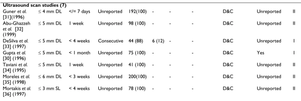
Ultrasound scan studies (7) Guner et al.
[31](1996)
≤ 4 mm DL </= 7 days Unreported 192(100) - - - D&C Unreported II
Abu-Ghazzeh et al. [32] (1999)
≤ 5 mm DL 1 week Unreported 98 (100) - - - D&C Unreported II
DeSilva et al. [33] (1997)
≤ 5 mm DL < 4 weeks Consecutive 44 (88) 6 (12) - - D&C Unreported I
Gupta et al. [30] (1996)
≤ 5 mm DL < 1 month Unreported 75 (100) - - - D&C Yes I
Taviani et al. [34] (1995)
≤ 5 mm DL 1 week Unreported 41 (100) - - - D&C Unreported II
Moreles et al. [35] (1998)
≤ 6 mm DL < 3 weeks Unreported 200(100) - - - D&C Unreported II
Mortakis et al. [36] (1997)
≤ 3 mm SL < 4 weeks Unreported 78 (100) - - - D&C Unreported II
*Numbers calculated from initial proportion of patients within these groups before missing outcome data was excluded. †Other refers to proportion of women included in the study who did not have abnormal uterine bleeding as an indication for investigation. HRT, hormone replacement therapy; NS, not specified (refers to proportion of women included in the study where the type of abnormal uterine bleeding was not specified); DB, directed biopsy; D&C, dilatation and curettage; TCRE, transcervical resection of the endometrium; DL, double layer endometrial thickness; SL, single layer endometrial thickness.
Effect of delayed verification on the diagnostic accuracy of miniature endometrial biopsy and transvaginal ultrasound in detect-ing endometrial hyperplasia
Figure 2
Effect of delayed verification on the diagnostic accuracy of miniature endometrial biopsy and transvaginal ultrasound in detect-ing endometrial hyperplasia. Pooled diagnostic odds ratios (dOR) for studies with immediate and delayed verification.
Table 1: Study characteristics and methodological quality. (Continued)
2000 1000 500 200
100 50 20
10
DOR (logarithmic scale) Delayed verification
Endometrial biopsy (n=8)
Ultrasound scan (n=7)
Total (n=11) Total (n=16) Ultrasound scan (n=8) Endometrial biopsy (n=8)
Immediate verification dOR (95% CI)
173.9 (20.0 – 1512.5)
30.6 (9.1 – 102.6)
67.2 (21.7 – 208.8)
24.9 (6.6 – 93.7)
15.6 (7.1 – 34.1)
immediately confirmed due to resource and other impli-cations. Thus diagnostic evaluations carried out in routine practice may mask the accuracy of tests.
Competing interests
None declared.
Table 2: Accuracy stratified by time delay between test performance and confirmation by chosen reference test histology.
Device (no. evaluations) & study (year published)
+ve test (sensitivity) -ve test (1-specificity) Odds ratio (95% CI)
IMMEDIATE VERIFICATION (</= 24 HOURS) (16 studies) Endometrial biopsy studies (8)
Sun-Kuie et al. [15](1992) 2/4 (0.5) 0/42 (0.0) 85.0 (3.2–2289.6) Goldberg et al. [16] (1981) 5/5 (1.0) 0/25 (0.0) 561.0 (10.0–31463.1) Sonnendecker et al. [17] (1981) 2/2 (1.0) 0/21 (0.0) 215.0 (3.5–13408.5) Kufahl et al. [18] (1997) 14/15 (0.9) 4/143 (0.03) 486.5 (50.8–4658.5) Kufahl et al. [18](1997) 3/15 (0.2) 15/154 (0.1) 2.3 (0.6–9.1) Goldschmit et al. [19] (1993) 11/14 (0.8) 1/135 (0.01) 491.3 (47.1–5127.3) Kavak et al. [20] (1996) 4/4 (1.0) 0/57 (0.0) 1035.0 (18.3–58563.1) Goldberg et al. [16] (1981) 6/6 (1.0) 0/25 (0.0) 663.0 (12.0–36690.0) Ultrasound scan studies (8)
Botsis et al. [21] (1992) 10/10 (1.0) 4/92 (0.04) 413.0 (20.8–8221.1) Garuti et al. [22] (1999) 44/46 (0.96) 196/313 (0.63) 13.1 (3.1–55.2) Haller et al. [23] (1996) 14/16 (0.88) 34/49 (0.69) 3.1 (0.6–15.3) Grigoriou et al. [24] (1996) 45/45 (0.98) 30/181 (0.17) 452.0 (27.1–7538.5) Karlsson et al. [25] (1993) 10/10(1.0) 21/78 (0.27) 56.2 (3.2–1000.5) Malinova et al. [26] (1996) 11/11 (1.0) 32/74 (0.43) 30.1 (1.7–529.5) Wolman et al. [27] (1996) 10/11(0.91) 12/39 (0.31) 22.5 (2.6–196.1) Malinova et al. [28] (1995) 7/7 (1.0) 19/54 (0.35) 27.31 (1.5–504.1) DELAYED VERIFICATION (> 24 HOURS) (11 studies)
Endometrial biopsy studies (4)
Stovall et al. [29] (1989) 8/16 (0.5) 4/149 (0.03) 36.3 (9.0–146.3) Krampl et al. [3] (1997) 14/35 (0.4) 24/284(0.0) 7.2 (3.3–16.0) Gupta et al. [30] (1996) 6/10 (0.6) 1/44 (0.02) 64.5 (6.1–677.6) Stovall et al. [29] (1989) 7/7 (1.0) 7/55 (0.13) 97.0 (5.0–1879.9) Ultrasound scan studies (7)
Guner et al. [31] (1996) 31/31 (1.0) 61/142 (0.43) 83.5 (5.0–1391.3) Abu-Ghazzeh et al. [32] (1999) 2/2 (1.0) 58/95 (0.61) 3.2 (0.2–68.6) DeSilva et al. [33] (1997) 2/3 (0.67) 10/44 (0.23) 6.8 (0.6–83.0) Gupta et al. [30] (1996) 4/4 (1.0) 22/68 (0.32) 18.6 (1.0–360.7) Taviani et al. [34] (1995) 2/2 (1.0) 16/37 (0.43) 6.5 (0.3–145.1) Moreles et al. [35] (1998) 33/37 (0.9) 37/143 (0.26) 23.6 (7.8–71.2) Mortakis et al. [36] (1997) 4/5 (0.8) 26/66 (0.39) 6.2 (0.7–58.2)
Table 3: Results of meta-regression analysis.
Outcome Explanatory variables Univariable analysis Multivariable analysis
Ratio dOR (95% CI)† P value Ratio dOR (95% CI)† P value
Endometrial hyperplasia
▪ Delay in verification of diagnosis (> 24 hours vs. < 24 hours) 0.31 (0.08–0.84) 0.089 0.26 (0.07–0.99) 0.048
▪ Study quality (category II vs. category I) 1.36 (0.34–5.53) 0.664 3.46 (0.79–15.0) 0.098
▪ Test (Endometrial biopsy vs. ultrasound endometrial thickness) 3.22 (0.83–12.55) 0.091 5.0 (1.2–20.7) 0.027
Authors' contributions
TJC and KSK conceived and designed the study with input from GtR. TJC conducted the systematic review and acquired all data. AC conducted the statistical analyses with input from KSK and GtR. TJC wrote all versions of the manuscript. KSK and GtR critically revised the manuscript for important intellectual content. All authors read and approved the final manuscript.
References
1. Fox H: Endometrial hyperplasia: a conceptual and practical approach.Gynaecology Forum 1996, 1:7-9.
2. Kurman RJ, Kaminski PF, Norris HJ: The behaviour of endome-trial hyperplasia: a long-term study of "untreated" hyperpla-sia in 170 patients.Cancer 1985, 56:403-412.
3. Krampl E, Soby B, Istre O: How representative are pipelle endometrial biopsies? A retrospective analysis of 324 biop-sies followed by transcervical resection of the endometrium or hysterectomy.Gynaecol Endosc 1997, 6:277-281.
4. Lijmer JG, Mol BW, Heisterkamp S, Bonsel GJ, Prins MH, Van der Meulen JHP, Bossuyt PM: Empirical evidence of design-related bias in studies of diagnostic tests.JAMA 1999, 282:1061-1066. 5. Reid MC, Lachs MS, Feinstein AR: Use of methodological
stand-ards in diagnostic test research. Getting better but still not good.JAMA 1995, 274:645-651.
6. Sheps SB, Schechter MT: The assessment of diagnostic tests: a survey of current medical research.JAMA 1984, 252:2418-2422. 7. Gupta JK, Chien PF, Voit D, Clark TJ, Khan KS: Ultrasonographic endometrial thickness for diagnosing endometrial pathology in women with postmenopausal bleeding: a meta-analysis.
Acta Obstet Gynecol Scand 2002, 81:799-816.
8. Clark TJ, Mann CH, Shah N, Song F, Khan KS, Gupta JK: Accuracy of outpatient endometrial biopsy in the diagnosis of endome-trial hyperplasia: a systematic quantitative review.Acta Obstet Gynecol Scand 2001, 80:784-793.
9. Cochrane Methods Working Group on Systematic Reviews of Screening and Diagnostic Tests: Recommended Methods
[http://www.cochrane.org/cochrane/sadtdoc1.htm]
10. Dunn G, Everitt B: Clinical Biostatistics. London: Edward Arnold 1995. 11. Khan KS, ter Riet G, Popay J, Nixon J, Kleijnen J: Study quality
assessment. In Undertaking Systematic Reviews of Research on Effec-tiveness. CRD's Guidance for Carrying Out or Commissioning Reviews 2nd edition. Edited by: Khan KS, Ter Riet G, Glanville J, Sowden AJ, Klei-jnen J. York: NHS Centre for Reviews and Dissemination (CRD), Uni-versity of York; 2001.
12. Sankey S, Weisfiels L, Fine M, Kapoor W: An assessment of the use of the continuity correction for sparse data in meta analysis.Comm Stat Sim Comput 1996, 25:1031-1056.
13. Sterne JAC, Egger M, Davey Smith G: Investigating and dealing with publication and other biases. In Systematic Reviews in Health Care: Meta-analysis in Context Edited by: Egger M, Davey Smith G, Alt-man DG. London: BMJ Publishing Group; 2001:189-208.
14. Song F, Sheldon T, Sutton A, Abrams K, Jones D: Methods for exploring heterogeneity in meta-analysis.Eval Health Prof 2001,
24:126-151.
15. Sun-Kuie T, Sian-Ann T, Ka-Mui C, Soo-Kim L: The diagnostic value and patient acceptability of outpatient endometrial sampling with Gynoscann. Aust NZ J Obstet Gynaecol 1992,
32:73-76.
16. Goldberg GL, Tsalacopoulos GDDA: A comparison of the Accu-rette and Vabra aspirator and uterine curettage.S Afr Med J 1982, 61:114-116.
17. Sonnendecker EW, Sevitz H, Hofmeyr GJ: Diagnostic accuracy of the accurette endometrial sampler. S Afr Med J 1982,
61:109-113.
18. Kufahl J, Pederson I, Eriksen PS, Helkjaer PE, Larsen LG, Jensen KL, de Nully P, Philipsen T, Wahlin A.: Transvaginal ultrasound, endometrial cytology sampled by Gynoscann and histology obtained by Uterine Explora Curette compared to the his-tology of the uterine specimen. A prospective study in pre-and postmenopasual women undergoing elective hysterectomy.Acta Obstet Gynecol Scand 1997, 76:790-796.
19. Goldchmit R, Katz Z, Blickstein I, Caspi B, Dgani R: The accuracy of endometrial Pipelle sampling with and without sonographic measurement of endometrial thickness.Obstet Gynecol 1993,
82:727-730.
20. Kavak Z, Ceyhan N, Pekin S: Combination of vaginal ultrasonog-raphy and pipelle sampling in the diagnosis of endometrial disease.Aust N Z J Obstet Gynaecol 1996, 36:63-66.
21. Botsis D, Kassanos D, Pyrgiotis E, Zourlas PA: Vaginal sonography of the endometrium in postmenopausal women. Clin Exp Obstet Gynecol 1992, 19:189-192.
22. Garuti G, Sambruni I, Cellani F, Garzia D, Alleva P, Luerti M: Hyster-oscopy and transvaginal ultrasonography in postmenopausal women with uterine bleeding. Int J Gynaecol Obstet 1999,
65:25-33.
23. Haller H, Matejcic N, Rukavina B, Krasevic M, Rupcic S, Mozetic D:
Transvaginal sonography and hysteroscopy in women with postmenopausal bleeding.Int J Gynecol Obstet 1996, 54:155-159. 24. Grigoriou O, Kalovidouros A, Papadias C, Antoniou G, Antonaki V,
Giannikos L: Transvaginal sonography of the endometrium in women with postmenopausal bleeding.Maturitas 1996, 23:9-14. 25. Karlsson B, Granberg S, Wikland M, Ryd W, Norstrom A: Endovag-inal scanning of the endometrium compared to cytology and histology in women with postmenopausal bleeding.Gynecol Oncol 1993, 50:173-178.
26. Malinova M, Pehlivanov B: Transvaginal sonography and proges-terone challenge for identifying endometrial pathology in postmenopausal women.Int J Gynecol Obstet 1996, 52:49-53. 27. Wolman I, Sagi J, Ginat S, Jaffa AJ, Hartoov J, Jedwab G: The
sensi-tivity and specificity of vaginal sonography in detecting endometrial abnormalities in women with postmenopausal bleeding.J Clin Ultrasound 1996, 24:79-82.
28. Malinova M, Pehlivanov B: Transvaginal sonography and endometrial thickness in patients with postmenopausal uterine bleeding.Eur J Obstet Gynecol Reprod Biol 1995, 58:161-165. 29. Stovall TG, Solomon SK, Ling FW: Endometrial sampling prior to
hysterectomy.Obstet Gynecol 1989, 73:405-409.
30. Gupta JK, Wilson S, Desai P, Hau C: How should we investigate women with postmenopausal bleeding? Acta Obstet Gynecol Scand 1996, 75:475-479.
31. Guner H, Tiras MB, Karabacak O, Sarikaya H, Erdem M, Yildirim M:
Endometrial assessment by vaginal ultrasonography might reduce endometrial sampling in patients with postmenopau-sal bleeding: A prospective study.Aust N Z J Obstet Gynaecol 1996, 36:175-178.
32. Abu-Ghazzeh Y, Shakoury WA, Barqawi R: Comparative study of transvaginal hysterosonography and biopsy for the evalua-tion of post-menopausal bleeding. Ann Saudi Med 1999,
19:116-119.
33. De Silva BY, Stewart K, Steven JD, Sathanandan M: Transvaginal ultrasound measurement of endometrial thickness and endometrial pipelle sampling as an alternative diagnostic procedure to hysteroscopy and dilatation and curettage in the management of post-menopausal bleeding. J Obstet Gynaecol 1997, 17:399-402.
34. Taviani A, Braccini S, Toniazzi P, Pantani P, Costamagna V, Gambini G, Pancanti V: Transvaginal echography in patients with post-menopausal metrorrhagia.Minerva Ginecol 1995, 47:369-372. 35. Morales FJ, Dualde D, Marinaro A: Value of vaginal ultrasound in
the diagnosis of postmenopausal metrorrhagia. Radiologia 1998, 40:255-262.
36. Mortakis AE, Mavrelos K: Transvaginal ultrasonography and hysteroscopy in the diagnosis of endometrial abnormalities.
J Am Assoc Gynecol Laparosc 1997, 4:449-452.
Pre-publication history
The pre-publication history for this paper can be accessed here: