R E S E A R C H
Open Access
APLP1 as a cerebrospinal fluid biomarker
for
γ
-secretase modulator treatment
Simon Sjödin
1*, Kerstin K. A. Andersson
1, Marc Mercken
2, Henrik Zetterberg
1,3, Herman Borghys
2, Kaj Blennow
1and Erik Portelius
1Abstract
Introduction:Alzheimer’s disease brains are characterized by extracellular plaques containing the aggregated amyloidβ42(Aβ42) peptide and intraneuronal tangles containing hyperphosphorylated tau. Aβ42is produced by sequential processing of the amyloid precursor protein (APP) byβ-secretase followed byγ-secretase. Substantial efforts have been put into developing pharmaceuticals preventing the production or increasing the clearance of Aβ42. However, treatments inhibitingγ-secretase have proven disappointing due to off-target effects. To circumvent these effects,γ-secretase modulators (GSMs) have been developed, which rather than inhibitingγ-secretase shift its preference into producing less aggregation-prone shorter Aβpeptides. Belonging to the same family of proteins as APP, amyloid-like protein 1 (APLP1) is also a substrate forγ-secretase. Herein we investigated whether the GSM E2012 affects APLP1 processing in the central nervous system by measuring APLP1 peptide levels in cerebrospinal fluid (CSF) before and after E2012 treatment in dogs.
Methods:An in-house monoclonal APLP1 antibody, AP1, was produced and utilized for immunopurification of APLP1 from human and dog CSF in a hybrid immuno-affinity mass spectrometric method. Seven dogs received a single dose of 20 or 80 mg/kg of E2012 in a randomized cross-over design and CSF was collected prior to and 4, 8 and 24 hours after dosing.
Results:We have identified 14 CSF APLP1 peptides in humans and 12 CSF APLP1 peptides in dogs. Of these, seven were reproducibly detectable in dogs who received E2012. We found a dose-dependent relative increase of the CSF peptides APLP1β17, 1β18 and 1β28 accompanied with a decrease of 1β25 and 1β27 in response to E2012 treatment. All peptides reverted to baseline over the time of sample collection.
Conclusion:We show an in vivo effect of the GSM E2012 on the processing of APLP1 which is measurable in CSF. These data suggest that APLP1 peptides may be used as biomarkers to monitor drug effects of GSMs on
γ-secretase processing in clinical trials. However, this requires further investigation in larger cohorts, including studies in man.
Introduction
Alzheimer’s disease (AD) is a progressive neurodegener-ative disorder and the most prevalent form of dementia [1]. It is characterized by extracellular plaques, containing aggregated amyloid-β(Aβ) peptides [2], and intraneuronal tangles consisting of hyperphosphorylated tau [3]. In the amyloid cascade hypothesis, it is postulated that there is an imbalance in the production and/or clearance of Aβ
which is believed to cause a series of events including microglial activation, oxidative stress, neuronal dysfunc-tion, formation of tangles and inevitable neurodegenera-tion [4]. For these reasons, targeting the producneurodegenera-tion of the aggregation prone and potentially toxic 42 amino acid residue-long variant of Aβ(Aβ42) has been subject to
ex-tensive research with the aim of developing disease modi-fying compounds that inhibits or modulates the enzymes responsible for the formation of Aβ (see elaborating re-views elsewhere [5–7]).
The amyloid precursor protein (APP) is subjected to proteolytic processing by three secretases: α-secretase, β-secretase and γ-secretase. The proteolytic activity of * Correspondence:simon.sjodin@neuro.gu.se
1Institute of Neuroscience and Physiology, Department of Psychiatry and Neurochemistry, The Sahlgrenska Academy at the University of Gothenburg, Sahlgrenska University Hospital, Mölndal S-431 80, Sweden
Full list of author information is available at the end of the article
α-secretase is likely attributed to the protein disintegrin and metalloproteinase domain-containing protein 10 (ADAM10) [8]; β-secretase has been identified as β-site APP-cleaving enzyme (BACE) [9]; whereasγ-secretase is a multisubunit protein complex consisting of nicastrin (NCSTN), anterior pharynx-defective 1 (APH-1), prese-nilin enhancer 2 (PEN2) and the N- and C-terminal fragments of presenilin 1 or 2 (PS1 or PS2) [10]. Aβ42is
produced through the amyloidogenic pathway where APP is cleaved by β-secretase [9] and subsequently by γ -secretase [11]. Alternatively Aβ42production is prevented
in the non-amyloidogenic pathway where α-secretase ra-ther thanβ-secretase cleaves APP within the Aβsequence generating shorter Aβpeptides [12].
Secretases are an appealing target to alter the meta-bolism of Aβ. However, recent clinical trials where AD patients were treated withγ-secretase inhibitors failed to reach their primary clinical endpoints; the cognitive de-cline was even worse in the treatment arm than in placebo [13, 14]. A diverse array of transmembrane proteins have been identified as γ-secretase substrates [15], including Notch [16, 17], and the problems associated with γ -secretase inhibitor treatment may be explained by the physiological functions of these substrates [15].
To overcome the possible negative effects of inhibiting γ-secretase, γ-secretase modulators (GSMs) have been developed to shift the production from the amyloidogenic Aβ42to less aggregation-prone peptides (e.g., Aβ38)
with-out affecting the release of the intracellular domains of APP and otherγ-secretase substrates such as Notch1, cad-herins, Erb4 and EphB2 [18]. Thus, these compounds modulate the cleavage pattern of Aβ without inhibiting, for example, Notch signaling. The effect of GSMs on APP processing has previously been shown to be measurable in vivo in cerebrospinal fluid (CSF) from, for example, monkeys [19] and guinea pigs [20], where the level of Aβ42 decreased and Aβ37 or Aβ38 increased [19, 20]. In
agreement with these studies, we recently showed that dogs treated with the GSM E2012 displayed a distinct shift in the CSF Aβ peptide pattern with decreased levels of Aβ42 as well as Aβ39 and Aβ40 accompanied with
in-creased levels of Aβ37[21]. The effects of different GSMs
on Aβproduction have been summarized in a review by Crump et al. [22].
Amyloid-like protein 1 (APLP1) and APP share, to a large extent, similar structural domains and are both suggested to be involved in neurite outgrowth, cell adhe-sion and neuronal migration [23]. APLP1 and APP are also known to form heterodimers with possible implica-tions in cell adhesion and synaptogenesis [24, 25]. APLP1 has been suggested to be processed by the same enzymatic activities as APP includingγ-secretase andα-secretase, gen-erating a p3-like peptide called ALP-1 [24, 26], similar to the putative p3 fragment identified as a possible proteolytic
product from APP [27]. While it is established that γ -secretase is responsible for the C-terminal processing of APLP1 [28–30], the secretase cleaving at the N-terminus has been suggested to be either α- [26] or β -secretase [28, 31].
Recently, it was shown that APLP is processed into short Aβ-like peptides (APLP1β25, 1β27 and 1β28) [28] and that the concentrations of CSF APLP1β25 and 1β27 were decreased while the concentration of CSF APLP1β28 was unchanged in four different PS1 mutation carriers [32] leading to an increased APLP1β28 to total APLP1β ratio [28]. In addition, cells treated with the Aβ42raising
GSM S2474, or cells harboring certain PS1 mutations were also found to have an increased APLP1β28 to total APLP1β ratio and it was suggested that this ratio may reflect the Aβ42 production in the brain [28]. However,
how the S2474 treatment or PS1 mutations affected the APLP1β28 concentrations were not reported. Whether APLP1 peptides can be used as CSF AD biomarkers re-flecting Aβ42production needs to be investigated in large
clinical studies, including early clinical stage of AD, AD dementia and control subjects, in which other CSF and imaging biomarkers are included.
In order to characterize APLP1 in CSF, we developed a monoclonal antibody directed against APLP1. We then used hybrid immuno-affinity-based enrichment of CSF APLP1 peptides from dogs followed by mass spectrometry (MS) analysis. In the present study we tested the hypoth-esis that peptides derived from APLP1 can be used as in vivo biomarkers reflecting treatment with the second generation GSM E2012.
Methods
Chemicals
The γ-secretase modulator E2012 (cas no. 870843-42-8, (3E)-1-[(1S )-1-(4-fluorophenyl)ethyl]-3-[[3-methoxy-4- (4-methyl-1H-imidazol-1-yl)phenyl]methylene]-2-piperidi-none) was synthesized according to the described proced-ure [33].
Experimental animals
polypropylene tubes, immediately placed on dry ice and stored at−80 °C until analysis. The method for implanting and collecting CSF via the cannula in dogs has been de-scribed elsewhere [34]. In brief, a hole was drilled in the cranium of the dogs into which the cannula was screwed. The cannula was positioned at the following coordinates: 38–40 mm rostral to the occipital protuberance and 8 mm lateral to the center of the sagittal crest. The study was con-ducted according to protocol 2009-252-SD MECHPHA, approved by the Ethical Committee on Laboratory Animal Testing (Ethische Commissie Dierproeven, Johnson & Johnson Pharmaceutical Research and Development, Beerse, Belgium).
Generation of anti-APLP1 antibody
A monoclonal antibody against APLP1, named AP1, was generated by immunization of 8-week-old Balb/c mice with the KLH-conjugated peptide APLP1 (amino acid 568–579, [UniProtKB:P51693]; Caslo ApS, Lyngby, Denmark) in complete Freund’s adjuvant (Sigma-Aldrich Co., St Louis, MO, USA). After three dosages (approximately 75 μg/ mouse), the spleen was removed and B cells were fused with the myeloma cell line SP2/0 following standard pro-cedure. Approximately 10 days after fusion, cell media were screened for APLP1 antibodies using APLP1 full length and neurogranin (Caslo ApS) as a negative control. Clones which reacted with the APLP1 protein were further grown, subcloned and subsequently frozen in liquid nitro-gen. The isotype was determined using a commercially available kit (Pierce Rapid Isotyping Kit-Mouse; Thermo Fisher Scientific Inc., Waltham, MA, USA). Finally, anti-bodies were purified using a protein G column (GE Healthcare Bio-Sciences AB, Uppsala, Sweden).
Immuno-affinity mass spectrometry
CSF collected prior to dosing and 4, 8 and 24 hours after dosing with 20 or 80 mg/kg E2012 was analyzed by hy-brid immuno-affinity MS as described previously with some modifications [21]. In brief, 4 μg of the in-house developed mouse monoclonal anti-APLP1 antibody AP1 was incubated with 25 μl magnetic beads conjugated to sheep anti-mouse IgG (Dynabeads M280, Invitrogen, Thermo Fisher Scientific Inc.). Beads with the AP1 anti-body complex were then washed in phosphate-buffered saline and added to 600 μl CSF containing 0.0025 % Tween, to a total volume of 625 μl. CSF with added beads was incubated over night at +8 °C on a rocking platform. Elution of APLP1 was performed in a five-step procedure using a KingFisher mL Magnetic Particle Processors system (Thermo Fisher Scientific Inc.). The eluate was dried and redissolved in 5 μl 0.1 % formic acid in 20 % acetonitrile. The sample in solution was mixed in a 2:1 ratio with a sample matrix containing 15 g/l alpha-cyano-4-hydroxycinnamic acid and 0.1 %
trifluoroacetic acid in acetonitrile and spotted on an MTP 384 target plate polished steel TF (Bruker Daltonics GmbH, Bremen, Germany) with a seed layer containing 20 g/l alpha-cyano-4-hydroxycinnamic acid in 10 % me-thanol and 90 % acetone. The samples were analyzed as duplicates by matrix-assisted laser desorption ionization time-of-flight/time-of-flight (MALDI-TOF/TOF) using an UltrafleXtreme (Bruker Daltonics GmbH) mass spectrom-eter operating at positive reflector mode. The sample spectra acquired were processed using flexAnalysis v3.3 (Bruker Daltonics GmbH) by performing baseline subtrac-tion, smoothening and internal calibration based on the theoretical monoisotopic masses of APLP1 fragments 1β17 (1615.79m/z), 1β18 (1728.88m/z), 1β22 (2143.11m/
z), 1β25 (2328.19m/z), 1β27 (2472.24m/z) and 1β28 (2585.32m/z). Exported peak lists were subjected to pro-cessing by an in-house MATLAB (Mathworks Inc., Natick, Massachusetts, USA) program which, in short, relative to the monoisotopic peak, integrated the peak areas within the limits of−2 to +5m/z. Following this, the integrated peak areas in each specific spectrum were nor-malized as the percentage of the total area of all peaks measured. Duplicate measurements were averaged and then a fold-change was calculated for samples acquired at 4, 8 and 24 hours against the sample collected prior to dosing as baseline.
Tandem mass spectrometric identification of peptides
Employing collision-induced dissociation and the LIFT technology of the UltrafleXtreme, tandem MS (MS/MS) spectra of selected APLP1 peptides were acquired. These were processed and annotated in the BioTools software v3.2 (Bruker Daltonics GmbH) by employing an in-house Mascot Deamon server (v2.3.2; Matrix Science Ltd. London, UK) search against an in-house designated APLP1 [UniProtKB:P51693] database. The parameters for searching the database were set to singly charged ions, variable methionine oxidation, a mass tolerance of 200 ppm, MS/MS tolerance of 0.5 Da and a MALDI-TOF/ TOF instrumentation. MS and MS/MS spectra were also acquired from human CSF using the same sample prepar-ation methodology as described above. CSF samples were supplied by clinical routine at the Clinical Neurochemistry Laboratory, The Sahlgrenska University Hospital, Mölndal, Sweden, in accordance with ethical approval from the re-gional ethics committee at the University of Gothenburg. The samples were de-identified by pooling.
Statistical analysis
Significant results were considered differences with a calculatedp-value of <0.05.
Results
Identification of APLP1 peptides in CSF
By combing the selectivity of the in-house mouse mono-clonal antibody, AP1, and MS, we here for the first time show that several APLP1 derived peptides may be used as biomarkers for target engagement of the GSM E2012 in vivo.
The amino acid sequence for APLP1 for Canis
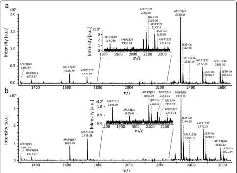
famil-iaris and Homo sapiens has a 100 % homology [Uni-protKB:J9JHP8 versus UniprotKB:P51693]. In total, 14 APLP1 peptides were identified in human CSF and 12 in dog CSF of which nine were confirmed by MS/MS (see Fig. 1 and Tables 1 and 2 for all identified peptides). All confirmed peptides started with the aspartic acid found at amino acid 568 of APLP1 [UniprotKB:P51693] and
included peptides ranging between APLP1β13 up to APLP1β28. An additional peak that had the mass of the peptide plus 16 Da, corresponding to oxidation of methio-nine at position 21, was also observed for several of the APLP peptides (Fig. 1). Fragment ion mass spectra used for the identification of APLP1β17 and APLP1β28 are shown in Fig. 2a and b, respectively.
Modulation of APLP1 processing by E2012 in dogs
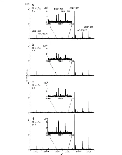
In total, seven APLP1 peptides were reproducibly de-tected in all dog CSF samples (APLP1β17, 1β18, 1β21, 1β22, 1β25, 1β27 and 1β28) and their response to treat-ment with E2012 was further investigated. See Fig. 3 for representative mass spectra from a dog treated with 80 mg/kg of the GSM E2012.
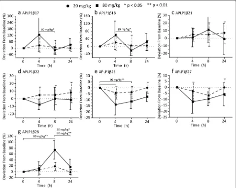
Four hours after administration of the GSM E2012 (80 mg/kg), we found higher levels of APLP1β17 (p= 0.027) and APLP1β18 (p= 0.016) compared to 8 hours
post-treatment. The increases reverted to baseline levels at 8 hours post-treatment and remained at baseline levels 24 hours post-treatment (Fig. 4a and b). No signi-ficant differences were observed for the two peptides after the 20 mg/kg dose (Fig. 4a and b). After adminis-tration of 80 mg/kg, the levels of APLP1β25 were signifi-cantly decreased both at 4 hours (p= 0.0032) and 8 hours (p= 0.0032) post-treatment (Fig. 4e). The 20 mg/kg dose showed a similar trend without reaching statistical signi-ficance (Fig. 4e). APLP1β28 showed a dose-dependent increase at 8 hours post-treatment which was significant for 80 mg/kg (p= 0.0032) but not 20 mg/kg (Fig. 4g).
However, the increase at 8 hours was significant relative to 24 hours post-treatment for both doses (p= 0.0032 and p= 0.047 for 80 mg/kg and 20 mg/kg, respectively; Fig. 4g).
APLP1β21 and APLP1β22 did not show any significant deviations from baseline for either of the two doses (Fig. 4c and d). The level of APLP1β27 showed a trend towards decreased levels in a dose-dependent manner in response to treatment but it did not reach statistical sig-nificance for either of the doses (Fig. 4f ).
Discussion
In the present study, we show that APLP1 in CSF is present as a variety of short peptides. For the first time we also demonstrate that the levels of several CSF APLP1 peptides are markedly affected in response to treatment with the GSM E2012 suggesting that APLP1-derived pep-tides may be used as biomarkers to prove target engage-ment for this type of treatengage-ments. To our knowledge there are no studies of the effect of γ-secretase modulators on APLP1 processing in vivo to date.
Previously, using an explorative proteomics approach, we identified eight endogenous APLP1 peptides in hu-man CSF including APLP1β17, 1β18, 1β25 and 1β27 [35]. By employing the targeted approach described herein we have identified and confirmed the presence of the previously identified peptides in CSF [28, 35] and have now extended the list of identified endogenous APLP1 peptides. A previous report showed an increase of APLP1β28 relative to the total amount of APLP1 pep-tides in CSF from AD subjects [28] while we recently showed that the levels of APLP1β25, 1β27 and 1β28 in Table 1APLP1 precursor ion identification in full mass spectrum
Sequence Theoretical
mass (Da)
Observed mass (Da) dogs
ppm dogs Observed mass (Da) human
ppm human Annotation
DELAPAGTGVSRE 1300.63 1300.60 −172 1300.63 39 APLP1β13
DELAPAGTGVSREA 1371.66 1371.65 −114 1371.67 20 APLP1β14
DELAPAGTGVSREAVSG 1614.78 1614.78 −8.5 1614.78 −0.058 APLP1β17
DELAPAGTGVSREAVSGL 1727.87 1727.87 13 1727.87 0.12 APLP1β18
DELAPAGTGVSREAVSGLL 1840.95 1840.96 12 1840.95 −8.2 APLP1β19
DELAPAGTGVSREAVSGLLI 1954.04 1954.04 8.5 1954.04 −8.5 APLP1β20
DELAPAGTGVSREAVSGLLIM 2085.08 2085.08 −2.4 2085.08 1.1 APLP1β21
DELAPAGTGVSREAVSGLLIMG 2142.10 2142.10 −7.6 2142.10 −0.46 APLP1β22
DELAPAGTGVSREAVSGLLIMGA 2213.14 – – 2213.13 −13 APLP1β23
DELAPAGTGVSREAVSGLLIMGAG 2270.16 – – 2270.16 8.5 APLP1β24
DELAPAGTGVSREAVSGLLIMGAGG 2327.18 2327.18 3.3 2327.18 1.0 APLP1β25
DELAPAGTGVSREAVSGLLIMGAGGG 2384.20 2384.19 −33 2384.19 −65 APLP1β26
DELAPAGTGVSREAVSGLLIMGAGGGS 2471.23 2471.23 −0.53 2471.23 −0.91 APLP1β27
DELAPAGTGVSREAVSGLLIMGAGGGSL 2584.32 2584.32 0.72 2584.32 0.31 APLP1β28
Full mass spectra of amyloid-like protein 1 (APLP1) immunoprecipitated from dog and human cerebrospinal fluid were acquired by MALDI-TOF/TOF. The Table shows the amino acid sequences, the observed masses in Da in dogs and human and their deviation from the theoretical peptide mass in ppm for the peptides identified
Table 2Tandem mass spectrometric identification of APLP1 peptides
Peptide Observedm/z ppm Ion score Expect a/b/y ions
APLP1β14 1372.71 30.4 48 1.6x10⁻⁵
–/4/6 APLP1β17 1615.83 23.7 79 1.3x10⁻⁸ 4/6/11
APLP1β18 1728.91 21.9 65 3.0x10⁻⁷
–/8/10 APLP1β19 1842.06 56.7 18 1.7x10⁻²
–/5/4 APLP1β21 2086.19 48.1 24 3.9x10⁻³
–/6/– APLP1β23 2214.14 −2.91 15 3.3x10⁻²
–/4/– APLP1β25 2328.51 138 79 1.3x10⁻⁸ 7/6/11
APLP1β27 2472.31 27.8 100 9.4x10⁻¹¹ 9/5/9
APLP1β28 2585.40 28.3 93 5.3x10⁻¹⁰ 9/11/10
CSF from Down’s syndrome patients are decreased as compared to healthy controls [36]. However, whether CSF APLP1β28 can be used as an AD or Down’s syn-drome biomarker needs to be investigated in large clin-ical studies.
Of the 12 identified APLP1 peptides in dog CSF, we show that several are affected in response to treatment with the GSM E2012 and that some peptides are affected in a dose-dependent manner as the effect was more pro-nounced with the higher dose (80 mg/kg). We found
Fig. 2Fragment ion mass spectra of APLP1 peptides. Fragment mass spectra generated by tandem mass spectrometry of the peptides
increased relative levels of APLP1β17, 1β18 and 1β28 in response to treatment accompanied by decreased rela-tive levels of APLP1β25 and 1β27. The changes in the levels for all of the peptides seem to revert towards base-line levels over time. For APLP1β21 and 1β22 we found no significant difference. These findings suggest that the modulation of the APLP1 processing affects the production over the range of peptides present, not only a single peptide or exclusively shorter or longer peptides. Furthermore, the transmembrane region of APLP1 ranges from position 581–603. Thus all identified APLP1 peptides, except APLP1β13, are located within this region and are accessible forγ-secretase processing.
Previously we have shown that the CSF levels of Aβ37
increases and Aβ42 decreases in dogs treated with the
GSM E2012 [21]. Here we show in the very same mater-ial that the levels of several APLP1 peptides are affected in response to E2012 treatment. Thus investigating the level of APLP1 peptides in GSM treatment trials may be highly informative and possibly increase our understand-ing of the mechanistics and dynamics of APLP process-ing in the disease.
One limitation with the current study is that the epi-tope of the APLP1 antibody used (AP1) has not been de-termined. However MS analysis, including MS/MS, confirms that the peptides studied in the present study
Fig. 4Modulation ofγ-secretase processing of APLP1 in dogs. APLP1 was immunoprecipitated from cerebrospinal fluid collected from dogs prior to and 4, 8 and 24 hours after receiving 20 and 80 mg/kg of theγ-secretase modulator E2012. Using MALDI-TOF/TOF, full mass spectra were recorded in which the intensity of the peak corresponding to a peptide was normalized against the intensity of all the peaks measured in the spectrum. Shown is the percent deviation from the sample collected prior to dosing (baseline) for the peptidesaAPLP1β17,bAPLP1β18,cAPLP1β21,
indeed are APLP1 derived. It should also be noted that the ratio between the different endogenous APLP1 pep-tides detected cannot be interpreted as a direct reflection of their absolute abundance in the CSF. This is because the ionization efficiency might be different for the differ-ent peptides. Phase I trials in man are needed to further study the potential of CSF APLP1 peptides to verify tar-get engagement of GSM drug candidates as well as long-term clinical trials to examine if these biomarkers also may predict a beneficial clinical treatment effect.
With GSMs, one of the benefits is to avoid the poten-tial inhibition of processing of non-intended substrates such as Notch [16, 17] which has been one of the main concerns when using γ-secretase inhibitors. However, GSMs have also shown an effect on Notch processing but in respect to cleavage site preference and not inhib-ition [37]. How GSMs affect the processing of the sub-strates is to a large extent unclear. It is likely dependent on the particular modulator used and the target with which it interacts [22]. First generation GSMs have been hypothesized to interact with the substrate, the secre-tase, or both [22], and it has been further speculated that, in the instance of modulators targeting the APP and Aβsequences, this might affect the processing by adjust-ing the position of the amino acid sequence in relation to the membrane [38]. Second-generation GSMs, to which E2012 belongs, have been shown rather to interact with the subunits of γ-secretase [22]. Specifically, E2012 has been indicated to bind to the N-terminal fragment of PS1 in a competitive manner [39, 40].
Conclusions
We have identified a number of APLP1 peptides in human and dog CSF. Furthermore we have shown that γ-secretase modulation, using the GSM E2012, affects APLP1 proteolytic processing in vivo and that this is reflected in the relative levels of a number of APLP1 peptides. The soluble nature of the APLP1 peptides [24, 28] and the fact that APLP1 is a γ-secretase sub-strate, as indicated herein and shown elsewhere [28–30], suggests that secreted APLP1 fragments may be inform-ative when investigating γ-secretase cleavage products in AD. Our findings show that APLP1 peptides can be used as in vivo biomarkers for E2012 treatment, which may be translated to other second-generation GSMs.
Abbreviations
Aβ:Amyloidβ; AD: Alzheimer’s disease; APLP1: Amyloid-like protein 1; APP: Amyloid precursor protein; CSF: Cerebrospinal fluid; GSM:γ-Secretase modulator; MALDI-TOF/TOF: Matrix-assisted laser desorption ionization time-of-flight/time-of-flight; MS: Mass spectrometry; MS/MS: Tandem mass spectrometry; PS: Presenilin.
Competing interests
HB and MM are employed by Janssen. SS, KKAA, HZ, KB and EP declare that they have no competing interests.
Authors’contributions
SS performed the hybrid immuno-affinity mass spectrometry experiments, statistical analyses and drafted the manuscript. HB designed the study, facilitated the animal treatment regime and CSF collection and revised the draft for important intellectual content. KKAA produced the monoclonal antibody and revised the draft for important intellectual content. MM designed the study and revised the draft for important intellectual content. HZ designed the study and revised the draft for important intellectual content. KB designed the study, designed the production of anti-APLP antibodies and revised the draft for important intellectual content. EP designed the study and drafted the manuscript. All authors interpreted the data. All authors agree with the presented findings and read and approved the final manuscript.
Acknowledgements
This study was supported by grants from the Research Council, Sweden (grant #14002), Hjärnfonden, and the Torsten Söderberg Foundation at the Royal Academy of Sciences.
Author details
1Institute of Neuroscience and Physiology, Department of Psychiatry and Neurochemistry, The Sahlgrenska Academy at the University of Gothenburg, Sahlgrenska University Hospital, Mölndal S-431 80, Sweden.2Janssen Research and Development, Beerse, Belgium.3UCL Institute of Neurology, University College London, London, UK.
Received: 21 August 2015 Accepted: 28 October 2015
References
1. Mayeux R, Stern Y. Epidemiology of Alzheimer disease. Cold Spring Harb Perspect Med. 2012;2:1–18. doi:10.1101/cshperspect.a006239.
2. Masters CL, Simms G, Weinman NA, Multhaup G, McDonald BL, Beyreuther K. Amyloid plaque core protein in Alzheimer disease and Down syndrome. Proc Natl Acad Sci U S A. 1985;82:4245–9.
3. Grundke-Iqbal I, Iqbal K, Tung YC, Quinlan M, Wisniewski HM, Binder LI. Abnormal phosphorylation of the microtubule-associated protein tau (tau) in Alzheimer cytoskeletal pathology. Proc Natl Acad Sci U S A.
1986;83:4913–7.
4. Hardy JA, Higgins GA. Alzheimer’s disease: the amyloid cascade hypothesis. Science. 1992;256:184–5.
5. De Strooper B. Lessons from a failed gamma-secretase Alzheimer trial. Cell. 2014;159:721–6. doi:10.1016/j.cell.2014.10.016.
6. Mangialasche F, Solomon A, Winblad B, Mecocci P, Kivipelto M. Alzheimer’s disease: clinical trials and drug development. Lancet Neurol. 2010;9:702–16. http://dx.doi.org/10.1016/S1474-4422(10)70119-8 (accessed 2015-11-09). 7. Citron M. Alzheimer’s disease: strategies for disease modification. Nat Rev
Drug Discov. 2010;9:387–98.
8. Kuhn PH, Wang H, Dislich B, Colombo A, Zeitschel U, Ellwart JW, et al. ADAM10 is the physiologically relevant, constitutive alpha-secretase of the amyloid precursor protein in primary neurons. EMBO J. 2010;29:3020–32. doi:10.1038/emboj.2010.167.
9. Vassar R, Bennett BD, Babu-Khan S, Kahn S, Mendiaz EA, Denis P, et al. Beta-secretase cleavage of Alzheimer’s amyloid precursor protein by the transmembrane aspartic protease BACE. Science. 1999;286:735–41. 10. Kimberly WT, LaVoie MJ, Ostaszewski BL, Ye W, Wolfe MS, Selkoe DJ.
Gamma-secretase is a membrane protein complex comprised of presenilin, nicastrin, Aph-1, and Pen-2. Proc Natl Acad Sci U S A. 2003;100:6382–7. doi:10.1073/pnas.1037392100.
11. De Strooper B, Saftig P, Craessaerts K, Vanderstichele H, Guhde G, Annaert W, et al. Deficiency of presenilin-1 inhibits the normal cleavage of amyloid precursor protein. Nature. 1998;391:387–90. doi:10.1038/34910.
12. Esch FS, Keim PS, Beattie EC, Blacher RW, Culwell AR, Oltersdorf T, et al. Cleavage of amyloid beta peptide during constitutive processing of its precursor. Science. 1990;248:1122–4.
13. Doody RS, Raman R, Farlow M, Iwatsubo T, Vellas B, Joffe S, et al. A phase 3 trial of semagacestat for treatment of Alzheimer’s disease. N Engl J Med. 2013;369:341–50. doi:10.1056/NEJMoa1210951.
phase 2 study of mild to moderate Alzheimer disease. Arch Neurol. 2012;69:1430–40. doi:10.1001/archneurol.2012.2194.
15. McCarthy JV, Twomey C, Wujek P. Presenilin-dependent regulated intramembrane proteolysis andγ-secretase activity. Cell Mol Life Sci. 2009;66:1534–55. doi:10.1007/s00018-009-8435-9.
16. De Strooper B, Annaert W, Cupers P, Saftig P, Craessaerts K, Mumm JS, et al. A presenilin-1-dependent gamma-secretase-like protease mediates release of Notch intracellular domain. Nature. 1999;398:518–22. doi:10.1038/19083.
17. Struhl G, Greenwald I. Presenilin is required for activity and nuclear access of Notch in Drosophila. Nature. 1999;398:522–5. doi:10.1038/19091. 18. Barthet G, Georgakopoulos A, Robakis NK. Cellular mechanisms of
gamma-secretase substrate selection, processing and toxicity. Prog Neurobiol. 2012;98:166–75. doi:10.1016/j.pneurobio.2012.05.006. 19. Ling IF, Golde TE, Galasko DR, Koo EH. Modulation of Abeta42 in vivo by
gamma-secretase modulator in primates and humans. Alzheimers Res Ther. 2015;7:55. doi:10.1186/s13195-015-0137-y.
20. Portelius E, Appelkvist P, Strömberg K, Höglund K. Characterization of the effect of a novelγ-secretase modulator on Aβ: a clinically translatable model. Curr Pharm Des. 2014;20:2484–90.
21. Portelius E, Van Broeck B, Andreasson U, Gustavsson MK, Mercken M, Zetterberg H, et al. Acute effect on the Abeta isoform pattern in CSF in response to gamma-secretase modulator and inhibitor treatment in dogs. J Alzheimers Dis. 2010;21:1005–12. doi:10.3233/JAD-2010-100573. 22. Crump CJ, Johnson DS, Li YM. Development and mechanism of
gamma-secretase modulators for Alzheimer’s disease. Biochemistry. 2013;52:3197–216. doi:10.1021/bi400377p.
23. Jacobsen KT, Iverfeldt K. Amyloid precursor protein and its homologues: a family of proteolysis-dependent receptors. Cell Mol Life Sci. 2009;66:2299–318. doi:10.1007/s00018-009-0020-8.
24. Minogue AM, Stubbs AK, Frigerio CS, Boland B, Fadeeva JV, Tang J, et al. gamma-secretase processing of APLP1 leads to the production of a p3-like peptide that does not aggregate and is not toxic to neurons. Brain Res. 2009;1262:89–99. doi:10.1016/j.brainres.2009.01.008.
25. Baumkotter F, Wagner K, Eggert S, Wild K, Kins S. Structural aspects and physiological consequences of APP/APLP trans-dimerization. Exp Brain Res. 2012;217:389–95. doi:10.1007/s00221-011-2878-6.
26. Eggert S, Paliga K, Soba P, Evin G, Masters CL, Weidemann A, et al. The proteolytic processing of the amyloid precursor protein gene family members APLP-1 and APLP-2 involves alpha-, beta-, gamma-, and epsilon-like cleavages: modulation of APLP-1 processing by n-glycosylation. J Biol Chem. 2004;279:18146–56. doi:10.1074/jbc.M311601200.
27. Haass C, Schlossmacher MG, Hung AY, Vigo-Pelfrey C, Mellon A, Ostaszewski BL, et al. Amyloid beta-peptide is produced by cultured cells during normal metabolism. Nature. 1992;359:322–5. doi:10.1038/359322a0.
28. Yanagida K, Okochi M, Tagami S, Nakayama T, Kodama TS, Nishitomi K, et al. The 28-amino acid form of an APLP1-derived Abeta-like peptide is a surrogate marker for Abeta42 production in the central nervous system. EMBO Mol Med. 2009;1:223–35. doi:10.1002/emmm.200900026.
29. Naruse S, Thinakaran G, Luo JJ, Kusiak JW, Tomita T, Iwatsubo T, et al. Effects of PS1 deficiency on membrane protein trafficking in neurons. Neuron. 1998;21:1213–21.
30. Walsh DM, Fadeeva JV, LaVoie MJ, Paliga K, Eggert S, Kimberly WT, et al. gamma-Secretase cleavage and binding to FE65 regulate the nuclear translocation of the intracellular C-terminal domain (ICD) of the APP family of proteins. Biochemistry. 2003;42:6664–73. doi:10.1021/bi027375c. 31. Li Q, Sudhof TC. Cleavage of amyloid-beta precursor protein and
amyloid-beta precursor-like protein by BACE 1. J Biol Chem. 2004;279:10542–50. doi:10.1074/jbc.M310001200.
32. Tagami S, Okochi M, Yanagida K, Kodama T, Arai T, Kuwano R, et al. Relative ratio and level of amyloid-beta 42 surrogate in cerebrospinal fluid of familial Alzheimer disease patients with presenilin 1 mutations. Neurodegener Dis. 2014;13:166–70. doi:10.1159/000355258.
33. Kimura T, Kawano K, Doi E, Kitazawa N, Shin K, Miyagawa T, et al. inventors; Cinnamide compound patent PCT Int. Appl. WO2005115990(A1). 2005. 34. Wilsson-Rahmberg M, Olovson SG, Forshult E. Method for long-term
cerebrospinal fluid collection in the conscious dog. J Invest Surg. 1998;11:207–14. 35. Holtta M, Zetterberg H, Mirgorodskaya E, Mattsson N, Blennow K, Gobom J.
Peptidome analysis of cerebrospinal fluid by LC-MALDI MS. PLoS One. 2012;7:e42555. doi:10.1371/journal.pone.0042555.
36. Portelius E, Holtta M, Soininen H, Bjerke M, Zetterberg H, Westerlund A, et al. Altered cerebrospinal fluid levels of amyloid beta and amyloid precursor-like protein 1 peptides in Down’s syndrome. Neuromolecular Med. 2014;16:510–6. doi:10.1007/s12017-014-8302-1.
37. Wanngren J, Ottervald J, Parpal S, Portelius E, Stromberg K, Borgegard T, et al. Second generation gamma-secretase modulators exhibit different modulation of Notch beta and Abeta production. J Biol Chem. 2012;287:32640–50. doi:10.1074/jbc.M112.376541.
38. Kukar TL, Ladd TB, Bann MA, Fraering PC, Narlawar R, Maharvi GM, et al. Substrate-targeting gamma-secretase modulators. Nature. 2008;453:925–9. doi:10.1038/nature07055.
39. Ebke A, Luebbers T, Fukumori A, Shirotani K, Haass C, Baumann K, et al. Novelγ-secretase enzyme modulators directly target presenilin protein. J Biol Chem. 2011;286:37181–6. doi:10.1074/jbc.C111.276972.
40. Pozdnyakov N, Murrey HE, Crump CJ, Pettersson M, Ballard TE, Am Ende CW, et al. Gamma-secretase modulator (GSM) photoaffinity probes reveal distinct allosteric binding sites on presenilin. J Biol Chem. 2013;288:9710–20. doi:10.1074/jbc.M112.398602.
Submit your next manuscript to BioMed Central and take full advantage of:
• Convenient online submission
• Thorough peer review
• No space constraints or color figure charges
• Immediate publication on acceptance
• Inclusion in PubMed, CAS, Scopus and Google Scholar
• Research which is freely available for redistribution


