104 |
P a g e
International Journal of Pharmaceutical
Development & Technology
e ISSN - 2248 - 910X
www.ijpdt.com
Print ISSN - 2248 - 9096NON-AQUEOUS PARENTERAL PREPARATION OF AN ANTICANCER
AGENT – A REVIEW
Pragya Sharma
1*, Raghvendra
1, Nidhi Dhama
1, Gaurav Kumar
21
Department of Pharmaceutics, Aligarh College of Pharmacy, Aligarh (U.P.) India 202001.
2B L Life Science Pvt. Ltd. Kasna, Greater Noida, (U.P.) India 201306.
ABSTRACT
Cancer is second leading cause of death worldwide after heart diseases. The three proven methods of treating cancer are surgery, radiation therapy and chemotherapy. Chemotherapy uses powerful drugs to kill cancer cells, control their growth, or relieve pain symptoms, it is systemic; it works throughout the body, so the metastatic tumors can also be treated. Antimitotic drugs show a wide spectrum of activity against various cancers. This article comprises reviews of formulation of an antimitotic drug of plant alkaloids which is approved for treating human malignancies mainly breast cancer, non-small cell lung cancer, prostate cancer, gastric cancer, head and neck cancer by inhibiting microtubules, a cell protein helpful in cell division this causes the death of cancer cells or apoptosis. Being non polar the drug causes irritation to gastrointestinal tract when given orally, hence parenteral formulation is suitable for administering the drug substance systemically. The drug is also very sensitive to heat and moisture, so as to avoid the drug degradation by moisture it is formulated as non-aqueous formulation utilizing water free solvents. The lipophilic nature makes it difficult to formulate in any formulation. Hence different pharmaceutical methodologies are applied to make the drug suitable for administration. As the drug substance is highly toxic it is given as iv infusion to reduce the toxicity (by diluting the drug), to overcome dehydration, to build up depleted blood volumes and to serve as an aid for the administration of medication.
Keywords: Non-Aqueous, Parenteral preparation, Anticancer Agent, Antimitotic.
INTRODUCTION
Cancer is a leading cause of death group worldwide and accounted for 7.4 million deaths (around 13% of all deaths) and there were 12.7 million new cases of cancer diagnosed in 2008. The main types of cancer are: Lung (1.3 million deaths/year), Stomach (803,000 deaths), Colorectal (639,000 deaths), Liver (610,000 deaths), Breast (519,000 deaths). Deaths from cancer worldwide are projected to continue rising, with an estimate 11.5 million deaths in 2030. Cancer is emerging as major public health problems in India also. Based on the cancer registry data it is estimated that there will be about 800,000 new cancer cases in India every year. In men the top three cancers are lung (16%), prostate (14%) and bowel (10%). In women the top three cancers are breast (23%), bowel (10%) and cervix (10%)[1].
CANCER
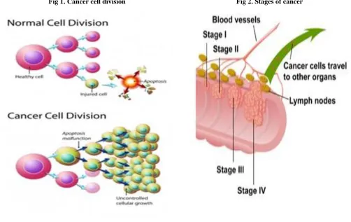
"Cancer" is the term we give to a large group of diseases that vary in type and location but have one thing in common: abnormal cell divisions. Cancer occurs when cells
in the body become abnormal and grow out of control and they have the ability to spread to other parts of the body
(metastasis)[2]. The development of cancer starts with
genetic changes in the cells. The time from genetic change to development of cancer is called the latency period. The latency period can be as long as 30 years or more Uncontrolled cell division results in the accumulation of abnormal cells in a mass of cells called a tumor[3].A tumor can be either benign or malignant. Benign tumors are non-cancerous; do not metastasize to other parts of the body. They usually can be removed by surgery (Papilloma, Adenoma, Lipoma, Osteoma, Angioma). While the Malignant tumors are cancer and can metastasize to other tissues forms secondary tumor. (Carcinomas, Sarcomas, Lymphoma, Leukemia)[4].
STAGES OF CANCER
The stage indicates basically the condition or the severity of the cancer. It also indicates the extent the cancer has spread it is used to determine a patient’s treatment and prognosis [5-6].
105 |
P a g e
Stage 0: It is the pre-cancerous or the initial stage i.e. cancer is not invaded to other tissues. The tumors can be completely eliminated in this phase by surgery.
Stage I: The cells start behaving abnormally but generally restrict to a particular area. Tumor size is less than 2 cm and can be treated by surgery.
Stage II: Tumor size can be 2 to 5 cm and can be eliminated by surgical methods. In this cancer is bigger than stage I but has not invaded the neighbouring tissues.
Stage III: This is the phase where the tumors exhibit a tendency to metastasize and the cell abnormality gets severe. Tumor size is more than 5 cm, surgical methods may be effective if done along with chemical or radiation treatment.
Stage IV: This is the final stage of cancerous infection and the chances of eliminating tumors are quite less. Harsh treatments like radiation and chemical therapy need to be undertaken to treat the tumors. It means the cancer has spread from where it started to another body organ. This is also called secondary or metastatic cancer.
CAUSES OF CANCER
The most common cancer risk factors are:
Tobacco smoke, Alcohol consumption, Genetic
predisposition, Estrogen exposure (women), Ionizing radiation, Ultraviolet radiation, Carcinogenic chemicals, Carcinogenic foods, Unhealthy diet, Free radicals. The disease is lifestyle related, has a long latent period and needs specialised infrastructure and human resource for treatment. Roughly one person in eight in the world would die of cancer[7].
TREATMENT OPTIONS
The methods of cancer treatment include
surgery, radiation therapy, chemotherapy, and
immunotherapy, either as a single therapy or in
combination. Transplantation with stem cells, or bone
marrow, is another method of treatment, which is commonly used in haematology patients[8]. To start any therapy, cancer is first diagnosed either by Biopsy, Ultrasound, Computed tomography (CT), Magnetic resonance imaging (MRI). The choice of therapy depends upon the cancer type and the stage of the disease, as well as the general condition of the patient. Complete removal of the affected tissue with least harm to the rest of the body is the treatment goal[9]. The surgery alone is effective mainly in the cases of benign tumors (their cells do not have the ability to invade the surrounding tissue, and stay clustered together in a single mass). In contrast, the malignant tumors have the tendency to invade adjacent tissue or to spread through the bloodstream and lymphatic vessels to form metastasis at other sites of the body, which often limits surgery effectiveness. In such cases the surgery is combined with other methods, and frequently with chemotherapy [10].
CHEMOTHERAPY
Chemotherapy is used with the intention of
cure, for the prolongation of life, or for palliation. Anti-cancer drugs destroy Anti-cancer cells by stopping growth or multiplication at some point in their life cycles as the drug altered the synthesis and function of deoxyribonucleic acid (DNA). Drugs may be administered intravenously (into a vein), orally (by mouth), by injection into a muscle, topically (applied to the skin) or in other ways, depending on the drug and the type of cancer. Chemotherapy is often given in cycles of alternating treatment and rest periods. In cancer treatment, surgery and radiation therapy have significant limitations, but the drugs offer the only approach to treat cases where the cancer has spread (metastasised) through the body [11]
The anticancer drugs can be administered to patients alone or in combination with other anticancer drugs. They can also be given before, during or after a patient receives surgery to kill any hidden cancer cells that remain in the body. Anticancer drugs are used not only for treating cancer, but also for relieving symptoms of the cancer. The way in which the other cells are affected by anticancer molecules determines the side-effects of the individual drugs[12]. Anticancer drugs significantly reduce the mortality rates and give longer overall patient survival times. The drugs used to combat cancer belong to one of two broad categories, the first is cytotoxic (cell killing) drugs and the second is cytostatic (cell stabilising drugs). Cytotoxic drugs work by interfering with DNA replication. While the cytostatic drugs are not designed to kill the cell involved, but simply to prevent it from reproducing.
A wide variety of chemotherapeutic agents are used in the treatment of malignancies. There are several categories of anticancer molecules which having different mode of action but the ultimate goal is same to destroy the cancerous cell. They vary in their cellular targets, mechanism of action and types of cancer they are used to treat [13].
A large number of anticancer drugs and they may be separated into four distinct types: alkylating agents, anti-tumour antibiotics, plant alkaloids and anti-metabolites. Among these the plant alkaloids (natural product drugs) have played a dominant role in pharmaceutical care for the treatment of cancer. It is an antimitotic agent and is used in combination with other agents for the treatment of a wide variety of cancers. Plant alkaloid includes- Vinca alkaloids, podophyllotoxins and taxanes types of agent, out of which taxanes are widely used due to its wide spectrum as antitumor agent and through a unique mechanism over different types of cancer [14].
ANTIMITOTIC DRUGS
106 |
P a g e
with antimitotics results in stabilisation of the microtubulesleading to cell cycle arrest in the G2/M phase[15]. Antimitotics are widely used in the treatment of ovarian and breast carcinomas as well as in standard therapies for lung, digestive and prostate cancers. Other solid tumours that these drugs have been effective in treating include carcinomas of the head and neck and bladder[16].
Treatment with antimitotics causes the stabilization of microtubules against depolymerization and the formation of abnormal bundles of microtubules. These abnormal bundles resist physiologic disassembly, accumulate within tumor cells and inhibit cell proliferation leading to cell cycle arrest, apoptosis, cytotoxicity and cell death, hence are active against a broad range of human malignancies. These undergo hepatic oxidative metabolism by the cytochrome P450 family[17].
NONAQUEOUS PARENTERAL FORMULATION According to I.P “parenterals are sterile pyrogen free preparations intended for administration by injection, infusion or implantation in to the body.” Parenteral administration bypasses the skin and gastrointestinal tract or this route of administration bypasses the normal body defence mechanisms. Parenteral route is best, when oral route is not suitable.
Mainly antimitotics are highly lipophilic in nature which makes it difficult to formulate in any formulation. Hence different pharmaceutical methodologies are applied to make the drug suitable for administration. Oral chemotherapy with lipophilic drug shares the two main drawbacks i.e., bioavailability and compliance. The oral bioavailability of these drugs is very poor but intravenous administration shows 100% bioavailabilty. So the i.v administration is used to increase the dose precision. Individual differences in absorption and drug metabolism with oral and parenteral route account for a great variability in plasmatic level and area under the curve. Considering the narrow therapeutic index of the drug, variability in plasmatic levels could increase toxicity[18].
Parenteral administration often permits dose reduction which is very much essential in order to reduce its
toxicity by exhibiting complete and immediate
bioavailability in case of intravenous infusion. Parenteral route is best, when oral route is not suitable but the lipophilic nature of the drug substance made it difficult to formulate in parenteral form also. The problem associated with parenteral dosage forms are decreased shelf life, stability in solution state, dissolution draw backs[20]. In order to overcome the above listed draw backs, non-aqueous formulation of the drug substance was proposed to and developed which showed improved stability shelf-life. Non aqueous formulation of the drug substance seems to be suitable preparation to prolong their shelf life, enhance their solubility and stability. Use of surfactant is one of the most frequently used method for this preparation, but the instability of the drug in surfactant and their harsh effects made this formulation difficult to
stabilize for efficacy, quality and safety point of view. Non aqueous injectable preparation need to be formulated in the non aqueous environment using water free solvents and excipients [21].
Literature Review
Shah JC et al. Etoposide an anticancer drug has low and erratic oral bioavailability which is due to low aqueous solubility, slow dissolution rate and instability in acidic pH. Etoposide solid-solid dispersions were prepared by coprecipitating the drug with polyethylene glycols (PEG) of different molecular weights in various ratios [22].
Beijnen JH et al. The chemical stability of the anticancer drug Etoposide in aqueous solution has been investigated utilizing a stability-indicating reversed-phase high-performance liquid Chromatography assay with ultraviolet detection [23].
Kumar D et al. The degradation of Docetaxel drug substance and its injection formulation has been investigated. The majority of impurities were observed in a base degradation study and all five degradation products were characterized. These impurities were isolated, enriched and were subjected to mass and NMR spectral studies [24].
Kearney AS et al. To evaluate the feasibility of formulating topotecan as a ready-to-use injectable solution, the pH-solubility profile was generated over a pH range of 2.5 to 4.5 at 25°C, and the pH-stability profile was generated over a pH range of 2 to 4, a temperature range of 60- 80°C, and at an ionic strength of ~ 0.15[25].
Kenneth CS et al. The flocculation-deflocculation behavior of cefazolin sodium (I) in nonaqueous media and the effect of surfactants as measured by zeta potential, sedimentation, and porosity were studied. The area under the plasma concentration versus time curve and urinary recovery indicated that cefazolin was 100% bioavailable from this nonaqueous preparation [26].
Krishna G et al. To develop and manufacture a stable parenteral formulation of VNP40101M (1,2-Bis(methylsulfonyl)-1-(2-chloroethyl)-2- [(2-methylamino) carbonyl] hydrazine), a novel antitumor agent. Solubility studies suggested that VNP40101M exhibited poor aqueous solubility but showed appreciable solubility in nonaqueous solvents. The solubility and stability of the drug was determined based on preformulation observations. A parenteral formulation containing 10 mg/mL of drug was prepared in a solvent system consisting of 30% ethyl alcohol and 70% polyethylene glycol-300 (PEG-300). Rubber closures, filter membranes, and liquid transfer tubing were selected on the basis of compatibility studies and absence of loss of drug due to adsorption of these component [27].
107 |
P a g e
of paclitaxel having good aqueous solubility and at the sametime free of any side effects. Various approaches employed so far include cosolvents, emulsions, micelles, liposomes, microspheres nanoparticles, cyclodextrins, pastes, and implants etc [28].
Thiesen J et al. Stability of reconstituted Docetaxel solutions were studied after reconstitution of the injection concentrate and also further dilution in two commonly used vehicle-solutions, 0.9% sodium chloride and 5% dextrose, in PVC bags and polyolefine containers. Chemical stability was measured by using a stability-indicating HPLC assay with ultraviolet detection. Physical stability was determined by visual inspection [29].
Dong Wai et al. N-Epoxymethyl-1, 8-naphthalimide (ENA)
is a novel antiproliferative drug candidate with potent anticancer and antifungal activity. It has an aqueous solubility and also exhibits hydrolytic instability. To minimize hydrolysis, ENA is proposed to be formulated as either lyophilized powders or nonaqueous solutions
followed by solubilization/reconstitution prior to
administration. In contrast, a combination of 70% Cremophor EL and 30% ethanol (v/v) proved effective in solubilizing ENA at 4 mg/mL, which exhibited good physical and chemical stability on storage at both 4 °C and room temperature over 4 months. The approach used in this work could serve as a useful reference in formulating nonpolar drugs with hydrolytic instability [30].
Fig 1. Cancer cell division Fig 2. Stages of cancer
ACKNOWLEDGEMENT
The corresponding author is highly thankful to management of Aligarh College of Pharmacy, Aligarh for giving guidance and moral support.
REFERENCES
1. WHO. Information on the cancer burden, 2010. NMH Fact Sheet [Online]. Available at:
http://www.who.int/mediacentre/factsheets/fs297/en/.
2. Ruddon RW. Cancer Biology, Oxford University Press: New York. 3rd ed., 1995, 502-522.
3. Barlow-Stewart K, Kirk J, Tucker K. Clinical Practice Guidelines Familial aspects of cancer: a guide to clinical practice.
National Health and Medical Research Council.1999.;[Online]. Available from: http://www.nhmrc.gov.au/
publications/synopses/cp67syn.htm.
4. Memmler R, Wood D. The Human Body in Health and Disease: Lippincott Williams & Wilkins, 7th ed., 2000, 229-234.
108 |
P a g e
6. Khanse S. Stages of cancer: Medical Knowledge for the Non Medical People, World press. 2011. Available at:
http://mediconweb.com/cancer/stages-of-cancer/
7. Westcott SA. Journey Into Cancer’s Causes. Anchorage (AK): Alaska Native Health Board. Alaska, 5th edn., 1999, 11-12.
8. DeVita VT, Chu E. Cancer Chemotherapy Research, 68, 2008, 864-65.
9. Kelloff GJ, Charles WB, Winfred FM, Steele VE. Cancer Cheomoprevention, Wattenberg L, Lipkin M, Boone CW, Kelloff
GJ, CRC Press, Florida. 1992, 41-56.
10. Ward DE. The Cancer Handbook: A Guide for the Nonspecialist, Cushing-Malloy: Michigan. 1999, 4th Edn,214.
11. Peterson DE & Lalla R. Overview of complications of systematic chemotherapy. In: Davies A & Epstein JB. (eds.) Oral complications of cancer and its management. 54(4), 2010, 318-322.
12. Riddick DS, Lee C, Ramji S, Chinje EC, Cowen RL, Williams KJ, Patterson AV, Stratford IJ, Morrow CS, Townsend AJ,
Jounaidi Y, Chen CS, Su T, Lu H, Schwartz PS, Waxman DJ. Cancer chemotherapy and drug metabolism. Drug Metab
Dispos, 33(8), 2005, 1083-96.
13. Skeel RT. Antineoplastic Drugs and Biologic Response Modifiers: Classification, Use, and Toxicity of Clinically Useful Agents. In: Skeel, R., T. (ed.) Handbook of Cancer Chemotherapy. Philadelphia: Lippincott William and Wilkins, 7th edn., 2010, 232-240.
14. Suffness M, Cordell GA.Antitumor Alkaloids in the Alkaloids, Brossi, A.; Ed.; Academic Press: New York, 25, 1985,
235-269.
15. Vaishampayan U. Taxanes: an overview of the pharmacokinetics and pharmacodynamics. Urology, 54(6A Suppl), 1999,
22-29.
16. Gligorov J and Lotz JP. Preclinical pharmacology of the taxanes: implications of the differences. Oncologist, 9(2), 2004, 3-8.
17. Ringel I, Horwitz SB. Studies with RP 56976 (Taxotere): A semisynthetic analogue of taxol. J Natl. Cancer Inst, 83, 1991,
288.
18. Denny William A. The Design and Development of Anticancer Drugs; Chemical Processes in New Zealand, 1st ed., 2, 2001,
223-29.
19. Beck S. Impact of a Systematic Oral Care Protocol on Stomatitis after Chemotherapy. Cancer Nursing, 2, 1979,185-99.
20. Kingston DGI. History and Chemistry in Paclitaxel in Cancer Treatment, McGuire WP, Rowinsky EK, Marcel Dekker, 1, 1995, 1-33.
21. Wessjohann L. Epothilones .Promising Natural Products with Taxol-like Activity. Angew. Chem. Int. Ed. Engl, 36(7), 1997,
715-18.
22. Shah JC, Chen JR, Chow D. Preformulation study of Etoposide: Increased solubility and dissolution rate by solid-solid dispersions. Int J Pharm, 113(1), 1995, 103-11.
23. Beijnen JH, Holthuis JJM, Kerkdijk HG, Van der Houwen OAGJ, Paalman ACA, Bult A. Degradation kinetics of Etoposide
in aqueous solution. Int J Pharm, 1988, 41(1-2), 169-178.
24. Kumar D, Tomar RS, Deolia SK, Mitra M, Mukherjee R, Burman AC. Isolation and characterization of degradation impurities in Docetaxel drug substance and its formulation. J Pharm and Biomed Ana, 43(4), 2007, 1228-1235.
25. Kearney AS, Patel K, Palepu NR. Preformulation studies to aid in development of ready-to-use injectable solution of the antitumor agent, topotecan. Int J Pharm, 127, 1996, 229-37.
26. Kenneth CS, Su ES, Quay JF, Campanale K M., John FS. Nonaqueous cephalosporin suspension for parenteral
administration. J Pharm Sci, 73(11), 1984, 1602-1606.
27. Krishna G, Hodnick WF, Lang W, Lin X, Karra S, Mao J. Pharmaceutical Development and Manufacturing of a Parenteral Formulation of a Novel Antitumor Agent. AAPS PharmSciTech, 2(3), 2001, 1-9.
28. Singla AK, Garg A, Aggarwal D. India Paclitaxel and its formulations. Int Pharm, 235(1-2), 2002, 179-192.
29. Thiesen J, Kramer I. Physico-chemical stability of Docetaxel premix solution and Docetaxel infusion solutions in PVC bags
and polyolefine containers. Pharm World Sci, 21(3), 1999, 137-141.
30. Dong Wai Kiong Ng, Uttam S, Reginald BH. Tan. Solubilization & Preformulation of poorly water soluble and hydrolysis