R E S E A R C H
Open Access
A panel of glycoproteins as candidate
biomarkers for early diagnosis and
treatment evaluation of B-cell acute
lymphoblastic leukemia
Marcio de Souza Cavalcante
1, José Camilo Torres-Romero
2,3, Marina Duarte Pinto Lobo
2,3,
Frederico Bruno Mendes Batista Moreno
2, Leonardo Primo Bezerra
2, Diego Silva Lima
4, Jesamar Correia Matos
5,
Renato de Azevedo Moreira
2and Ana Cristina de Oliveira Monteiro-Moreira
2*Abstract
Background:Acute lymphoblastic leukemia is the most common malignant cancer in childhood. The signs and symptoms of childhood cancer are difficult to recognize, as it is not the first diagnosis to be considered for nonspecific complaints, leading to potential uncertainty in diagnosis. The aim of this study was to perform proteomic analysis of serum from pediatric patients with B-cell acute lymphoblastic leukemia (B-ALL) to identify candidate biomarker proteins, for use in early diagnosis and evaluation of treatment.
Methods:Serum samples were obtained from ten patients at the time of diagnosis (B-ALL group) and after induction therapy (AIT group). Sera from healthy children were used as controls (Control group). The samples were subjected to immunodepletion, affinity chromatography withα-D-galactose-binding lectin (fromArtocarpus incisa seeds) immobilized on a SepharoseTM4B gel, concentration, and digestion for subsequent analysis with nano-UPLC tandemnano-ESI-MSE. The programExpressionEwas used to quantify differences in protein expression between groups.
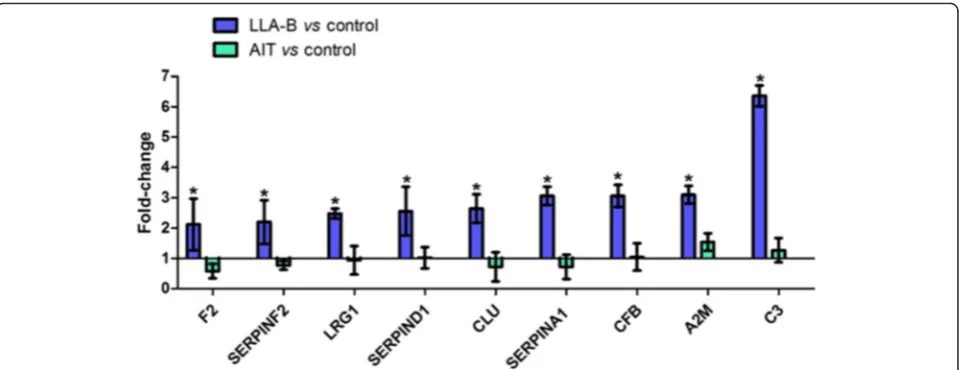
Results:A total of 96 proteins were identified. Leucine-rich alpha-2-glycoprotein 1 (LRG1), Clusterin (CLU), thrombin (F2), heparin cofactor II (SERPIND1), alpha-2-macroglobulin (A2M), alpha-2-antiplasmin (SERPINF2), Alpha-1 antitrypsin (SERPINA1), Complement factor B (CFB) and Complement C3 (C3) were identified as candidate biomarkers for early diagnosis of B-ALL, as they were upregulated in the B-ALL group relative to the control and AIT groups. Expression levels of the candidate biomarkers did not differ significantly between the AIT and control groups, providing further evidence that the candidate biomarkers are present only in the disease state, as all patients achieved complete remission after treatment.
Conclusion:A panel of protein biomarker candidates has been developed for pre-diagnosis of B-ALL and also provided information that would indicate a favorable response to treatment after induction therapy.
Keywords:Acute lymphoblastic leukemia, Biomarker, Lectin, Frutalin, Mass spectrometry
* Correspondence:acomoreira@unifor.br
2Center of Experimental Biology (NUBEX), University of Fortaleza (UNIFOR),
Fortaleza, Ceará, Brazil
Full list of author information is available at the end of the article
Background
Acute lymphoblastic leukemia (ALL) is the most com-mon malignant cancer in childhood, and is responsible for approximately 25 % of all childhood cancers and 72 % of all cases of pediatric leukemia [1]. The current standards for diagnosis of ALL integrate the study of cell morphology, immunophenotyping, and genetics/cyto-genetics, as described in the classification of lymphoid cancers published by the World Health Organization (WHO) in 2008 [2]. Of lymphoid cancers, as designated using the most recent WHO classification, the purely leukemic presentation, B-lineage ALL (85 %) is the most common [3]; it will be addressed in this study. The signs and symptoms of childhood cancer are very challenging to identify, as it is not the first diagnosis to be consid-ered for nonspecific complaints, leading to potential un-certainty in diagnosis. Moreover, children showing the first signs of cancer frequently do not appear severely ill, which may delay diagnosis. In addition, childhood can-cer can mimic other common childhood diseases and even normal developmental physiological processes [4]. Measures to improve cancer incidence rates in adults in-clude prevention of exposure to known carcinogenic risk factors, such as smoking, but environmental factors play a very small role in the development of childhood can-cers. Thus, primary prevention measures are not effect-ive in averting the development of cancer in this age group, and secondary prevention, i.e. early diagnosis, is therefore essential [5]. In the specific case of ALL, early diagnosis and treatment increase the chances of a cure [4]. Affinity chromatography withα-D-galactose-binding lectin from Artocarpus incisa immobilized on a Sephar-ose™4B gel, combined with identification and quantifica-tion of glycoproteins by mass spectrometry, are excellent tools for comparative serum studies. The biomarker pipeline is commonly viewed as a series of preclinical phases: biomarker discovery, and verification before the final clinical evaluation. The comparative analysis results in a list of hundreds of proteins that are differentially-expressed between healthy and diseased samples [6]. In this study, the preclinical phase of biomarker discovery was applied and a proteomic analysis of serum samples from pediatric patients with B-ALL was performed, to analyze levels of glycoprotein expression, with the aim of identifying biomarkers to aid in the early diagnosis of B-ALL and to assess the response to induction therapy.
Methods
Patients and samples
Serum samples were collected from ten pediatric pa-tients with B-ALL at diagnosis and after induction therapy. These patients were diagnosed based on mor-phological, immunophenotypic, and genetic tests. The study population was composed mainly of children from
the lower middle class, who attended a reference hos-pital for the diagnosis and treatment of childhood can-cers in the State of Ceará -Brazil. The mean age of the patients was 6.15 years (n= 10); the average age of fe-male patients was 3.8 years (n= 6) and that of male pa-tients was 3.5 years (n= 4). All patients were assigned as low risk (LR) when aged from 1 to 9 years old, with a white blood cell (WBC) count <50 × 109/L, and no CNS involvement. The risk stratification and chemotherapy were performed according to the protocol of the Brazilian Group for Treatment of Childhood Leukemia (GBTLI-LLA-2009). All patients experienced complete clinical and morphological remission and are presently surviving. The serum samples used as controls were obtained from ten non-leukemic pediatric patients. All samples were stored at −80 °C until use and concentrations were determined using a Nanovue Plus™apparatus (GE Healthcare, Uppsala, Sweden). The study was conducted with the approval of the Research Ethics Committee at the Hospital Infantil Albert Sabin, associated with the Secretary of Health of the State of Ceará.
Serum preparation
For each sample, high-abundance human serum albumin (HSA) and immunoglobulin G (IgG) proteins were de-pleted using a HiTrap™ Albumin & IgG Depletion col-umn (GE Healthcare) on an ÄKTA purifier 10 Fast protein liquid chromatography (FPLC) system (GE Healthcare) according to the manufacturer’s instructions. After removal of high-abundance proteins, the flow-through fractions were collected and stored at −80 °C until use.
Lectin affinity chromatography
The serum-free high-abundance proteins were thawed, centrifuged for 15 min at 12,000 ×g at 8 °C, filtered through a 0.22-μM membrane (Vertipure™PVDF syringe filters, Veritical) and applied to a 5-mL column packed with Sepharose-Frutalin, prepared as mentioned previ-ously, in a XK16 column on an ÄKTA purifier 10 FPLC system (GE Healthcare). The column was washed with five CV of buffer A (20 mM Tris–HCl, pH 7.4), and the lectin-bound proteins were eluted with four CV of elu-tion buffer B (20 mM Tris–HCl, pH 7.4, with 0.2 M gal-actose). The eluted protein solution was dialyzed and concentrated by spinning at 8000 ×g(Vivaspin® 6, with a molecular weight cut-off of 3 kDa, GE Healthcare), and used for further analyses.
Proteomic analysis
trypsin (Promega, Madison, WI, USA). At the end of this process, the samples were centrifuged and the super-natant was transferred to new vials, to which 5μL of in-ternal standard, alcohol dehydrogenase (ADH, 50 fmol, access code P00330 in SwissProt) and 85 μL of 3 % acetonitrile solution with formic acid 0.1 % were added. The final glycoprotein and ADH concentrations were es-timated to be 250 ng/μL and 25 fmol/μL, respectively, in a final volume of 200 μL. The quantitative and qualita-tive nano-UPLC tandem nano-ESI-MSE experiments were performed on digested samples using peptide reversed-phase chromatography with 3 to 40 % (v/v) of acetonitrile containing 0.1 % formic acid for 90 min, main-tained at a flow rate of 600 nL/min for 100 min in a nanoACQUITY UPLC core system. A nanoACQUITY C18 UPLC BEH 1.7μm, 100μm × 10 cm reversed-phase column was used in conjunction with an SCX 5 μm, 180 μm × 23 mm pre-column. All analyses were per-formed with electrospray ionization in positive ion ESI(+) mode and a NanoLockSpray source. Data-independent ac-quisition (MSE) was performed with a SYNAPT HDMS G1 mass spectrometer (Waters, Manchester, UK), pro-grammed to automatically switch between standard MS (3 eV) and high-energy collision MSE(12–50 eV), applied to trap‘T-wave’CID (collision-induced dissociation) cells, in the presence of argon gas. The transfer from the colli-sion cell was adjusted to 1 eV, with 1 s, at both low and high energy. After the time of flight analysis (TOF), m/z 50 to 3000 spectra were collected. However, the RF offset of the quadrupole was set for efficient acquisition of LC/ MS data from m/z 300–3000, ensuring that any mass less than m/z 300 observed in the LC/MSE data stemmed only from the collision cell. Thus, the low-mass values were known CID fragmentation products, not the result of source fragmentation. For processing of spectra and data-base searches, the Protein Lynx Global Server (PLGs) package v. 2.4, containing the ExpressionEv. 2.4 program, was used. The PLGs used a new algorithm to process raw data obtained by MS using ion properties, i.e. retention time, intensity of precursor/productions, and exact mass. A label-free proteomic approach was used for the quanti-tative analysis. Then the PLGs generated a list of all pre-cursors and products. This list contained the mass of the precursor and product ions for each peptide, to be searched against the non-redundant proteins database, UniProtKB/Swiss-Prot 57.1, under search conditions based on taxonomy [Human (Homo sapiens)]. For each protein, the program ExpressionEselected all correspond-ing peptides from the samples and compared the inten-sities of these for relative protein quantification. Using the intensity of a peptide of known quantity, ADH, the pro-gram performed self-standardization of data sets. Lists of proteins were then filtered to show only those present in all three repeated injections of each sample, from which
an output table was created. This table showed the names, access codes, and expression levels of the proteins, and in-dicated whether they were up-regulated ≥2-fold, down-regulated≤0.5-fold, or whether they did not show signifi-cant differences between the groups (unchanged), 0.5< ex-pression level <2.
Protein interaction network analysis
The differentially expressed proteins were used for path-way analysis. Swiss-Prot accession numbers were inserted into the Search Tool for the Retrieval of Interacting Genes/Proteins (STRING) software, version 9.05, which is available at http://string.embl.de/, with the following ana-lysis parameters: Homo sapiens, confidence level 0.400– 0.900, using the active prediction method [7].
Results and discussion
Patient characteristics
The demographic and clinical data for the patients are summarized in Table 1. To form the panel of candidate proteins for early biomarkers and demonstrate their ex-pression profiles, the pediatric patients were evaluated at two different times: at diagnosis (B-ALL Group; n= 10) and after induction therapy (AIT Group; n= 10). Sam-ples of healthy children (Control Group; n= 10) were obtained for comparison.
Reduction of dynamic range
The depletion of high-abundance proteins in serum, HSA and IgG, followed by affinity chromatography with the plant lectin Frutalin immobilized on Sepharose™ 4B (Fig. 1), reduced the dynamic range and increased the capacity to identify lower-abundance proteins. The retained fraction (FR) peak containing the protein of interest was concentrated and digested, for later analysis by nano-LC-MS/MS.
Proteomic analysis
Table 1Summary of characteristics of patients with B-ALL
Code Assigned
Gender Age at Diagnosis
FAB Classification
Immunophenotypical Classification
Karyotype Risk
Group
MRD Treatment Outcome
P1 M 3 L1 Common Absence of metaphases LR _ CR
P2 F 3 L1 Pre-B Absence of metaphases LR _ CR
P3 F 5 L1 Common 56,XX,+X,+4,+6,+8,+10,+11,+14,+17,+21,+mar/
46,XX
LR _ CR
P4 M 2 L1 Common 46,XY LR _ CR
P5 F 2 L1 Common 46,XX LR _ CR
P6 F 3 L1 Common 54,XX,+X,+6,+15,+15,+17,+18,+21,+21/46,XX LR _ CR
P7 F 5 L1 Common 47,XY,+21 c LR _ CR
P8 M 6 L1 Common Absence of metaphases LR _ CR
P9 F 5 L1 Pre-B 46, XX LR _ CR
P10 M 3 L1 Pre-B 46,XY LR _ CR
Code: Internal register assigned for the study;MMale,FFemale,LRLow Risk,MRDMinimum Residual Disease,CRComplete remission
LRG1 was 2.16-fold upregulated in the B-ALL group compared to the Control group. LRG1 is involved in cell adhesion and development, protein-protein interaction, and signal transduction. Studies have shown that it may be a biomarker for ovarian cancer, pancreatic cancer, oral squamous cell carcinoma [8, 9], and non-small cell lung cancer [10]. CLU, also called apolipoprotein J, was 2.64-fold upregulated in the B-ALL group compared to the control. CLU is expressed in a variety of tissues, with a ubiquitous pattern of expression. It has a high sensitiv-ity to external stress stimuli. It has been reported to function in complement inhibition, clearance of cellular debris, folding of denatured proteins (chaperone), and regulation of cell death [11, 12]. Several studies have shown that it is over-expressed in many serious physio-logical conditions, including degenerative kidney disease and various neurodegenerative conditions. CLU also plays an important role in tumorigenesis and progres-sion of many human cancers [13]. Higher levels of this protein have been observed in the progression of pros-tate cancer and renal carcinoma compared to normal tissues [14, 15] and it was shown to be over-expressed in anaplastic large cell lymphomas [16], ovarian cancer [17], esophageal cancer [18], and colorectal cancer; it has also been implicated in pre-diagnosis of colorectal cancer [19]. The accumulation of CLU protein is correlated with the aggressiveness of certain breast tumors [20].
Our results also confirm the important relationship between cancer and phenomena associated with blood coagulation. Several studies have reported that approxi-mately 50 % of patients with malignant disease and more than 90 % of those that evolve to metastasis present evi-dence of abnormalities in coagulation and/or fibrinolysis [21–25]. Several of the proteins identified in this study,
protein was 3.1-fold upregulated in the serum of B-ALL patients, and its level of expression decreased signifi-cantly after induction therapy. Nevertheless, compared with serum samples from control patients, no significant changes in expression levels of A2M were observed. This shows that A2M is in fact upregulated in patients with B-ALL and can be considered both as a candidate bio-marker of disease as well as for evaluation of the re-sponse to induction therapy. Moreover, serum analysis is less invasive than bone marrow biopsy [39].
Alpha-1 antitrypsin (SERPINA1) was 3.06-fold upregu-lated compared to controls. SERPINA1 is a serum glyco-protein that is synthesized primarily in the human liver and in macrophages. It belongs to the serpin family, which plays a central role in the control of degradation of tissues through its inhibitory effect on neutrophil elastase and other serine proteases, including trypsin, chymotrypsin, cathepsin G, plasmin, thrombin, tissue kallikrein, and activated factor X (FXa) [40–42]. Many types of cancer cells have been shown to express and se-crete alpha-1 antitrypsin [43–45]. In addition, high serum levels of this protein have been reported in a number of inflammatory diseases and in various malignancies, such as hepatocellular carcinoma, multiple myeloma, pancre-atic carcinoma, prostate carcinoma, primary carcinoma of the lung, cervical carcinoma, gastric cancer, laryngeal can-cer, nasopharyngeal carcinoma, breast cancan-cer, and colorec-tal cancer [45–52]. Some studies reported that levels of this protein were correlated with cancer stage [47, 53–56]. A previous study of lung and prostate cancers, showed a direct correlation between high serum levels of SERPINA1 and cancer stage. It has been also suggested that the main
source of the increase in blood levels of this protein in cancer patients is the growth of cancer cells [47]. In a re-cent survey, also of lung cancer and prostate cancer, a de-crease in the serum levels of SERPINA1 was observed several weeks after treatment, suggesting that it may be an important indicator of the effectiveness of cancer treat-ment [57].
Complement factor B (CFB) and Complement C3 (C3), which are both associated with the complement system, were found to be 3.06- and 6.36-fold upregu-lated. The complement system is an essential part of the innate immune response that participates in the elimin-ation of altered cells from the body. Due to the large number of genetic and epigenetic alterations associated with carcinogenesis, neoplastic transformation can in-crease the ability of malignant cells to activate this sys-tem [58], which may explain the over-expression of these proteins, in agreement with other studies that re-port the over-expression of complement proteins in pul-monary cells [59, 60]. Although malignant cells often develop mechanisms for protection against and resistance to complement, recent research on the mechanisms regu-lating complement activity demonstrated that the over-expressed proteins showed inhibitory activity [61–63]. Therefore, the proteins found to be upregulated in this study may be candidate biomarkers of B-ALL, because the alteration in expression was observed only in cancer pa-tients. This result is corroborated by the absence of changes in expression levels of these proteins in the serum of patients who underwent induction therapy and who ex-perienced complete remission, as well as in healthy indi-viduals from the control group.
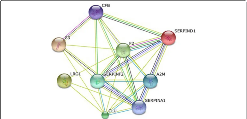
All nine proteins identified found to be upregulated in the B-ALL group showed evidence of interaction, as shown by the protein interaction network generated by the STRING interaction database (http://string-db.org/) (Fig. 3). Some studies have shown that increased expres-sion of thrombin is associated with leukemia, as well as several other diseases [64, 65]. Similarly, as previously mentioned, the increase in blood levels of SERPINA1 in cancer patients is proportional to the growth of cancer cells; a decrease is observed several weeks after chemo-therapy. Because these proteins lack specificity for the diagnosis of B-ALL if used in isolation, we suggest the creation of panel of biomarker.
Conclusion
LRG1, CLU, F2, SERPIND1, A2M, SERPINF2, SER-PINA1, CFB, and C3 were identified as candidate bio-markers for early diagnosis of B-ALL; all were over-expressed in the B-ALL group compared to the control and AIT groups. The AIT group did not display any sig-nificant changes in the expression levels of these pro-teins, compared to the control group. All patients in the AIT group achieved complete remission after treatment; this indicates that these biomarkers are only present in the disease state. These candidate biomarkers may im-prove the pre-diagnosis of B-ALL, which is currently dif-ficult to diagnose in the early stages; the biomarkers may also provide key information on the response to treat-ment after induction therapy. Further clinical studies are necessary to determine whether over-expression of these proteins is associated with development of acute symp-toms in patients with suspected ALL. These markers may help to determine response to treatment and to im-prove the survival of children with this disease.
Abbreviations
ALL:Acute lymphoblastic leukemia; WHO: World Health Organization; LR: Low Risk; WBC: White Blood Cell; HSA: Human Serum Albumin; IgG: Immunoglobulin G; FPLC: Fast protein liquid chromatography; ADH: alcohol dehydrogenase; STRING: Search Tool for the Retrieval of Interacting Genes/Proteins; LRG1: Leucine-rich alpha-2-glycoprotein 1; CLU: Clusterin; F2: thrombin; SERPIND1: heparin cofactor II; A2M: alpha-2-macroglobulin; SERPINF2: alpha-2-antiplasmin; SERPINA1: Alpha-1 antitrypsin; CFB: Complement factor B; C3: Complement C3; TAT: Thrombin-antithrombin complex.
Competing interests
The authors declare no conflict of interest.
Authors’contributions
MSC organized the research plan, performed the experiments and drafted the manuscript; JCTR performed data analysis and participated in manuscript writing; MDPL, FBMBM and LPB performed MS and chromatography experiments; DSL and JCM provided samples and clinical data; RAM contributed to the experimental design and provided research facilities; ACOMM coordinated the study, participated to its design and contributed to writing. All authors read and approved the manuscript.
Acknowledgements
FINEP, CNPq, RENORBIO-UNIFOR, ALBERT SABIN HOSPITAL
Author details
1Northeast Network of Biotechnology (RENORBIO), State University of Ceará,
Fortaleza, Ceará, Brazil.2Center of Experimental Biology (NUBEX), University of Fortaleza (UNIFOR), Fortaleza, Ceará, Brazil.3Department of Biochemistry and Molecular Biology, Federal University of Ceará, Fortaleza, Ceará, Brazil. 4Development and Technological Innovation in Drug Program, Federal
University of Ceará, Fortaleza, Ceará, Brazil.5Reference Center at Children’s Cancer Diagnosis and Adolescents Dr. Murilo Martins, Albert Sabin Hospital, Fortaleza, Ceará, Brazil.
Received: 30 September 2015 Accepted: 4 January 2016
References
1. Scheurer ME, Bondy ML, Gurney JG. Epidemiology of Childhood Cancer. In: Pizzo PA, Poplack DG, editors. Principles and practice of pediatric oncology. 6th ed. Philadelphia: Lippincott Williams and Wilkens; 2011. p. 2–16. 2. Vardiman JW, Thiele J, Arber DA, Brunning RD, Borowitz MJ, Porwit A, et al.
The 2008 revision of the World Health Organization (WHO) classification of myeloid neoplasms and acute leukemia: rationale and important changes. Blood. 2009;114:937–51.
3. Chiaretti S, Zini G, Bassan R. Diagnosis and subclassification of acute lymphoblastic leukemia. Mediterr J Hematol Infect Dis. 2014;6:e2014073. 4. Rodrigues KE, Camargo B. Diagnóstico precoce do câncer infantil:
responsabilidade de todos. Rev Assoc Med Bras. 2003;49:29–34.
5. Michalowski MB, Lorea CF, Rech A, Santiago P, Lorenzoni M, Taniguchi A, et al. Diagnóstico precoce em oncologia pediátrica: uma urgência médica. Bol Cient Pediatr. 2012;01:13–8.
6. Parker CE, Borchers CH. Mass spectrometry based biomarker discovery, verification, and validation e Quality assurance and control of protein biomarker assays. Mol Oncol. 2014;8(4):840–58.
7. Jensen LJ, Kuhn M, Stark M, Chaffron S, Creevey C, Muller J, et al. STRING 8– a global view on proteins and their functional interactions in 630 organisms. Nucleic Acids Res. 2009;37:D412–16.
8. Andersen JD, Boylan KL, Jemmerson R, Geller MA, Misemer B, Harrington KM, et al. Leucine-rich alpha-2-glycoprotein-1 is upregulated in sera and tumors of ovarian cancer patients. J Ovarian Res. 2010;3:21.
9. Tung CL, Lin ST, Chou HC, Chen YW, Lin HC, Tung CL, et al. Proteomics-based identification of plasma biomarkers in oral squamous cell carcinoma. J Pharm Biomed Anal. 2013;75:7–17.
10. Liu Y, Luo X, Hu H, Wang R, Sun Y, Zeng R, et al. Integrative proteomics and tissue microarray profiling indicate the association between overexpressed serum proteins and non-small cell lung cancer. PLoS One. 2012;7:e51748. 11. Park S, Mathis KW, Lee IK. The physiological roles of apolipoprotein J/
clusterin in metabolic and cardiovascular diseases. Reviews in Endocrine and Metabolic Disorders. 2014;15:45–53.
12. Antonelou MH, Kriebardis AG, Stamoulis KE, Trougakos IP, Papassideri IS. Apolipoprotein J/clusterin in human erythrocytes is involved in the molecular process of defected material disposal during vesiculation. PLoS One. 2011;6:e26033.
13. Trougakos IP, So A, Jansen B, Gleave ME, Gonos ES. Silencing expression of the clusterin/apolipoprotein j gene in human cancer cells using small interfering RNA induces spontaneous apoptosis, reduced growth ability, and cell sensitization to genotoxic and oxidative stress. Cancer Res. 2004;64: 1834–42.
14. Parczyk K, Pilarsky C, Rachel U, Koch-Brand C. Gp80 (clusterin:TRPM-2) mRNA levels is enhanced in human renal clear cell carcinomas. J Cancer Res Clin Oncol. 1994;120:186–8.
15. Steinberg J, Oyasu R, Lang S, Sintich S, Rademaker A, Lee C, et al. Intracellular levels of SGP-2 (clusterin) correlate with tumor grade in prostate cancer. Clin Cancer Res. 1997;3:1707–11.
16. Wellmann A, Thieblemont C, Pittaluga S, Sakai A, Jaffe ES, Siebert P, et al. Detection of differentially expressed genes in lymphomas using cDNA arrays: identification of clusterin as a new diagnostic marker for anaplastic large-cell lymphomas. Blood. 2000;96:398–404.
17. Hough CD, Cho KR, Zonderman AB, Schwartz DR, Morin PJ. Coordinately up-regulated genes in ovarian cancer. Cancer Res. 2001;61:3869–76. 18. Guo W, Ma X, Xue C, Luo J, Zhu X, Xiang J, et al. Serum clusterin as a tumor
19. Bertuzzi M, Marelli C, Bagnati R, Colombi A, Fanelli R, Saieva C, et al. Plasma clusterin as a candidate pre-diagnosis marker of colorectal cancer risk in the Florence cohort of the European Prospective Investigation into Cancer and Nutrition: a pilot study. BMC Cancer. 2015;15:56.
20. Redondo M, Villar E, Torres-Muñoz J, Tellez T, Morell M, Petito C. Overexpression of clusterin in human breast carcinoma. Am J Pathol. 2000;157:393–9. 21. Kwon H-C, Oh SY, Lee S, Kim S-H, Han JY, Koh RY, et al. Plasma levels of
prothrombin fragment F112, D-dimer and prothrombin time correlate with clinical stage and lymph node metastasis in operable gastric cancer patients. Jpn J Clin Oncol. 2008;38:2–7.
22. Bick RL. Coagulation abnormalities in malignancy: a review. Semin Thromb Hemost. 1992;18:353–72.
23. Luzzatto G, Schafer A. The prethrombotic state in cancer. Semin Oncol. 1990;17:147–59.
24. O’Connor N, Gozzard DI, Love EM, Lu Yin JA, Turck WPG. Haemostatic abnormalities and malignant disease. Lancet. 1986;8:303–4. 25. Hillen HF. Thrombosis in cancer patients. Ann Oncol. 2000;11:273–6. 26. Wojtukiewicz MZ, Tang DG, Ciarelli JJ, Nelson KK, Walz DA, Diglio CA, et al.
Thrombin increases the metastatic potential of tumor cells. Int J Cancer. 1993;54:793–806.
27. Wojtukiewicz MZ, Sierko E, Zacharski LR, Zimnoch L, Kudryk B, Kisiel W. Tissue factor-dependent coagulation activation and impaired fibrinolysis in situ in gastric cancer. Semin Thromb Hemost. 2003;29:291–300. 28. Gunji Y, Lewis J, Gorelik E. Fibrin formation inhibits the in vitro cytotoxic
activity of human natural and lymphokine-activated killer cells. Blood Coagul Fibrinolysis. 1990;1:663–72.
29. Bruhn HD, Zurborn KH. Influences of clotting factors (thrombin, factor XIII) and of fibronectin on the growth of tumor cells and leukemic cells in vitro. Blut. 1983;46:85–8.
30. Wojtukiewicz MZ, Sierko E, Rak J. Contribution of hemostatic system to angiogenesis in cancer. Semin Thromb Hemost. 2004;30:5–20.
31. Di Micco P, Romano M, Niglio A, Nozzolillo P, Federico A, Petronella P, et al. Alteration of haemostasis in non-metastatic gastric cancer. Dig Liver Dis. 2001;33:546–50.
32. Rahr HB, Sorensen JV, Larsen JF, Jensen FS, Bredahl C. Markers of coagulation and fibrinolysis in portal blood from patients with and without gastric malignancy. Scand J Gastroenterol. 1994;29:516–21.
33. Abbasciano V, Tassinari D, Sartori S, Trevisani L, Arcudi D, Bianchi MP, et al. Usefulness of coagulation markers in staging of gastric cancer. Cancer Detect Prev. 1995;19:331–6.
34. Ikeda M, Furukawa H, Imamura H, Shimizu J, Ishida H, Masutani S, et al. Poor prognosis associated with thrombocytosis in patients with gastric cancer. Ann Surg Oncol. 2002;9:287–91.
35. Lee JH, Ryu KW, Kim S, Bae JM. Preoperative plasma fibrinogen levels in gastric cancer patients correlate with extent of tumor.
Hepatogastroenterology. 2004;51:1860–3.
36. Yamashita H, Kitayama J, Nagawa H. Hyperfibrinogenemia is a useful predictor for lymphatic metastasis in human gastric cancer. Jpn J Clin Oncol. 2005;35:595–600.
37. Yamashita H, Kitayama J, Kanno N, Yatomi Y, Nagawa H.
Hyperfibrinogenemia is associated with lymphatic as well as hematogenous metastasis and worse clinical outcome in T2 gastric cancer. BMC Cancer. 2006;6:147.
38. Yamashita H, Kitayama J, Ishikawa M, Nagawa H. Tissue factor expression is a clinical indicator of lymphatic metastasis and poor prognosis in gastric cancer with intestinal phenotype. J Surg Oncol. 2007;95:324–31. 39. Braoudaki M, Lambrou GI, Vougas K, Karamolegou K, Tsangaris GT,
Tzortzatou-Stathopoulou F. Protein biomarkers distinguish between high-and low-risk pediatric acute lymphoblastic leukemia in a tissue specific manner. J Hematol Oncol. 2013;6:52.
40. Gettins PG. Serpin structure, mechanism, and function. Chem Rev. 2002;102: 4751–804.
41. Carrell RW. Alpha 1-Antitrypsin: molecular pathology, leukocytes, and tissue damage. J Clin Invest. 1986;78:1427–31.
42. Carrell RW, Jeppsson JO, Laurell CB, Brennan SO, Owen MC, Vaughan L, et al. Structure and variation of human alpha 1-antitrypsin. Nature. 1982;298: 329–34.
43. Higashiyama M, Doi O, Kodama K, Yokouchi H, Tateishi R. An evaluation of the prognostic significance of alpha-1-antitrypsin expression in
adenocarcinomas of the lung: an immunohistochemical analysis. Br J Cancer. 1992;65:300–2.
44. Poblete MT, Nualart F, del Pozo M, Perez JA, Figueroa CD. Alpha 1-antitrypsin expression in human thyroid papillary carcinoma. Am J Surg Pathol. 1996;20:956–63.
45. Yamaguchi N, Yamamura Y, Koyama K, Ohtsuji E, Imanishi J, Ashihara T. Characterization of new human pancreatic cancer cell lines which propagate in a protein-free chemically defined medium. Cancer Res. 1990; 50:7008–14.
46. El-Akawi ZJ, Al-Hindawi FK, Bashir NA. Alpha-1 antitrypsin (alpha1-AT) plasma levels in lung, prostate and breast cancer patients. Neuro Endocrinol Lett. 2008;29:482–4.
47. El-Akawi ZJ, Abu-Awad AM, Sharara AM, Khader Y. The importance of alpha-1 antitrypsin (alphaalpha-1-AT) and neopterin serum levels in the evaluation of non-small cell lung and prostate cancer patients. Neuro Endocrinol Lett. 2010;31:113–6.
48. Trachte AL, Suthers SE, Lerner MR, Hanas JS, Jupe ER, Sienko AE, et al. Increased expression of alpha-1-antitrypsin, glutathione S-transferase pi and vascular endothelial growth factor in human pancreatic adenocarcinoma. Am J Surg. 2002;184:642–8.
49. Solakidi S, Dessypris A, Stathopoulos GP, Androulakis G, Sekeris CE. Tumour-associated trypsin inhibitor, carcinoembryonic antigen and acute-phase reactant proteins CRP and alpha1-antitrypsin in patients with gastrointestinal malignancies. Clin Biochem. 2004;37:56–60.
50. Hong WS, Hong SI. Clinical usefulness of alpha-1-antitrypsin in the diagnosis of hepatocellular carcinoma. J Korean Med Sci. 1991;6:206–13.
51. Bernacka K, Kuryliszyn-Moskal A, Sierakowski S. The levels of alpha 1-antitrypsin and alpha 1-antichymotrypsin in the sera of patients with gastrointestinal cancers during diagnosis. Cancer. 1988;62:1188–93. 52. Tountas Y, Sparos L, Theodoropoulos C, Trichopoulos D. Alpha 1-antitrypsin
and cancer of the pancreas. Digestion. 1985;31:37–40.
53. Li Y, Krowka MJ, Qi Y, Katzmann JA, Song Y, Mandrekar SJ, et al. Alpha1-antitrypsin deficiency carriers, serum alpha 1-Alpha1-antitrypsin concentration, and non-small cell lung cancer survival. J Thorac Oncol. 2011;6:291–5. 54. Millan J, Senra A, Lorenzo A, Romero J, Rodriguez P, Gallurt P. Biological
changes in serum antiproteases as related to tumor progression. Tumour Biol. 1988;9:15–20.
55. Varela AS, Lopez Saez JJ. Utility of plasmatic levels of alpha-1-antiprotease (A1AP) as a cancer marker. Cancer Lett. 1995;89:15–21.
56. Thompson DK, Haddow JE, Smith DE, Ritchie RF. Elevated serum acute phase protein levels as predictors of disseminated breast cancer. Cancer. 1983;51:2100–4.
57. El-Akawi ZJ, Abu-Awad AM, Khouri NA. Alpha-1 antitrypsin blood levels as indicator for the efficacy of cancer treatment. World J Oncol. 2013;4:83–6. 58. de Rodriguez Cordoba S, Diaz-Guillen MA, Heine-Suner D. An integrated
map of the human regulator of complement activation (RCA) gene cluster on 1q32. Mol Immunol. 1999;36:803–8.
59. Niehans GA, Cherwitz DL, Staley NA, Knapp DJ, Dalmasso AP. Human carcinomas variably express the complement inhibitory proteins CD46 (membrane cofactor protein), CD55 (decay-accelerating factor), and CD59 (protectin). Am J Pathol. 1996;149:129–42.
60. Nishioka K, Kawamura K, Hirayama T, Kawashima T, Shimada K. The complement system in tumor immunity: significance of elevated levels of complement in tumor bearing hosts. Ann NY Acad Sci. 1976;276: 303–15.
61. Varsano S, Frolkis I, Rashkovsky L, Ophir D, Fishelson Z. Protection of human nasal respiratoryepitheliumfromcomplement-mediatedlysisbycell-membraneregulators of complement activation. Am J Respir Cell Mol Biol. 1996;15:731–7.
62. Varsano S, Rashkovsky L, Shapiro H, Ophir D, Mark-Bentankur T. Human lung cancer cell lines express cell membrane complement inhibitory proteins and are extremely resistant to complement mediated lysis; a comparison with normal human respiratory epithelium in vitro, and an insight into mechanism(s) of resistance. Clin Exp Immunol. 1998;113:173–82. 63. Pio R. Control of complement activation by cancer cells and its
implications in antibody-mediated cancer immunotherapy. Inmunologia. 2006;25:173–87.
64. Ginsberg JS, Demers C, Brill-Edwards P, Johnston M, Bona R, Burrows RF, et al. Increased thrombin generation and activity in patients with systemic lupus erythematosus and anticardiolipin antibodies: evidence for a prothrombotic state. Blood. 1993;81:2958–63.