Published 31.xii.2017 Volume 57(2), pp. 351–380 ISSN 0374-1036 http://zoobank.org/urn:lsid:zoobank.org:pub:29090109-53D4-48D3-B926-6DE5D3F79ADE
https://doi.org/10.1515/aemnp-2017-0081
Taxonomic changes within Imatidiini and Hybosispini
(Coleoptera: Chrysomelidae: Cassidinae)
Lukáš SEKERKA
Department of Entomology, National Museum, Cirkusová 1740, CZ-193 00, Praha 9 – Horní Počernice, Czech Republic; e-mail: sagrinae@gmail.com
Abstract. Type specimens of various taxa of Imatidiini and Hybosispini were examined to verify their identity. The following taxonomic changes are proposed based on these comparisons. New combinations: Cephaloleia apertura (Staines, 2013) comb. nov. (from Aslamidium Borowiec, 1984), Cephaloleia jataiensis (Pic, 1923) comb. nov. (from Xenispa Baly, 1859), Pseudimatidium bicoloricornis (Pic, 1926) comb. nov. (from Windsorispa Sekerka, 2014), Xanthispa miniacea (Blanchard, 1843) comb. nov. (from Homalispa Baly, 1859), Xenispa aeneipennis (Baly, 1859) comb. nov. (from Cephaloleia Chevrolat, 1836), Xenispa gilvipes (Uhmann, 1930) comb. nov. (from Cephaloleia), Hybosispa claripes (Pic, 1923) comb. nov. (from Solenispa Weise, 1905), Hybosispa delectabilis (Staines, 1996) comb. nov. (from Cephaloleia), Hybosispa sulciceps (Baly, 1885) comb. nov. (from Cephaloleia), and Hybosispa truncatipennis (Baly, 1869) comb. nov. (from Cephaloleia). New synonyms: Cephaloleia impressa Uhmann, 1930 = Demothispa clermonti Pic, 1934 syn. nov.; Xenispa atra (Pic, 1926) = Cephaloleia cyanea Staines, 1996, syn. nov. Change in status: Cephaloleia fernandoi (Bondar, 1940) stat. restit. is removed from synonymy with C. opaca Baly, 1859. The monotypic genus Serratispa Staines, 2002 assign. nov. is transferred from Sceloenoplini to Imatidiini based on morphological characters. Three species are described: Ce-phaloleia fouquei sp. nov. from Bolivia and Peru, C. renei sp. nov. from Ecuador, and Xenispa fouquei sp. nov. from Venezuela. Keys to Cephaloleia basalis Weise, 1910 species-group, Venezuelan species of Xenispa Baly, 1859, and species of Hybosispa Weise, 1905 are proposed to simplify identifi cation of respective taxa. Most of the taxa discussed herein are provided with colour photographs.
Introduction
Imatidiini is a large Neotropical tribe of cassidine beetles presently containing 401 descri-bed species classifi ed in 22 genera and distributed from southern United States to northern Argentina. Species in this tribe are primarily associated with monocotyledonous plants, mainly belonging to Arecaceae, Bromeliaceae, Orchidaceae, Poaceae and nearly all families of Neotropical Zingiberales. Larval stadia are onisciform in shape and the fi nal one enclo-sing the pupa. Adult beetles are characterized by usually smooth dorsum without costae on elytra and pronotum with a setigerous pore bearing a long seta situated in anterior corners or along anterior margin. I have recently revised the genera of Imatidiini and summarized all available information on morphology and biology of the tribe (SEKERKA 2014). The paper also included numerous taxonomic changes, mainly transfers from one genus to another due to wrong interpretation of the type species designation of Demotispa Baly, 1859. I verifi ed most types of involved taxa, however, some placements remained tentative and based on primary descriptions as the respective type material was unavailable to me at that time. Since 2014 I had opportunity to examine most of the types not previously studied revealing additional misplaced taxa. The present contribution deals with these taxa and their proper placement.
I also reviewed types of several species of Cephaloleia Chevrolat, 1836 and found that they actually belong to Hybosispini. Originally this small tribe contained only fi ve species placed in a single genus Hybosispa Weise, 1910. They are superfi cially very similar morphologically to Imatidiini but clearly differ in not bearing any setigerous pore on pronotum and having acute and more or less projecting inner margin of eye. With the transfers proposed here the tribe contains presently two genera and 18 species. Since the number of species in Hybosispa doubled due to new transfers I propose here a key to that genus.
Lastly, I describe three new, recently discovered species of Imatidiini. Two species were previously identifi ed as Cephaloleia kolbei Weise, 1910 but clearly differ from the type. The third species is a high montane Xenispa Baly, 1859 from Venezuela, which is similar only to X. pulchella Baly, 1859, the type species of Xenispa. All three species are dedicated in loving memory to René Fouquè (1980–2016), who perished in a car accident in November 2016 and to whom this issue of Acta Entomologica Musei Nationalis Pragae is dedicated. We shall never forget René.
Material and methods
‘HOLOTYPUS [or PARATYPUS respectively] | [Genus name] [♂ or ♀ symbol] | [species name] sp. nov. | L. Sekerka des. 2017’.
A Canon EOS 550D camera with Canon MP-E 65 mm lens at the aperture 5.4 were used to take numerous separate images at different focal planes and later composed for optimum focus using Helicon Focus software.
Specimens examined in this study are deposited in following collections:
BMNH Natural History Museum, London, United Kingdom (Max Barclay & Michael Geiser); BPBM Bishop P. Bernice Museum, Honolulu, Hawaii, USA (Jim Boone);
CMNC Canadian Museum of Nature, Ottawa, Canada (François Génier); DSCC Davide Sassi collection, Castelmarte, Italy;
DWPC Donald Windsor collection, Ciudad de Panamá, Panama;
HNHM Hungarian Natural History Museum, Budapest, Hungary (Ottó Merkl); LSPC Lukáš Sekerka collection, Prague, Czech Republic;
MCSNM Museo Civico di Storia Naturalle di Milano, Milan, Italy (Fabrizio Rigato); MDVC Mauro Daccordi collection, Verona, Italy;
MNHN Muséum National dʼHistoire Naturelle, Paris, France (Antoine Mantilleri);
MNKM Museo de Historia Natural ‘Noel Kempff Mercado’, Santa Cruz de la Sierra, Bolivia (Julieta Ledezma); MNRJ Museu Nacional, Universidade Federal, Rio de Janeiro, Brazil (Marcela and Miguel Monné);
NHMW Naturhistorisches Museum, Vienna, Austria (Harald Schilhammer);
SEMC Snow Entomological Museum, University of Kansas, Lawrence, Kansas, USA (Michael Engel, Zack Falin); SDEI Senckenberg Deutsches Entomologisches Institut, Müncheberg, Germany (Konstiantyn Nadein); USNM National Museum, Smithsonian Institution, Washington D.C., USA (Alexander Konstantinov).
Systematic part
Imatidiini Hope, 1840
Cephaloleia apertura (Staines, 2013) comb. nov. (Fig. 1)
Aslamidium (Neoaslamidium) apertura Staines, 2013: 286 (original description incl. black-and-white photograph). Type locality. Venezuela, Aragua State, Rancho Grande Biological Station, 1550 m a.s.l.
Type material examined. HOLOTYPE: glued, ‘VENEZUELA: Aragua | Rancho Grande Biol. Stn., 1550 m | 10°21’38”N,
67°41’38”W | 12-14 MAY1998; J. Ashe,R.Brooks,P.Hanley | VEN1ABH98 029 ex: fl ight intercept trap [w, p, cb] || [bar code] | SM0128228 | KUNHM-ENT [w, p, cb] || HOLOTYPE | Aslamidium | (Neoaslamidium) | apertura
Staines | des. C. L. Staines 2013 [r, p, cb]’ (SEMC).
Remarks. STAINES (2013) described this species based on a single specimen and placed it in the subgenus Neoaslamidium Borowiec, 1998 of the genus Aslamidium Borowiec, 1984 based on antennomere I being longer than antennomere II.
The genus Aslamidium is characteristized by pronotum with distinctly explanate margin with conspicuous latero-basal impression on each side. The subgenus Neoaslamidium differs in pronotum being much narrower than the base of elytra and at least basally parallel-sided while the nominotypical subgenus has pronotum as broad as base of elytra and semicircular (SEKERKA 2014).
longer than II but this is not a unique diagnostic character for the genus as many other taxa among Imatidiini have this character. Moreover A. apertura also has antennomere III very long, somewhat shorter than antennomere I while all species of Aslamidium have antennomere I shorter than III. The present species has subquadratic pronotum with lateral margins very narrowly widened only in anterior half and lateral slopes of the disc without any impression; elytra with narrowly explanate margin and smooth outer edge. Based on above mentioned characters I fi nd the species belongs to the genus Cephaloleia and I hereby transfer A. aper-tura to that genus.
Cephaloleia apertura has body size 4.3 mm, densely punctate vertex, head brown infuscate with black, black pronotum with anterior 1/5 and lateral widened margin yellow, and elytra yellow with black diffused pattern forming 16 more or less isolated spots (Fig. 1). Such pattern is not present in any other described species as all similarly sized species have elytra either with only two to eight spots or vittae. Cephaloleia apertura also has antennomere I with short apical projection on lower side and is distinctly longer than III and the latter is twice as long as II while small species of Cephaloleia with yellow-black dorsum has antennomere I shorter than III and not expanded on apex.
The coordinates on the locality label do not accurately represent the collection site as they are at elevation of 930 m a.s.l. and situated in the Carabobo State. Therefore I do not mention them in the type locality.
Distribution. Venezuela: Aragua (STAINES 2013).
Cephaloleia atripes (Pic, 1926) comb. nov. (Fig. 2)
Amplipalpa atripes Pic, 1926b: 8 (original description).
Type locality. Original type locality ‘Brésilʼ specifi ed by DESCARPENTRIES & VILLIERS (1959) to Brazil, Goiás, Rio
Verde based on the label data of the holotype.
Type material examined. HOLOTYPE: glued, ‘GOYAZ | RIO VERDE [w, p, cb] || coll Pic [w, hw by Pic, s] || n sp
prés | caerulea [w, hw by Pic, s] || type [y, hw by Pic, s] || TYPE [r, p, cb] || atripes | n sp [w, hw by Pic, s]’ (MNHN).
Remarks. PIC (1926b) described this species presumably based on a single specimen as the description is written in singular and only a single length measurement was given. Therefore I consider the specimen holotype by monotypy. Pic placed the species in the genus Ampli-palpa Harold, 1875 (= OedioAmpli-palpa Baly, 1859) and compared it to A. foveipennis Pic, 1923. Amplipalpa atripes since its description has been listed only in catalogues (e.g. UHMANN 1957a, 1964).
has also quite unusual shape of pronotum as it has anterior corners obliquely truncate (Fig. 2) while all uniformly metallic blue species of Cephaloleia have anterior corners rounded.
Distribution. Brazil: Goiás (DESCARPENTRIES & VILLIERS 1959).
Cephaloleia fernandoi (Bondar, 1940) stat. restit. (Fig. 3)
Himatidium fernandoi Bondar, 1940: 38 (original description).
Cephaloleia opaca (misidentifi cations): UHMANN (1968): 125 (partim, material from Bahia); STAINES & GARCÍA-RO -BLEDO (2014): 239 (partim, material from Bahia).
Type locality. Brazil, Bahia, ‘municípios de Jequié e Nazarethʼ.
Type material examined. PARATYPES: 8 spec., glued in pairs and pinned on one pin ‘cotipo [r, s, hw] || Himatidium
2861 | fernandoi Bond. | n. sp. [w, hw, cb, bf] || 395 [w, hw, cb]ʼ (MNRJ); 1 spec., same data (LSPC); 1 spec., glued ‘Brasil | Bahia | Bondar [w, hw, cb] || Himatidium | fernandesi [sic!] | Bondar [w, hw by Monrós, cb] || cotipo [r, hw, s] || Himatidium | fernandoi | Bondar [w, hw, s] || Cephaloleia [hw] | opaca [hw] | Baly [hw] | F. Monrós det. 19[p]24[hw] [w, p + hw by Monrós, cb] || F.Monros | Collection | 1959 [w, p, cb] || NOT A TYPE | Described from one | specimen deposited | in the British Museum. | C.L. Staines 1987 [w, hw by Staines, s, red frame]ʼ (USNM); 4 spec., glued on separate cards pinned on one pin, ‘BRASIL | BAHIA | Bondar leg. [w, hw, cb] || F.Monros | Collection | 1959 [w, p, cb]ʼ (USNM).
Remarks. BONDAR (1940) described H. fernandoi based on a long series of specimens collected in Jequié and Nazareth in Brazilian state of Bahia. He collected the material on infl orescence of Calathea ovata (Nees & Mart.) Lindl. and C. virginalis Linden ex Regel. (Maranthaceae). Bondar did not specify the exact number of specimens but explicitly stated that the type is in his collection and cotypes in BMNH and MNRJ. I did not have the opportunity to study the collection of Bondar to see how he labelled the type. But since he explicitly mentioned type and cotypes I consider for now the specimens in MNRJ as paratypes. It is also unclear, which of the two municipalities is the actual type locality, therefore I cite both in the original spelling.
in MNRJ). Monrós certainly remounted the material but I have no doubts that all specimens belong to the original type series. See additional information under C. opaca.
Recently I had opportunity to study types of both taxa and without doubt H. fernandoi belongs to the genus Cephaloleia, however in my opinion it is distinct from C. opaca and thus I hereby restore its species rank. These two species form a distinct and unique species-group of Cephaloleia characterized by moderately large body (ca. 6 mm), distinctly impressed rows of punctures and elevated intervals while all other species of Cephaloleia lack markedly elevated intervals. Cephaloleia fernandoi can be easily separated by markedly thick antennae. See Table 1 for morphological differences among the two species.
Distribution. Brazil: Bahia (BONDAR 1940).
Table 1. Distinguishing characters between C. fernandoi (Bondar, 1940) and C. opaca Baly, 1859. character / species Cephaloleia fernandoi
Fig. 3
Cephaloleia opaca Fig. 4
antennae short and thick long and slender
antennomeres III–XI as wide as long longer than wide
anterior corners of pronotum
obtuse and moderately projecting
anterad angulate and strongly projecting anterad antero-lateral margin of
pronotum forming weak but distinct angle
forming weak angle, more distinct in males
lateral sides of pronotum distinctly canaliculate weakly canaliculate
length/width ratio of elytra 1.4 1.6
ventral colouration yellow thorax laterally black
Cephaloleia fouquei sp. nov. (Fig. 9)
Cephaloleia kolbei (misidentifi cations): STAINES & GARCÍA-ROBLEDO (2014): 198 + Fig. 170 (partim, redescription,
faunistics); JALINSKY et al. (2014): Figs 2C–D (noted); CHABOO & STAINES (2015): 389 (faunistics).
Type locality. Bolivia, Beni Department, Rurrenabaque.
Type material. HOLOTYPE: ♂, glued ‘BOLIVIA Beni | Rurrenabaque | 18.-21.xi.1998 | V. Tichý lgt. [w, p, cb]’
(NMPC). PARATYPES: BOLIVIA: BENI: 2 ♂♂ 3 ♀♀, same data as holotype (LSPC, 1♀ NMPC); 4 ♂♂ 4 ♀♀,
‘BO-LIVIA C.Tello | Beni | I-II-2000 | Rurrenabaque [w, p, cb]’ (MCSNM); 4 ♂♂ 1 ♀, ‘BO‘BO-LIVIA C.Tello | Beni | VI-2000 | Rurrenabaque [w, p, cb]’ (MCSNM, 1 ♂ LSPC); 17 ♂♂ 11 ♀♀, ‘BOLIVIA C.Tello | Beni | VII-2000 | Rurrenabaque [w, p, cb]’ (MCSNM, 3 ♂♂ 3 ♀♀ LSPC, 1 ♀ MNHN, 1 ♂ 1 ♀ SDEI); 1 ♂ 2 ♀♀, ‘BOLIVIA C.Tello | Beni | IX-2000 | Rurrenabaque [w, p, cb]’ (MCSNM); 9 ♂♂ 10 ♀♀, ‘BOLIVIA | Rurrenabaque | VII-VIII-05 C.Tello [w, p, cb]’ (MCSNM, 1 ♂ 1 ♀ BMNH, 1 ♂ 1 ♀ BPBM, 1 ♂ LSPC). COCHABAMBA: 1 ♂ 2 ♀♀, ‘BOLIVIA | CHAPARE |
Limbo 2000 m | I.952 | Coll. A. Martinez [w, hw, cb] || F. Monros | Collection | 1959 [w, p, cb]’ (USNM); 1 ♂,‘Bo-livia | Chapare Limbo | 2000 m I-1952 | A. Martinez [w, hw, cb]’ (USNM); 1 ♂, ‘Bo♂,‘Bo-livia | Dto Cochabamba | p.
(USNM). PERU: CUZCO: 1 ♀, ‘Marcapata | Peru [w, p, cb] || Cephal. prope [hw] | kolbei Ws. [hw] | UHMANN
DET.5[p]6[hw] [w, p + hw by Uhmann, cb]’ (HNHM). MADREDE DIOS: 1 ♂, ‘PERU: Madre de Dios Dept. | CICRA
1999; R.Brooks | PERU1B99 051, ex: rolled Heliconia leaves [w, p, cb] || [barcode] | SM0142472 | KUNHM-ENT [w, p, cb] || Cephaloleia | kolbei Weise | det. C. Staines 2000 [w, p, cb]’ (SEMC); 1 ♀, same data as previous but ‘SM0142473’ and without identifi cation label (LSPC); 1 ♂, same data as previous but ‘SM0142474’ (LSPC); 1 ♂, same data as previous but ‘SM0142475’ (SEMC); 1♀, same data as previous but ‘SM0142476’ (SEMC); 1 ♂, same data as previous but ‘SM0142477’; without identifi cation label but with additional label ‘DUPLICATE RETAINED | FROM THE UNIVERSITY | OF KANSAS | COLLECTION [w, p, cb]’ (USNM). UCAYALI: 1 ♀, ‘PERU: Ucayali
Dept. | Tingo Maria-Pucallpa Rd. | Puente Chino, km 205 | 1300 m, 9°8’12”S,75°47’20”W | 14 OCT 1999; R.Brooks | PERU1B99 007B | ex: rolled Heliconia leaves [w, p, cb] || [barcode] | SM0187453 | KUNHM-ENT [w, p, cb] || Cephaloleia | kolbei Weise | det. C. Staines 2012 [w, p, cb]’ (SEMC); 1 ♀, same data as previous but ‘SM0187454’ and without det. label (LSPC); 1 ♀, same data as previous but ‘SM0187455’ (SEMC); 1 ♂, same data as previous but ‘SM0187456’ (SEMC); 1 ♂, same data as previous but ‘SM0187457’ (LSPC); 1 ♂, same data as previous but ‘SM0187458’ and without det. label (USNM).
Description. Body elongate, parallel-sided. Length 6.6–9.2 mm.
Body yellow, elytra with elongate black spot on each humerus and common transverse preapical black spot; apical 1/5 length of elytra yellow, lateral margins entirely yellow. Hu-meral spots always emarginate internally, preapical spot always emarginate centrally. Elytral pattern quite constant and only varies in depth of emargination of individual spots.
Frons with broad and low carina extending from clypeus and slightly projecting to ver-tex. Frons and vertex smooth, shiny, and moderately and sparsely punctate. Vertex fl at and transverse, with moderately large and oval basal impression. Head moderately constricted beyond eyes. Eyes normal, large, not projecting from dorsal outline of head. Gena large, smooth, shiny and fi nely punctate. Mouthparts typical, not modifi ed. Antennae moderately long, thick with two basal antennomeres sparsely pubescent, remainder densely pubescent. Antennomere I elongate, densely pubescent on ventral side and with apex slightly projecting anteriad on ventral side. Antennomere II distinctly triangular in males and globose in females. Antennomeres III–IV elongate with distinctly projecting apices on inner side, thus appearing subtriangular; V–VI with very minutely projecting apices, remaining antennomeres
unmodi-fi ed. In females all antennomeres from III on unmodifi ed with rounded apices. Length ratio of antennomeres: 100 : 54 : 59 : 52 : 54 : 41 : 51 : 45 : 54 : 56 : 87. Antennomere III 1.1× as long as II, antennomeres IV, V, VII, VIII, X subequal in length, VI and IX distinctly shorter. Antennomeres thicker from VIII onwards, VIII–XI subquadratic, 1.2× as long as wide. An-tennomere XI 0.8× as long as I and with rounded apex.
Scutellum subtriangular, smooth and distinctly less shiny than pronotum due to faint shagreen.
Base of elytra distinctly wider than base of pronotum. Basal margin smooth and bisinuate. Humeral angles broadly rounded and not projecting. Humeral calli distinctly convex, smooth, shiny and impunctate, not separated by explanate margin and thus forming anterior corners of elytra. Elytra overall regularly and weakly convex, smooth, moderately shiny with faint shagreen and without impressions. Punctation moderately large, arranged in ten mostly regular and not impressed rows plus scutellar row, apical 1/5 of elytra (yellow portion between apical margin and transverse black spot) irregularly punctate. Scutellar row long, formed by ca. 14 punctures and reaching to basal 1/4 of elytra. Punctures in rows more or less regularly arranged, interspaces as wide as puncture diameter or narrower. Internal rows similar, penultimate row with larger punctures and ultimate with smaller and denser punctures. Ultimate row regular without distinct vacancies. Intervals smooth and shiny, 1–2× as wide as rows, not elevated or impressed. Interval 5 and mainly 7 with several additional irregular punctures. Lateral margins narrow, barely explanate and canaliculate in central part 3/5 of length. Explanate part smooth and shagreen, impunctate. Outer margin moderately swollen and smooth, in apical 1/5 declivous. Elytral apices rounded and slightly emarginate at suture.
Prosternal collar smooth and shiny. Prosternal process broad, strongly expanded apically and with weak medial impression. Epipleura smooth, shagreen, and bare. Thoracic ventrites
fi nely and sparsely punctate, and shagreen. Abdominal ventrites densely micropunctate and pubescent with long semiadherent setae. Ventrites I and II fused, suture indicated only laterally.
Sexual dimorphism distinct, antennomeres II–III triangular in males and globose in fema-les. Apical margin of ventrite V emarginate in males and bisinuate with slightly pronounced central part in females.
Legs normal, unmodifi ed, last tarsomere elongate, projecting beyond sole of penultimate. Claws simple, divergent.
Variability. Specimens from lowlands always have antennomere I with long dense pubes-cence on underside, while specimens from mountains have short pubespubes-cence but otherwise are morphologically similar.
Differential diagnosis. Cephaloleia fouquei sp. nov. can be easily distinguished from the two similar species by densely pubescent ventral side of antennomere I while C. renei sp. nov. and C. kolbei Weise, 1910 have antennomere I only with fundamental short and adherent pubescence. Cephaloleia kolbei (Fig. 11) also differs in thick antennae with antennomeres II–IV strongly triangular and larger in size (above 10 mm). Cephaloleia fouquei has slender antennae with antennomeres II–III weakly triangular. Cephaloleia renei (Fig. 10) has similar size as C. fouquei but differs also (except antennomere I) in humeral spot on elytra always emarginate and antennomeres II–III more triangular. It also has coarser and denser punctation of pronotum.
Host plant. Maranthaceae: Calathea lutea (Aubl.) E. Mey. ex Schult. (JALINSKY et al. 2014, as C. kolbei).
Etymology. The species is dedicated in loving memory to René Fouquè (1980–2016), friend and enthusiastic entomologist, who was world specialist on the tenebrionid tribe Stenosini.
Brazil, Ecuador and Peru having size 7–10 mm, yellow body with black humeral and common preapical spot on elytra, and attributed them to C. kolbei, which was until that time known only from Colombia. However, they did not examine the holotype. I did study the holotype and compared it to additional specimens with similar pattern as well as to the part of material published by STAINES & GARCÍA-ROBLEDO (2014). The holotype clearly differs from other populations published by STAINES & GARCÍA-ROBLEDO (2014) as it has very robust antennae with antennomeres II–IV strongly triangular while the other populations have antennae slen-der and antennomeres II–III barely triangular, basically only II has more or less triangular shape. Cephaloleia kolbei is also stouter and larger (length above 10.0 mm vs. 7.0–8.5 mm, exceptionally exceeding 9.0 mm). Further comparison of specimens revealed that the material belongs to two similar yet distinct species here described as C. fouquei sp. nov. and C. renei sp. nov. Therefore all subsequent records of C. kolbei belong to one of the newly described species and C. kolbei seems to be restricted to Pacifi c versant of the Cordillera Occidental in Colombia.
Records from Bolivia and Peru belong to C. fouquei sp. nov. Subsequently the same specimens from Peru were again published by JALINSKY et al. (2014) and CHABOO & STAINES (2015). The record from Brazil is based on a single specimen from São Paulo de Olivença deposited in USNM, which belongs to a different species group. The specimen is most similar to C. alternans Waterhouse, 1881 but not identical.
Distribution. Bolivia: Beni, Cochabamba and Peru: Cuzco, Madre de Dios, Pasco, Ucayali.
Cephaloleia impressa Uhmann, 1930 (Figs 5–6)
Cephalolia impressa Uhmann, 1930a: 36 (original description).
Demothispa clermonti Pic, 1934: 2 (original description), syn. nov.
Type localities. Cephalolia impressa: ‘Brasilien: Saõ Pauloʼ. Demothispa clermonti: ‘Brésil: Hansaʼ.
Type material examined. Cephalolia impressa: HOLOTYPE: ♂, glued, ‘SAÕ PAULO | BRAS. MRÁZ LGT. | MUS.
PRAGENSE [w, p, cb, bf] || Collect. | Spaeth [w, p, cb] || HOLOTYPE [r, t, s] || Cephalolia | impressa n. sp. | Det. Uh-mann 28. [w, hw by UhUh-mann, cb]ʼ (NHMW). Demothispa clermonti: HOLOTYPE: ♂, glued, ‘Hansa | S.Catharina-Brésil
| Coll.J.Clermont [w, p, s] || type [y, hw by Pic, s] || TYPE [r, p, cb] || Clermonti | n sp [w, hw by Pic, s]’ (MNHN).
Remarks. Recently I had opportunity to compare types of Cephaloleia impressa (Fig. 5) and Demothispa clermonti (Fig. 6) and found that they are conspecifi c. The holotype of D. clermonti has slightly paler colouration thus its punctation appears coarser while the holotype of C. impressa has elytra deep pitch black and thus the punctation appears somewhat fi ner. Otherwise the two specimens have identical shape, size and proportions therefore I synony-mize D. clermonti with C. impressa.
Cephaloleia impressa belongs to a peculiar species-group with two more species – C. basalis (Weise, 1910) and C. jataiensis (Pic, 1923). All three share small size of body (length 3.5–4.1 mm), quite convex elytra with lateral constriction in the basal 1/3 of the disc, and minutely but distinctly serrate apico-lateral margins of elytra (at magnifi cation 40×). Cepha-loleia basalis and C. impressa share the same short shape and convexity of body while C. jataiensis is less convex and more elongate. All three species can be distinguished using the key below C. jataiensis.
Cephaloleia jataiensis (Pic, 1923) comb. nov. (Fig. 7)
Demothispa jataiensis Pic, 1923: 8 (original description).
Type locality. Original type locality ‘Brésilʼ specifi ed by DESCARPENTRIES & VILLIERS (1959) to Brazil, Goiás, Jatahy
based on the label data of the holotype.
Type material examined. HOLOTYPE: ♀, glued, ‘Jatahy | Prov.Goyas.Brésil [g, p, cb] || TYPE [w, p in red, cb] ||
Demothispa | jatahensis | m. [w, hw by Weise, s] || TYPE [w, p in red, cb] || jataiensis Pic | (1923) [w, hw by Pic, s]’ (MNHN).
Remarks. I have tentatively placed this species to Xenispa Baly, 1859 in my previous paper (SEKERKA 2014) because of very fi nely serrate apical margin of elytra and curious shape of body, however I was not entirely convinced and suggested it might belong also to Cephaloleia. Recently I had opportunity to compare the holotype of D. jataiensis with those of D. clermonti and C. impressa and found that they belong to the same species-group together with C. ba-salis (Weise, 1910), thus D. jataiensis is here transferred to Cephaloleia. The species can be separated using a key given below. For further comments see also remarks under C. impressa.
Distribution. Brazil: Goiás (DESCARPENTRIES & VILLIERS 1959).
Key to Cephaloleia basalis species-group
1 Anterior corners of pronotum strongly projecting anteriad. Antennomere I simple. Ver-tex sparsely and moderately densely punctate, not impressed. Elytra strongly convex, lateral impressions on disc deep. ... 2 – Anterior corners of pronotum barely projecting anteriad. Antennomere I with a small
acute tip projecting anteriad on verntral side. Vertex coarsely and densely punctate, dis-tinctly impressed. Elytra moderately convex, lateral impressions on disc shallow. ... ... C. jataiensis (Pic, 1923) 2 Base of elytra with large common red patch. Pronotum narrower, 1.3× as wide as long.
Vertex strongly shiny, only with macropunctation and micropunctation. ... ... C. basalis (Weise, 1910) – Elytra uniformly pitch black. Pronotum broader, 1.5× as wide as long. Vertex somewhat
opaque due to microreticulation. ... C. impressa Uhmann, 1930
Cephaloleia opaca Baly, 1859 (Fig. 4)
Cephaloleia opaca Baly, 1859: 62 (original description). UHMANN (1968): 125 (partim, material from Santa Catarina);
STAINES & GARCÍA-ROBLEDO (2014): 239 (partim, material from Rio de Janeiro and Santa Catarina).
Type locality. ‘Brazilʼ.
Type material examined. HOLOTYPE: pinned, ‘Type | H. T. [w, p, s, circle label with red frame] || Baly Coll. [w, p,
cb] || Cephaloleia | opaca | Baly | Brazil [g, hw by Baly, cb]’ (BMNH).
Additional material examined. BRAZIL: RIODE JANEIRO: Rio de Janeiro, 1883, 1 spec., P. Germain lgt. (USNM),
1945, 1 spec., Dr. Nick lgt. (USNM); Trinidad near Parati, 2.–10.xii.2000, 2 spec., A. Kudrna jr. lgt. (LSPC). SANTA
CATARINA: Blumenau, 1 spec. (USNM); Corupá ([formely] Hansa Humboldt), i.1947, 2 spec. (USNM, LSPC).
Rio de Janeiro, Santa Catarina, Peru and Venezuela. The material from Bahia belongs to C. fernandoi (see further Remarks under that species). Records from Rio de Janeiro and Santa Catarina belong to C. opaca and the material from Santa Catarina represents the dark form mentioned by UHMANN (1968). Specimens from Venezuela belong to Pseudostilpnaspis cf. curvipes (Uhmann, 1951). I did not examine the material from Peru but I question this record as C. opaca seems to be restricted to lowlands along the Brazilian Atlantic coast. Therefore I do not include Peru and Venezuela in distribution of C. opaca.
The material belonging to C. opaca published by STAINES & GARCÍA-ROBLEDO (2014) is repeated here as the authors did not state number of specimens, collectors, etc. The specimen from Blumenau was published as the record from Brazil without further data. The specimen collected by P. Germain was fi gured (STAINES & GARCÍA-ROBLEDO 2014: Fig. 200).
Distribution. Brazil: Rio de Janeiro and Santa Catarina (UHMANN 1968, STAINES & GARCÍA-ROBLEDO 2014, present paper).
Cephaloleia renei sp. nov. (Fig. 10)
Cephaloleia kolbei (misidentifi cation): STAINES & GARCÍA-ROBLEDO (2014): 198 (partim – Ecuadorian specimens).
Type locality. Ecuador, Napo Province, 2 km S of Puerto Misahuallí, 01°02′46″S, 77°39′23″W, 450 m a.s.l. Type material. HOLOTYPE: ♂, glued, ‘ECUADOR Napo prov. | 2 km S of Puerto Misahuallí | 01°02’46”S, 77°39’23”W,
450m | second growth 8.xii.2009 | MAR: Calathea sp. | L. Sekerka & K. Štajerová lgt. [g, p, cb]’ (NMPC). PARATYPES:
ECUADOR: NAPO: 13 ♂♂ 9 ♀♀, same data as holotype (LSPC, 1 ♀ NMPC); 2 ♂♂ 4 ♀♀,‘ECUADOR Napo
prov. | 2 km S of Puerto Misahuallí | 439m, 01°02’S / 77°39’W, 450m | 8 XII 2009 | leg. L. Borowiec [w, p, cb]’ (1 ♂ 1 ♀ BMNH, 2 ♀♀ MCSNM, 1 ♂ 1 ♀ SDEI); 4 ♂♂ 8 ♀♀,‘ECUADOR Napo prov. | Río Puno (10 km SE of | Puerto Misahuallí), 417m | 01°02‘S / 77°36‘W | 13 XII 2009 leg. L. Borowiec [w, p, cb]’ (LSPC, 1 ♂ 1 ♀ MNHN, 1 ♂ 1 ♀ SEMC); 2 ♂♂ 1 ♀, glued, ‘ECUADOR Napo prov. | Río Hollín 6.xii.2009 | Narupa-Loreto Rd. 1068m | 0°43‘04“S, 77°38‘19“W | MAR: Calathea sp. | L. Sekerka & K. Štajerová lgt. [g, p, cb]’ (LSPC). ORELLANA: 1
♀, glued, ‘ECUADOR: Napo | Huaticocha | 0°45.22‘S/77°27.84‘W | VIII-19-1997 | Fred G. Andrews [w, p, cb] || DUPLICATE | RETAINED FROM | THE CALF. DEPT. | FOOD & AGRIC. | COLLECTION [w, p, cb] ’ (USNM); 1 ♂, ‘ECUADOR Orellana prov. | Río Suyuno (7 km N of Loreto) | 426m, | 00°37‘S / 77°17‘W, | 10 XII 2009, leg. L. Borowiec [w, p, cb]’ (LSPC). SUCUMBÍOS: 2 ♂♂ 2 ♀♀, 4 spec., glued, ‘ECUADOR Sucumbíos pr. | Cascada San
Rafael | 00°06’15”S, 77°35’13”W | montane forest 1300m | MAR: Calathea sp. | D. Windsor lgt. 20.-25.vii.2008 [g, p, cb]’ (1 ♂ 1 ♀ BPBM, 1 ♂ 1 ♀ LSPC, 4 spec. DWPC); 1 ♀, glued, ‘ECUADOR: Napo | Limoncocha | 3 June 1977 | W. E. Steiner [w, p, cb] || *m [w, p, cb] ’ (USNM).
Description. Body elongate, parallel-sided. Length: 7.6–9.5 mm.
Body yellow, elytra with elongate black spot on each humerus and common transverse preapical black spot; apical 1/5 length of elytra yellow, lateral margins entirely yellow. Hu-meral spots not obviously emarginate, slightly vary in size. Transverse preapical spot sligthly to distinctly emarginate on anterior margin.
side. Antennomere II globose in both sexes. Antennomeres III–IV elongate with distinctly projecting apices on inner side, thus appearing subtriangular; V–VI with very minutely pro-jecting apices, remaining antennomeres unmodifi ed. In females all antennomeres from III on unmodifi ed with rounded apices. Length ratio of antennomeres: 100 : 69 : 75 : 68 : 66 : 62 : 62 : 65 : 69 : 69 : 112. Antennomere III 1.1× as long as II, antennomeres IV, V, VIII, IX, X subequal in length, VI and VII equally long. Antennomeres thicker from VI onwards, VII–XI subqadratic, 1.2× as long as wide. Antennomere XI 1.1× as long as I and with rounded apex.
Pronotum as wide as long, for most of length parallel-sided, basal corners sharp and slightly divergent, thus pronotum appears slightly wider at base than around midlength. Anterior 1/6 regularly narrowing anteriad and forming anterior corners. Anterior corners subacute, broad, and distinctly depressed thus separated from disc. Anterior margin deeply emarginate between corners, regularly convex. Setigerous pores not on tubercles, barely visible, situated anteriorly in curved points, and bearing single very long seta. Due to weakly delimited setigerous pores and quite fragile setae pronotum appears to lack setae in most of paratypes. Lateral margins not explanate, slightly swollen and forming narrow and barely canaliculate ridge. Disc weakly and regularly convex without any impressions, strongly shiny, fi nely and sparsely macropunctate. Surface smooth with faint and barely visible shagreen. Basal margin bisinuate and smooth.
Scutellum subtriangular, smooth and distinctly less shiny than pronotum due to faint shagreen.
Base of elytra distinctly wider than base of pronotum. Basal margin smooth and bisinuate. Humeral angles broadly rounded and not projecting. Humeral calli distinctly convex, smooth, shiny and impunctate, not separated by explanate margin and thus forming anterior corners of elytra. Elytra overall regularly and weakly convex, smooth, moderately shiny with faint shagreen and without impressions. Punctation moderately large, arranged in ten mostly re-gular and not impressed rows plus scutellar row, apical 1/5 of elytra (yellow portion between apical margin and transverse black spot) irregularly punctate. Scutellar row long, formed by ca. 14 punctures and reaching to basal 1/4 of elytra. Punctures in rows more or less regularly arranged, interspaces as wide as puncture diameter or narrower. First and ultimate rows with smaller and denser arranged punctures than remaining rows. Ultimate row regular without distinct vacancies. Intervals smooth and shiny, 1–2× as wide as rows, not elevated or impressed. Interval 5 and mainly 7 with several additional irregular punctures. Lateral margins narrow, barely explanate and canaliculate in central part 3/5 of length. Explanate part smooth and shagreen, impunctate. Outer margin moderately swollen and smooth, in apical 1/5 declivous. Elytral apices rounded and slightly emarginate at suture.
Prosternal collar smooth and shiny. Prosternal process broad, strongly expanded apically and with weak medial impression. Epipleura smooth, shagreen, and bare. Thoracic ventrites
fi nely and sparsely punctate, and shagreen. Abdominal ventrites densely micropunctate and pubescent with long semiadherent setae. Ventrites I and II fused, suture indicated only laterally.
Sexual dimorphism more or less distinct, antennomeres III–IV with slight projection on inner side of apex in males and globose in females. Apical margin of ventrite V emarginate in males and bisinuate with slightly pronounced central part in females.
Differential diagnosis. Cephaloleia renei sp. nov. is most similar to C. fouquei sp. nov. (Fig. 9) as both have similar size and shape. The latter differs in antennomere I with dense and long pubescence on ventral side (vs. without such pubescence), emarginate humeral spot on elytra (vs. not emarginate), antennomeres II–III weakly but distinctly triangular (vs. barely triangular), and pronotum moderately densely punctate with moderately coarse punctures (vs. sparsely punctate with much fi ner punctures). Cephaloleia kolbei (Fig. 11) differs in larger size, length above 10 mm (vs. 7.0–8.5 mm) and strongly triangular antennomeres II–IV (vs. only II–III barely triangular). See also differential diagnosis under C. fouquei.
Host plant. Maranthaceae: Calathea variegata (K. Koch) Linden ex Körn.
Biology. Specimens from the type locality were collected inside young rolled leaves of Calathea variegata. Remaining specimens were collected also in leaf-rolls of various species of Calathea. Adult beetles as well as larvae were clearly eating leaf surface when the leaf was unrolled.
Etymology. The species is dedicated in loving memory to René Fouquè (1980–2016), friend and enthusiastic entomologist, who was world specialist on the tenebrionid tribe Stenosini.
Remarks. Specimens of C. kolbei without locality data and those from Ecuador (all in USNM) published by STAINES & GARCÍA-ROBLEDO (2014) belong to C. renei sp. nov. The three specimens without locality labels are strongly damaged and thus are not included in the type series. The two from Ecuador are designated as paratypes. See also remarks under C. fouquei sp. nov.
Distribution. Ecuador: Napo, Orellana and Sucumbíos.
Pseudimatidium bicoloricornis (Pic, 1926) comb. nov.
Demothispa bicoloricornis Pic, 1926a: 14 (original description).
Type locality. Original type locality ‘Guyane Fr.ʼ specifi ed by DESCARPENTRIES & VILLIERS (1959) to French Guyana:
Saint-Laurent-du-Maroni based on the label data of the holotype.
Type material examined. HOLOTYPE: glued, ‘GUYANE FRANÇSE | St-Jean du Maroni | COLLECTION LE MOULT
[w, p, cb] || coll Pic [w, hw by Pic, s] || type [y, hw by Pic, s] || TYPE [r, p, cb] || Museum Paris | Coll. M. Pic [b, p, cb] || bicoloricornis | n sp [w, hw by Pic, s]’ (MNHN).
Remarks. When I was assembling the data for my previous paper on Imatidiini (SEKERKA 2014) I tentatively placed this species to Windsorispa Sekerka, 2014 because PIC (1926a) compared it to W. latifrons (Weise, 1910) and the type was on loan to another researcher. Last year I had opportunity to study the holotype and found that it in fact belongs to the genus Pseudimatidium Aslam, 1966 and is most similar to P. madoni (Pic, 1936), which differs in stronger punctation of elytra and pronotum.
Distribution. French Guyana: Saint-Laurent-du-Maroni (DESCARPENTRIES & VILLIERS 1959).
Serratispa quadricostata Staines, 2002 assign. nov. (Fig. 12)
Serratispa quadricostata Staines, 2002: 749 (original description). Type locality. Brazil, Amazonas, São Paulo de Olivença.
Type material examined. HOLOTYPE: glued, ‘S.to Paulo dʼOlivença | M. de Mathan | Juin.Juillet 1883. [w, p, cb, bf] ||
Remarks. Serratispa quadricostata is the only species of this genus and both taxa were described by STAINES (2002) based on a single specimen from the Upper Amazon Basin. Staines placed the genus to the tribe Sceloenoplini without any justifi cation. He stated only that ‘Serratispa differs from all other hispine genera by the following combination of characters: Antenna 11-segmented, fi liform; head with vertex depressed, coarsely punctate; pronotum with a pair of setae on anterior margin behind head, lateral margins serrate; elytra costate with lateral plicae, with 11 rows of punctures at base.’ These cha-racters unfortunately are not informative for tribal placement of Serratispa.
I have examined the holotype and in my opinion it does not belong to Sceloenoplini. The latter tribe is superfi cially similar to Chalepini in elytra with costae as remaining Neotropical tribes do not have primarily costate elytra. The costate elytra were probably one of the reasons why Serratispa was placed in Sceloenoplini, the other might have been presence of fi liform antennae, however, Sceloenoplini have completely different formation of antennae. They always have terminal four antennomeres (VIII–XI) differently shaped or structured from the remaining antennomeres. Serratispa has the antennomeres VIII–XI similar to the four previous ones. Four other Neotropical tribes have pronotum with a seta present only near or at anterior corners: Arescini, Chalepini, Hispoleptini, and Imatidiini. Arescrini clearly differs in transverse interantennal projection, large size, and enlarged middle coxae. Chalepini as well as Sceloenoplini differ in antennal insertions widely separated from mouthparts by a large clypeus. Hispoleptini have linear clypeus like Serratispa but differs in antennae inserted close to the vertex thus their base and entire antennomeres I are visible from above. In my opinion Serratispa belongs to Imatidiini as it has typical structure of the head for this tribe: vertex with longitudinal interantennal carina; antennae inserted close to the oral cavity thus their insertions and basal part of antennomere I are not visible from above, fi liform, 11-seg-mented with only two basal sparsely pubescent antennomeres, remaining densely pubescent. Therefore I hereby transfer Serratispa to Imatidiini.
Serratispa runs to the couplet 9 in my key to genera of Imatidiini (SEKERKA 2014: 264), which includes Pseudimatidium and curious stout species currently classifi ed in Cephaloleia. These clearly differ in smooth and convex vertex without carina and fi nely punctate elytra with entire margins while Serratispa has vertex deeply impressed on each side and divided by mid smooth carina and elytra very coarsely punctate with serrate margins.
Distribution. Brazil: Amazonas (STAINES 2002).
Xanthispa miniacea (Blanchard, 1843) comb. nov. (Fig. 13)
Cephaloleia miniacea Blanchard, 1843: pl. 24, Fig. 1 (original illustration). BLANCHARD (1847): 211 (description).
Type locality. ‘province de Chiquitos (Bolivia)’. Type material. Not studied, see Remarks.
Additional material examined. BOLIVIA: BENI: José Ballivián Province: Rurrenabaque, i.–ii.2000, 1 spec., C.
Tello lgt. (MCSNM). SANTA CRUZ: Andrés Ibáñez Province: Reserva privada Potrerillos del Guendà, 17°40.26′S,
Remarks. WEISE (1910) transferred C. miniacea to the genus Homalispa based on the original
fi gure and description, and stated that it seems to be most similar to Cephaloleia cimicoides Guérin-Méneville, 1844. BALY (1859) did not mention Cephaloleia miniacea but proposed the genus Xanthispa Baly, 1859 for C. cimicoides and differentiated it from Homalispa by the shape of labium and relative lengths of the three basal antennomeres. WEISE (1910: 67–68) in a footnote considered Xanthispa a subgenus of Homalispa and subsequent authors accepted this classifi cation (e.g. WEISE 1911, UHMANN 1957a, STAINES 2002). Most recently I have restored the genus status of Xanthispa based on morphological characters (SEKERKA 2014).
Cephaloleia miniacea was validated by the fi gure 1 on plate 24 published on 15th
De-cember 1843 and all names are attributed to Emile Blanchard (BLANCHARD 1843, SHERBORN & GRIFFIN 1934). A short description was published four years latter by the same author but unfortunately its utility is limited to the colouration of the species: ‘Aurantiaco-miniacea, infra dilutior; antennis nigris, articulo primo rufo; pedibus rufo-miniaceis, tarsis piceis; elytris immaculatis, striato-punctatis. Long. 7 mill.’ [= Orange-like, underside paler; antennae black, antennomere I red; legs red-like, tarsi pitch black; elytra immaculate, striato-punctate. Length 7 mm.] (BLANCHARD 1847: 211).
The material from the Voyage dans lʼAmérique méridionale by dʼOrbigny was deposited in the MNHN. Some specimens were incorporated in the main collection (nowadays in boxes labelled as ‘collection historique’) while many others remained in the original boxes and are provided only with rounded green labels with number referring to the register of the Orbigny collection from his voyage to South America. In November 2016 I have searched all respective boxes and unfortunately did not fi nd any specimen of C. miniacea. Nevertheless there is still possibility that the specimen was misplaced and exists somewhere in MNHN.
During recent research in Bolivia we found a species of Xanthispa feeding on palms of the genus Attalea (Windsor & Sekerka, unpubl. data), which has similar colouration, size and shape of body as the original fi gure of C. miniacea. The new material was also collected from the same region as the type of the latter species. Therefore I assume our specimens belong to C. miniacea and hereby transfer it to the genus Xanthispa.
Xanthispa miniacea and X. cimicoides are superfi cially quite similar but can be easily separated by the shape of pronotum, which is strongly trapezoidal in X. cimicoides (Fig. 14) and subquadratic and more or less parallel-sided at least in basal 2/3 in X. miniacea (Fig. 13). Also, X. cimicoides has broadly oval elytra in outline due to broader explanate margin of elytra while X. miniacea has narrower explanate margin of elytra and thus its body appears more ‘rectangular’.
Distribution. Bolivia: Beni: Santa Cruz (BLANCHARD 1847, present paper).
Xenispa aeneipennis (Baly, 1859) comb. nov. (Fig. 8)
Cephaloleia aeneipennis Baly, 1859: 59 (original description). Type locality. ‘Venezuela’.
Type material examined. HOLOTYPE: pinned, ‘Type | H. T. [w, p, cb, circle label with red frame] || Type [g, hw, s] ||
Remarks. BALY (1859) described this species based on a single specimen as he clearly indica-ted. Subsequently it was mentioned only in catalogues (e.g. WEISE 1911, BRYANT 1942, UHMANN 1957a). STAINES & GARCÍA-ROBLEDO (2014) provided redescription, colour photograph and reported additional specimen from Colombia without any further data.
I have studied the holotype and in my opinion the species belongs to the genus Xenispa as it possesses all principal characters of that genus, e.g. long antennae, serrate lateroapical margins of elytra, smooth and convex frons and vertex. Therefore I hereby transfer Cephaloleia aeneipennis to the genus Xenispa.
Xenispa aeneipennis is most similar to X. carinata (Pic, 1934) also known from Venezuela. The two species can be distinguished using the herein proposed key to Venezuelan species of Xenispa, see page 374.
I was not able to verify the identity of the specimen from Colombia (STAINES & GARCÍA-ROBLEDO 2014) as it was not found in the collections of NHMW (Harald Schillhammer, pers. comm. October 2017) and I doubt that the identifi cation was correct since the authors made numerous errors in species identifi cation. So the holotype remains the only specimen of this species known to me.
Distribution. Colombia? (STAINES & GARCÍA-ROBLEDO 2014) and Venezuela (BALY 1859).
Xenispa argentina (Monrós & Viana, 1947) (Fig. 19)
Demothispa argentina Monrós & Viana, 1947: 158 (original description). Type locality. ‘Argentina: Formosa, dto Capital: Isla de Oroʼ.
Type material examined. HOLOTYPE: glued (missing right midtarsi and left hind leg), ‘Formaso | M. Viana | I. 936 [w,
hw, cb] || 55 [red, typed on typing machine, cb] || Dibujado [w, hw, cb] || Demothispa [Uhmann hw] | sp. [Uhmann hw] | UHMANN DET.37 [w, p, s] || ARGENTINA | 1968 Colin. | J. Daguerre [w, p, cb] || Demothispa [Vianna hw] | argentina nov. [Vianna hw] | M. VIANA DETRM. [w, p, cb] || 9 [g, typed, cb] || Holotipo [r, hw, cb] || HOLOTYPE [r, p, cb, bb] || BLNO | 002832 [blue, p, cb, bb] || USNMENT | [data matrix barcode] | 00911217 [w, p, cb]ʼ (USNM).
Remarks. MONRÓS & VIANA (1947) described D. argentina based on two specimens and compared it with D. latifrons and D. bicoloricornis. STAINES (2009) transferred it to the genus Stilpnaspis Weise, 1905 without any explanation. Most recently, I have assigned it to Xenispa Baly, 1859 based on original fi gure but stated that the transfer is tentative as no type material was examined. Subsequently, I had opportunity to study the holotype of D. argentina (Fig. 19) and I can hereby confi rm the transfer to Xenispa as it has all the principal characters of that genus as mentioned in my previous paper (SEKERKA 2014).
Xenispa argentina has similar shape of body to many other species of that genus but differs at fi rst glance by uniformly yellow dorsum while all other species of Xenispa have at least elytra black or metallic blue. The species is so far known only from the two type specimens.
Distribution. Argentina: Formosa and Paraguay: Itapúa (MONRÓS & VIANA 1947).
Xenispa atra (Pic, 1926a) (Fig. 15)
Demothispa atra Pic, 1926a: 13 (original description).
Cephaloleia cyanea Staines, 1996: 25 (original description), syn. nov.
Type localities. Demothispa atra: ‘Vénézuelaʼ. Cephaloleia cyanea: Venezuela, Aragua State, Rancho Grande, 1100 m.
Type material examined. Demothispa atra: HOLOTYPE: glued, ‘Venézuela [w, hw by Pic, s] || 9 [w, hw, s] || Gestro
vidit [w, hw by Pic, s] || coll Pic [w, hw by Pic, s] || voi | aeneipennis | Baly [w, hw by Pic, s] || type [y, hw by Pic, s] || TYPE [r, p, cb] || Museum Paris | Coll. M. Pic [b, p, cb] || C. atra | n sp [w, hw by Pic, s]’ (MNHN).
Cephaloleia cyanea: HOLOTYPE: glued, ‘VENEZUELA: Aragua | Rancho Grande | 8 [hw] May 1973 1100 m. | Ginter
Ekis [w, p, cb] || Type Number | 105427 [hw] | U.S.N.M. [r, p, cb] || HOLOTYPE | Cephaloleia [hw] | cyanea Staines [hw] | Des. C. L. Staines [hw] | 1994 [hw] [r, p, cb] || USNM ENT | [data matrix code] | 00871337 [w, p, cb]’ (USNM). Additional material examined. BRAZIL: ‘Brasiliaʼ without further data, 1 spec. (BMNH, ex coll. A. Fry, register no. 43830). VENEZUELA: ARAGUA: Rancho Grande Biological Station, 10°21′N, 67°41′W, 1300 m, 12.–14.v.1998,
1 spec. (Malaise trap), J. Ashe, R. Brooks & R. Hanley lgt. (SEMC).
Remarks. PIC (1926a) described D. atra presumably based on a single specimen as the description is written in singular and only a single length measurement was mentioned, and compared it to D. jataiensis. I moved D. atra tentatively to Xenispa based on the primary description (SEKERKA 2014) as the holotype was on loan. In November 2016 I had opportunity to study the holotype and those observations indicate the species clearly belongs to Xenispa as it possesses all principal characters of that genus, e.g. long antennae, serrate apical margin of elytra, smooth and convex frons and vertex.
STAINES (1996) described Cephaloleia cyanea based on series of eleven specimens: holotype was collected in Rancho Grande in Venezuela, three paratypes in Caracas (USNM), one paratype near Arcabuco in Bocaya Department of Colombia (USNM), and remaining six paratypes in Monte Verde in Costa Rica (CMNC and collection of Henry Hespenheide, USA). I had oppor-tunity to study most of the type series and found none of the paratypes is conspecifi c with the holotype and each belongs to one of three different species. The original description is a mixture of characters of all four taxa but seems to be mostly based on specimens from Caracas. One of them was also fi gured in the original description (STAINES 1996). I have compared the holotype of C. cyanea with that of D. atra and found that they are conspecifi c and thus I synonymize C. cyanea with the latter species.
STAINES & GARCÍA-ROBLEDO (2014) published a redescription of C. cyanea but it was mostly based on the Colombian specimen, which they also fi gured. The material listed in ‘Specimens examined’ consists of actual paratypes designated in 1996. STAINES & GARCÍA-ROBLEDO (2014: 117) also recorded Cephaloleia coroicoana Uhmann, 1930 from Venezuela based on a single specimen. However, the specimen was misidentifi ed and belongs to X. atra and is given here in additional material examined. Therefore, Venezuela must be removed from the range of C. coroicoana. Xenispa atra is known to me based on four specimens only, the two types and two additional specimens listed here. The BMNH specimen has vague locality data ‘Brasilia’ and came from the collection of W. W. Saunders. I think it was quite likely mislabelled and the species does not occur in Brazil as the specimen is morphologically identical to the remaining specimens not displaying any morphological variability. According to the properly labelled specimens the species occurs in the central part of Cordillera de la Costa in Venezuela, which has a quite unique fl ora and fauna with high local endemism. Therefore I do not include Brazil in the species distribution.
Xenispa atra is a very characteristic species as it has narrow lateral margins of pronotum while most Xenispa species have wide lateral margins. This species can be easily separated from other Venezuelan species of Xenispa using the key on page 374.
Xenispa fouquei sp. nov. (Fig. 18)
Type locality. Venezuela, State of Mérida, Mérida-La Estación La Montaña, approx. 8°34.433′N, 71°6.879′W, 2500 m a.s.l.
Material examined. HOLOTYPE: ♂, glued, ‘VENEZUELA Es[t]ado Mérida | 2500m above La Montaña | S. above
Mérida | on bamboo 6 Aug 1977 | Adams/Bernard [w, p, cb] || Adams/Bernard | BMNH{E} | 1978-281 [w, p, cb]’ (BMNH). PARATYPES: VENEZUELA: 3 ♂♂ 6 ♀♀, same data as holotype (BMNH, 1 ♂ 1 ♀ LSPC, 1 ♂ NMPC).
2 spec., ‘VENEZUELA | Merida, Carbonera | 2000m 19.iv.1989 [w, p, s]’ (DSCC, LSPC); 1 spec., ‘Venezuela, Mérida | Carbonera, 2300 m | 17.VI.1993, leg. C. Bordón [w, p, cb]’ (MDVC); 2 spec., ‘Venezuela – Tachira | La Grita, 2400 m | 16.III.1983, leg. Bordon [w, p, cb]’ (LSPC); 2 spec., ‘PREGONERO, m. | 2500. Edo.Táchira [w, p, cb] || VENEZ. Bordón | leg. 2 XI 1978 [w, p, cb]’ (MDVC); 1 spec., ‘carret. Trujillo- | Boconé, 2500 m. [w, p, cb] || Edo.Trujillo.VENEZ | Bordón 17 XI 1981 [w, p, cb]’ (LSPC).
Description. Body broadly oval. Length: 5.8–7.2 mm.
Head, pronotum, pro- and mesothorax yellow. Scutellum, metathorax and abdomen black. Antennomere I brown, remaining black. Fore and middle legs yellow including trochanters and coxae and infuscate on knees. Hind legs infuscate brown-black, tarsi, trochanters and coxae pale brown. Elytra dark metallic blue.
Frons and vertex smooth, shiny, and impunctate. Vertex with moderately deep oval basal impression. Eyes large, gena clearly visible, smooth. Interantennal carina present, short and low. Mouthparts directing forwards but not visible from above. Antennae long, thin, with two basal sparsely pubescent antennomeres, remainder densely pubescent. Length ratio of antenno-meres: 100 : 103 : 208 : 164 : 155 : 145 : 139 : 143 : 132 : 117 : 200. Antennomere III as long as I and II combined, from IV antennomeres gradually shorter toward apex of antenna except XI, which is twice as long as I. Antennomere I globose, remaining antennomeres elongate, distinctly longer than broad. Tip of antennomere XI subacuminate. Antennomeres unmodifi ed.
Pronotum sub-trapezoidal, 1.75× as wide as long (measured at base), with rounded sides. Basal margin bisinuate, basal corners acute. Anterior margin deeply emarginate above head. Anterior corners projecting anterad, wide and broadly rounded. Anterior margin gradually bent towards corners forming obtuse angle. Tubercles with setigerous pores situated in the angle. Lateral margins moderately explanate, not distinctly bordered from disc. Outer edge moderately swollen, moderately canaliculate and slightly serrate in basal 1/2. Disc smooth,
fi nely and very sparsely micro-punctate. Lateral slopes regular, not impressed, each with circa 15 coarse and sparsely arranged punctures.
Scutellum subpentagonal, smooth, shiny, and microsculptured.
Intervals appear more or less elevated due to impressed rows of punctures. Explanate margin broad, approximately as wide as 1/4 of disc, appears slightly rugose due to irregular surface, shiny and impunctate. Outer margin moderately swollen, distinctly canaliculate and dentate from humeral angles to apex. Each denticle with short seta on tip. Apices conjointly rounded, slightly emarginate in females.
Prosternal collar smooth and shiny. Prosternal process broad, strongly expanded apically and impressed along whole length. Epipleura smooth, micro-reticulate and sparsely pubescent. Thoracic ventrites micro-reticulate, sparsely punctate and pubescent. Mesepisterna micro-re-ticulate, densely punctate and pubescent. Abdominal ventrites micro-remicro-re-ticulate, moderately densely punctate and pubescent with long semiadherent setae. Ventrites I and II centrally fused.
Sexual dimorphism indistinct. Ventrite V emarginate in males and truncate in females. Legs normal, last tarsomere elongate, projecting beyond sole of penultimate. Claws simple, divergent.
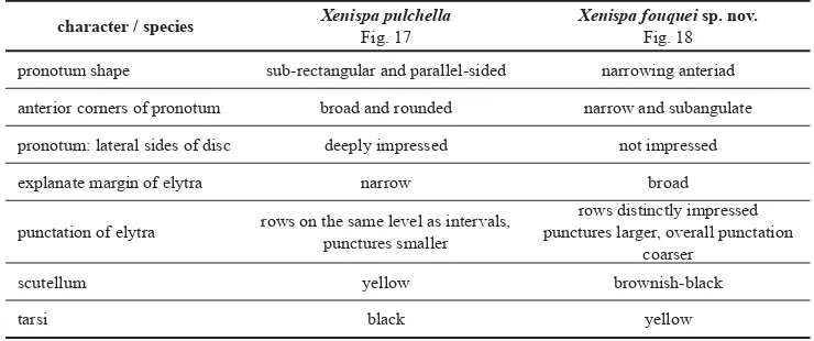
Comparative diagnosis. The new species is morphologically similar only to X. pulchella Baly, 1859, the type species of the genus. They both share similar shape of pronotum and body but can be distinguished by characters summarized in the Table 2. Venezuelan species of Xenispa can be distinguished using key on page 374.
Etymology. The species is dedicated in loving memory to René Fouquè (1980–2016), friend and enthusiastic entomologist, who was world specialist on the tenebrionid tribe Stenosini.
Biology. According to the label data the BMNH specimens were collected on bamboo; what is most likely correct as species of Xenispa are mainly associated with various Neotropical bambusoid Poaceae.
Remarks. According to the label data the BMNH specimens were collected about fi fty me-ters in elevation above La Montaña, which is the middle station of the cable tram connecting Barinitas and La Aguada. The coordinates are approximate and do not represent the actual spot where the specimens were collected. They were taken using GoogleEarth on the cable tram transect at 2500 m a.s.l. just to illustrate habitat.
Distribution. Venezuela: Mérida.
Table 2. Distinguishing characters between X. pulchella Baly, 1859 and X. fouquei sp. nov. character / species Xenispa pulchella
Fig. 17
Xenispa fouquei sp. nov. Fig. 18
pronotum shape sub-rectangular and parallel-sided narrowing anteriad
anterior corners of pronotum broad and rounded narrow and subangulate
pronotum: lateral sides of disc deeply impressed not impressed
explanate margin of elytra narrow broad
punctation of elytra rows on the same level as intervals, punctures smaller
rows distinctly impressed punctures larger, overall punctation
coarser
scutellum yellow brownish-black
Xenispa gilvipes (Uhmann, 1930) comb. nov. (Fig. 18)
Cephalolia gilvipes Uhmann, 1930b: 230 (original description).
Type locality. Costa Rica, San José Province, Santa María de Dota, approx. 9°39′N, 83°58′W, ca. 1500–1600 m. Type material examined. HOLOTYPE: pinned (both antennae missing, right elytron partly broken), ‘COSTA RICA |
F. NEVERMANN | 4.I.24 [g, p, cb, last row hw] || Sta. Maria de | Dota | an Blüten [verso of the previous label, hw] || Type No. | 54603 [hw] | USNM [r, p, cb] || HOLOTYPE [r, t, s] || Cephalolia | gilvipes n. sp. [w, hw by Uhmann, s] || USNM ENT | [data matrix code] | 00871395 [w, p, cb]’ (USNM).
Additional material examined. COSTA RICA: CARTAGO: Cañon, 2450 m, 28.xi.1994, 22 spec., H. Forster lgt.
(15 spec. NHMW, 6 spec. LSPC, 1 spec. NMPC).
Remarks. UHMANN (1930b) described this species based on a single specimen and stated that it was collected on fl owers. The holotype is strongly damaged missing both antennae from the antennomere II on and right elytron is broken due to the pin. In my opinion this species does not belong to Cephaloleia as it has conspicuously serrate apical margin of elytra, long antennae compared to the length of body and smooth convex frons and vertex. These cha-racters are typical for the genus Xenispa and C. gilvipes conforms to other species currently classifi ed in that genus. Therefore I hereby transfer C. gilvipes to Xenispa.
The species was so far reported only three times based on specimens collected in high mountains of central Costa Rica above 1500 m a.s.l. (FLOWERS & HANSON 2003, MCKENNA & FARELL 2005, STAINES & GARCÍA-ROBLEDO 2014). I had no opportunity to verify any of the recently published specimens, thus I cannot confi rm their identifi cations. The specimens reported here agree perfectly with the holotype, having the same shape, colouration and pun-ctation. Particularly characteristic is the shape of pronotum as it has only very narrow lateral margins, which are not explanate. A similarly shaped pronotum is known only in Xenispa uhmanni (Pic, 1934) from Colombia, which differs in having black antennae and legs while X. gilvipes has them pale brown. The locality Cañon is situated on the Pan-American Highway and is about 6 km air distance to NW from the type locality of X. gilvipes.
Distribition. Costa Rica: Cartago, Limón, Puntarenas (STAINES & GARCÍA-ROBLEDO 2014, present paper), Heredia (MCKENNA & FARELL 2005), and San José (FLOWERS & HANSON 2003).
Key to Venezuelan species of Xenispa
1 Pronotum of the same colour as elytra, legs black or metallic. Small species, length below 4 mm with narrow margin of elytra. ... 2 – Pronotum, head, fore- and mid-legs yellow. Large species, length 6.0–7.5 mm with
broadly explanate margin of elytra (Fig. 18). ... X. fouquei sp. nov.
2 Lateral margins of pronotum strongly swollen, broad and separated by a deep sulcus from the disc. Dorsum metallically coloured. ... 3 – Lateral margins of pronotum narrow and separated by shallow sulcus from the disc.
Dorsum pitch-black, pronotum somewhat paler coloured (Fig. 15). ... ... X. atra(Pic, 1926) 3 Pronotum narrower, 1.4–1.5× as wide as long. Swollen lateral margin of pronotum of the same
– Pronotum broader, 1.7× as wide as long. Swollen lateral margin of pronotum distinctly narrowed at base and anteriorly. Elytra very dark brown with bluish metallic tint. ... ... Xenispa sp.1
4 Pronotum shiny, microreticulation centrally vanished and more or less visible only late-rally. Elytra metallic antracite. ... X. carinata (Pic, 1934) – Pronotum subopaque due to more prominent microreticulation. Elytra very dark olive-green (Fig. 8). ... X. aeneipennis (Baly, 1859)
Hybosispini Weise, 1910
WEISE (1910) proposed the tribe for a single species Hybosispa melanura Weise, 1910 from Bolivia which is externally very similar to Cephaloleia but has no seta on pronotum while all Imatidiini have a seta at each anterior corner of pronotum or next to them. Hybosispa also has strongly convex body, which is nearly circular in cross-section, and head with prominent carina on inner margin of the eye. Imatidiini have more or less fl attened body, always with transverse cross-section and head without prominent carina along eyes.
Latter on, UHMANN (1933, 1939, 1940) described three more Hybosispa species from Bra-zil. Subsequently, UHMANN (1964: 405) and STAINES & GARCÍA-ROBLEDO (2014: 7) transferred Cephaloleia macella Pic, 1923 and C. bipartita Pic, 1926, respectively, to Hybosispa. Finally, SEKERKA (2014) revised genera of Imatidiini and found that Solenispa Weise, 1905 lack setae on pronotum and have similar morphology of the head to Hybosispa and thus transferred Solenispa to Hybosispini. WEISE (1905) when describing Solenispa mentioned the shape of the head and based on it differentiated Solenispa from Stenispa Baly, 1859, however, he did not notice that Solenispa lacks pronotal setae.
I have studied type material of various species of Cephaloleia and found three more species lack pronotal setae and have the shape of body and formation of the head similar to Hybosispa and are here transferred to the latter genus. Additionally, the only non-Andean member of the genus Solenispa – S. claripes Pic, 1923 is here transferred also to Hybosispa. Therefore Hybosispa presently contains ten species and they can be recognized using a key proposed on page 377.
Hybosispa claripes (Pic, 1923) comb. nov.
Solenispa claripes Pic, 1923: 10 (original description).
Type locality. Original type locality ‘Brésilʼ specifi ed by DESCARPENTRIES & VILLIERS (1959) to Brazil, Rio de Janeiro,
Theresópolis based on the label data of the type.
Type material examined. HOLOTYPE: glued, ‘Therezopolis [g, hw by Donckier, cb] || Solenispa | claripes m [w, hw
by Weise, s] || co-Type [w, p in red, cb] || type [y, hw by Pic, s] || TYPE [r, p, cb] || Sol. claripes | (1923) Pic [w, hw by Pic, s]’ (MNHN).
Remarks. According to the label data the specimen was studied by J. Weise who recognized it as a new species of Solenispa, however, he did not publish the description and PIC (1923) 1 This refers to the three paratypes of X. cyanea Staines, 1996 from Caracas, one of which was fi gured by STAINES
validated the name by very short and uninformative description, which is written in singular and Pic gave only a single length measurement suggesting that he had only a single specimen at his disposal. Afterward it was listed only in catalogues (e.g. UHMANN 1957a, 1964).
I have studied the holotype and in my opinion it belongs to the genus Hybosispa as it has typical shape of that genus and abdominal ventrites are not pubescent, while all known species of Solenispa have them densely pubescent. Therefore I hereby transfer S. claripes to Hybosispa. It is most similar to H. nitida Uhmann, 1939 and H. rufi ventris Uhmann, 1940 as all three share uniformly black dorsum. However, H. claripes differs in more impressed frons and vertex and pale coloured legs.
Distribution. Brazil: Rio de Janeiro (DESCARPENTRIES & VILLIERS 1959).
Hybosispa delectabilis (Staines, 1996) comb. nov. (Fig. 20)
Cephaloleia delectabilis Staines, 1996: 26 (original description incl. black-and-white photograph). Type locality. Mexico, Chiapas, Parque educativo Laguna Belgica, 16 km NW of Ocozocoautla.
Type material examined. HOLOTYPE: glued, ‘MEXICO, Chiapas, | Pq. Laguna Belgica, | 16kmNW Ocozocoautla |
14.VI.1989.H.Howden [w, p, cb] || Flight intercept | trap [w, p, cb] || HOLOTYPE | Cephaloleia [hw] | delectabilis [hw] | Staines 1994 [hw] | Des. C. L. Staines [hw] [r, p, bc] || [data matrix code] | Canadian Museum of | Musée canadien de la | NATURE | CMNEN 00012091 [w, p, cb, bf]’ (CMNC).
Remarks. STAINES (1996) described C. delectabilis based on single specimen and placed it in the genus Cephaloleia without further comment. Studying the black-and-white photograph, which accompanied the original description I doubted that the species belongs to the genus Hybosispa but essential characters were not visible or mentioned in the original description. I loaned the holotype and found that it has no seta on anterior margin of pronotum and the inner margins of eyes are produced, therefore I hereby transfer the C. delectabilis to Hybosispa. The species is so far known only from the holotype.
Distribution. Mexico: Chiapas (STAINES 1996).
Hybosispa sulciceps (Baly, 1885) comb. nov. (Fig. 21)
Cephaloleia sulciceps Baly, 1885: 26 (original description). Type locality. Panama, Chiriquí Province, Bugaba.
Type material examined. SYNTYPE: glued, ‘SYN- | TYPE [w, p, s, circular label with blue frame] || Bugaba, | Panama.
| Champion. [w, p, cb] || Godman-Salvin | Coll., Biol. | Centr.-Amer. [w, p, cb]’ (BMNH).
The exact position of the type localities is unknown. Bugaba without information on ele-vation usually refers to lowland (below 300–500 ft) part of the Bugaba District in the Western Chiriquí. The area is situated around present day city of La Concepción and the small pueblo Bugaba is situated ca. 3.7 km to south. Some other syntypes in BMNH and a syntype in USNM have on the label also elevation 800–1500 ft. This elevation, however might refer to much larger area extending from South-western slopes of Volcán Barú to Río Chiriquí Viejo. Based on CHAMPIONʼs (1907) itinerary he visited several places in this area but in the end specimens were provided just with label ‘Bugaba’ and elevation.
STAINES (1996) published additional 17 specimens of this species from Panamá and Costa Rica. These records were partly published again by STAINES & GARCÍA-ROBLEDO (2014) and mixed with several new records. Thus far I have had no opportunity to verfy these records.
Distribution. Costa Rica: Cartago, Limón, Punarenas and Panamá: Chiriquí, Panamá (BALY 1885, STAINES 1996).
Hybosispa truncatipennis (Baly, 1869) comb. nov. (Fig. 22)
Cephaloleia truncatipennis Baly, 1869: 371 (original description). Type locality. ‘Upper Amazons’.
Type material examined. HOLOTYPE: pinned, ‘Type | H.T. [w, p, cb, circular label with red frame] || Baly Coll. [w,
p, cb] || Cephaloleia | truncatipennis | Baly | Upper Amazons [g, hw by Baly, cb]’ (BMNH).
Remarks. BALY (1869) described C. truncatipennis presumably based on a single specimen as he gave just a single length and mentioned only a female. The species was only listed in catalogues since its description until STAINES & GARCÍA-ROBLEDO (2014) who included it in their revision of Cephaloleia and reported one specimen from Capella in Brazil. However based on the photograph presented in the revision (STAINES & GARCÍA-ROBLEDO 2014: 314, Fig. 258) the specimen was misidentifi ed and belonged to another species of Cephaloleia. The redescription of C. truncatipennis is also not in full agreement with the holotype and seems to combine characters of both specimens thus may not be reliable.
The holotype (Fig. 22) has anterior corners of pronotum clearly without any seta and head just like the other species of Hybosispa therefore I hereby transfer it to the latter genus. The species is thus far known only from the single specimen collected by H. W. Bates in the Upper Amazons. Unfortunately no precise data are available for it and thus whether it was captured in Brazil or in Peru is unknown.
Distribution. Amazonas state of Brazil or Region of Loreto in Peru.
Key to species of Hybosispa
1 Abdominal ventrites bare; antennae short and thick, distal antennomeres as wide as long. Lowland or mid-elevation species (usually below 1000 m) from southern Mexico to southern Brazil. ... Hybosispa Weise, 1910 ... 2 – Abdominal ventrites densely pubescent; antennae longer and slender, most of distal