
S H O R T G E N O M E R E P O R T
Open Access
Complete genome sequence of
Actinobacillus equuli
subspecies
equuli
ATCC
19392
T
Barbara F Huang, Andrew M Kropinski, Adina R Bujold and Janet I MacInnes
*Abstract
Actinobacillus equuli
subsp.
equuli
is a member of the family
Pasteurellaceae
that is a common resident of the oral cavity
and alimentary tract of healthy horses. At the same time, it can also cause a fatal septicemia in foals, commonly known
as sleepy foal disease or joint ill disease. In addition,
A. equuli
subsp.
equuli
has recently been reported to act as a
primary pathogen in breeding sows and piglets. To better understand how
A. equuli
subsp.
equuli
can cause disease,
the genome of the type strain of
A. equuli
subsp.
equuli,
ATCC 19392
T, was sequenced using the PacBio RSII sequencing
system. Its genome is comprised of 2,431,533 bp and is predicted to encode 2,264 proteins and 82 RNAs.
Keywords:
Actinobacillus equuli
subsp.
equuli
, Sleepy foal disease, Joint ill disease, Commensal, Equine
Introduction
Actinobacillus equuli
subsp.
equuli
, previously known as
‘
Bacillus viscosum-equi
’
, or
‘
Shigella equirulis
’
, is a
com-mon resident of the oral flora of healthy horses, as well
as that of the alimentary and genital tracts [1,2]. It has
also been reported to be present in other host species
such as mice, seemingly without ill effect [3] and on rare
occasions, has been transmitted through bite wounds to
humans [4].
A. equuli
subsp.
equuli
is the etiological
agent of sleepy foal disease, an acute form of fatal
septi-cemia in neonatal foals that may progress to a chronic
form, joint ill disease, producing lesions in the kidneys,
joints, and lungs [5-8]. Horses with
A. equuli
infection
can present with arthritis, bronchitis, pneumonia,
pleuri-tis, peritonipleuri-tis, sepsis, endocardipleuri-tis, pericardipleuri-tis, nephripleuri-tis,
meningitis, metritis, and abortion [7,9-12].
A. equuli
subsp.
equuli
was previously proposed to act as a
second-ary pathogen in foals; however, a recent study by Layman
and colleagues [13] has revealed that
A. equuli
subsp.
equuli
has the potential to act as a primary pathogen given
favourable conditions. Recently, it has been reported to
also be a primary pathogen in sows and piglets [14,15].
The hemolytic counterpart of this bacterium,
A. equuli
subsp.
haemolyticus,
is isolated more frequently from
the respiratory tract rather than the oral cavity. It can
also cause septicemia and sequelae such as arthritis and
meningitis, respiratory tract infections, and mare
repro-ductive loss syndrome [8,10,16].
The similar colonial morphology and biochemical
markers and shared 16S rRNA sequences make
differenti-ation of
A. equuli
from
Actinobacillus suis
difficult [8]. In
addition, little is known about the virulence factors of
A.
equuli
subsp.
equuli.
To be better able to identify and to
im-prove our understanding of the mechanism of
pathogen-host interactions [7], the genome of the type strain
A. equuli
subsp.
equuli
strain ATCC 19392
Twas sequenced. This
strain was isolated from foal blood and deposited in
the American Type Culture Collection by the Equine
Research Station (New Market, UK) in 1953 [17].
Organism information
Classification and features
As a member of the genus
Actinobacillus, A. equuli
subsp
. equuli
belongs to the family
Pasteurellaceae
, class
Gammaproteobacteria
[18] (Table 1). Phylogenetic
ana-lysis using 16S rRNA sequences suggests that
A. equuli
subsp
. equuli
is most closely related to
A. suis
and
A.
hominis
(Figure 1).
A. equuli
subsp.
equuli
is a small, Gram-negative,
non-motile, pleomorphic bacterium [15,16,19] (Figure 2). It is
NAD-independent, nonhemolytic, and CAMP negative
* Correspondence:[email protected]
Department of Pathobiology, University of Guelph, Ontario Veterinary College, Ontario N1G 2 W1, Canada
[15,20].
A. equuli
subsp.
equuli
produces large amounts
of extracellular slime that imparts sticky properties in
solid and liquid media cultures [19,31]
.
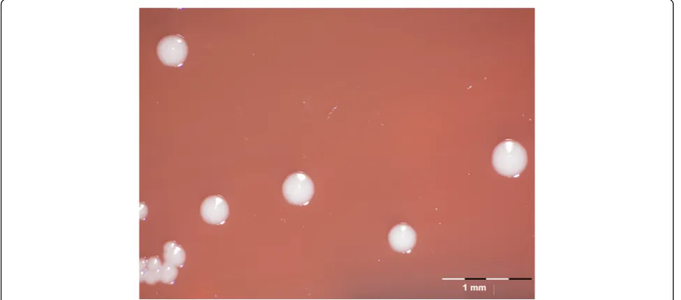
On nutrient or
blood agar, smooth, grayish-white, circular colonies are
produced with an average diameter of 1-2 mm after
growth for 24 h [35] (Figure 3). On initial culture from
clinical material, colonies are viscous and usually rough
but become smooth in successive subcultures [1,19].
Growth using liquid culturing methods has been
re-ported to increase viability in comparison to solid media
Table 1 Classification and features of
A. equuli subsp. equuli
ATCC 19392
TMIGS ID Property Term Evidence codea
Classification DomainBacteria TAS [21]
PhylumProteobacteria TAS [22]
ClassGammaproteobacteria TAS [23,24]
OrderPasteurellales TAS [25]
FamilyPasteurellaceae TAS [26,27]
GenusActinobacillus TAS [28,29]
SpeciesActinobacillus equuli TAS [28,30,31]
SubspeciesActinobacillus equulisubsp.equuli TAS [20]
Type strain ATCC 19392T
Gram stain Negative TAS [32]
Cell shape Rods (pleomorphic) TAS [33]
Motility Non-motile TAS [33]
Sporulation Non-sporulating TAS [33]
Temperature range Mesophile (20 - 44°C) TAS [33]
Optimum temperature 37°C TAS [20]
pH range 6.0–8.4 TAS [1]
Carbon source Saccharolytic TAS [19]
MIGS-6 Habitat Host, equine or swine upper respiratory tract, alimentary tract, and genital tract TAS [4,5,19]
MIGS-6.3 Salinity 0.5% NaCl NAS
MIGS-22 Oxygen requirement Facultative anaerobe TAS [19,33]
MIGS-15 Biotic relationship Commensal or opportunistic TAS [14,15]
MIGS-14 Pathogenicity Variable TAS [13]
MIGS-4 Geographic location New Market, UK TAS [17]
MIGS-5 Sample collection 1953 TAS [17]
MIGS-4.1 MIGS-4.2 Latitude Not reported
Longitude Not reported
MIGS-4.4 Altitude Not reported
a
Evidence codes - TAS: Traceable Author Statement; NAS: Non-traceable Author Statement (i.e., not directly observed for the living, isolated sample, but based on a generally accepted property for the species, or anecdotal evidence). These evidence codes are from the Gene Ontology project [34].
Figure 1Phylogenetic tree based on 16S rRNA sequences ofActinobacillus sensu strictospecies plusA. capsulatusandH. parasuisas outgroups.
cultures, and viscosity is retained upon repeated
subcul-turing [1,19]. The usual temperature range for growth of
this bacterium is 20-39°C, with an optimum at 37°C,
though some
A. equuli
subsp.
equuli
strains have been
shown to grow at temperatures as high as 44°C [33].
Acid but not gas is produced from sucrose, mannitol,
galactose, lactose, maltose, mannose, melibiose,
trehal-ose, raffintrehal-ose, and glycerol fermentation [19,20,33].
A.
equuli
subsp.
equuli
is capable of reducing nitrate and
produces
α-galactosidase,
α-glucosidase,
β-xylosidase,
ure-ase, and oxidase [19,20,33].
Genome sequencing information
Genome project history
A. equuli
subsp.
equuli
was selected for sequencing
be-cause of its importance to the horse industry as the
etio-logic agent of sleepy foal disease and joint ill disease [7].
Sequencing was done at the McGill University and
Génome Québec Innovation Centre (Montréal, QC,
Canada) using the PacBio RS II DNA Sequencing
System, and assembled using PacBio RS II software and
Celera Assembler.
A. equuli
subsp.
equuli
was
annota-ted using the NCBI Prokaryotic Genome Annotation
Figure 2Gram stain ofA. equulisubsp.equuliATCC 19392Tat 1000 X magnification.Pipeline. A summary of the project information and the
Minimum Information about a Genomic Sequence is
shown in Table 2 [38].
Growth conditions and genomic DNA preparation
A. equuli
subsp.
equuli
was grown from a frozen (-70°C)
seed stock on sheep blood agar plates overnight in an
atmosphere of 5% CO2
at 37°C. After subculture,
well-isolated colonies were used for genomic DNA isolation.
Cells were lysed using modified B1 (150 mM Tris · Cl,
50 mM EDTA, 0.5% Tween®-20, 0.5% Triton X-100,
pH 8.0) and B2 (750 mM NaCl, 50 mM MOPS, 15%
iso-propanol, 0.15% Triton X-100, pH 7.0) buffers. DNA
was then column purified using a QIAGEN Plasmid
Midi Kit (Qiagen, Germany) following manufacturer's
protocol for binding and elution. The resultant DNA
preparation was characterized using a NanoDrop model
ND1000 Spectrophotometer and was diluted to a
con-centration of ~0.47 mg/μl.
Genome sequencing and assembly
Single Molecule, Real-Time DNA sequencing (Pacific
Biosciences) [39] was done to obtain the genome sequence
of the
A. equuli
subsp.
equuli
ATCC 19392
T. A total of
133,616 raw subreads were generated with an average
length of 4,348 bp using two SMRT Cells in a PacBio RSII
sequencer. The resultant subread length cutoff value, 29.42,
was used in the Basic Local Alignment with Successive
Refinement step [40] where short reads were used to
cor-rect for errors on long reads [39]. The corcor-rected reads were
assembled into contigs according to the Hierarchical
Genome Assembly Process (HGAP) workflow using the
Celera Assembler and refined using BLASR to align raw
reads on contigs [39]. Final processing was conducted using
Quiver, a variant calling algorithm, to generate high quality
consensus sequences [39]. There were a total of 4,777
cor-rected reads with an average length of 7,804 bp and a final
product of one contig.
Genome annotation
Genes were identified using the NCBI Prokaryotic
Genome Annotation Pipeline. The prediction software,
GeneMark, is integrated into the pipeline and performs
unsupervised gene finding using heuristic Markov
Models [41]. Additional gene prediction analysis and
functional annotation was performed within the
Inte-grated Microbial Genomes (IMG) platform [42]
devel-oped by the Joint Genome Institute [43] (Table 3).
Genome properties
The genome of
A. equuli
subsp.
equuli
is a single circular
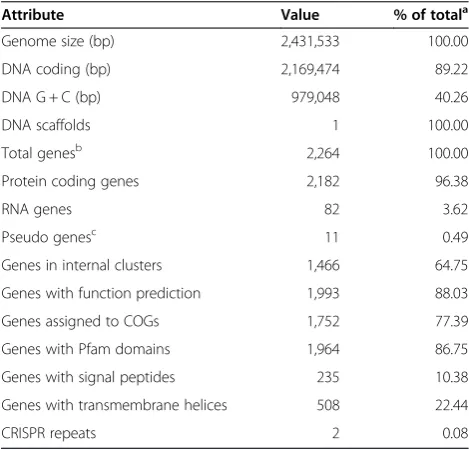
chromosome that is 2,431,533 bp in length with a G + C
content of approximately 40.3%. It is predicted to contain
2,264 genes, of which 2,182 code for proteins and 82
for RNA; 11 pseudogenes are also present (Table 3 and
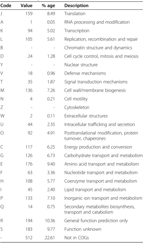
Figure 3). Approximately 3/4 of the predicted genes can
be assigned to one of 25 functional COG categories
(Table 4). Of particular note with regard to virulence
are several lipopolysaccharide genes predicted to
encode biosynthetic enzymes for the O-antigen and
lipid A components. Adhesins of different types were
Table 2 Project information and its association with MIGS
version 2.0 compliance [38]
MIGS ID Property Term
MIGS-31 Finishing quality Complete
MIGS-28 Libraries used SMRTbell library
MIGS-29 Sequencing platforms PacBio RS II
MIGS-31.2 Fold coverage 196x
MIGS-30 Assemblers PacBio RS II, Celera
MIGS-32 Gene calling method GeneMarkS+
Locus Tag ACEE
Genbank ID CP007715
GenBank Date of Release December 15, 2014
GOLD ID Gp0095186
BIOPROJECT PRJNA247050
MIGS-13 Source Material Identifier ATCC 19392T
Project relevance Equine and swine pathogenesis
Table 3 Genome statistics
Attribute Value % of totala
Genome size (bp) 2,431,533 100.00
DNA coding (bp) 2,169,474 89.22
DNA G + C (bp) 979,048 40.26
DNA scaffolds 1 100.00
Total genesb 2,264 100.00
Protein coding genes 2,182 96.38
RNA genes 82 3.62
Pseudo genesc 11 0.49
Genes in internal clusters 1,466 64.75
Genes with function prediction 1,993 88.03
Genes assigned to COGs 1,752 77.39
Genes with Pfam domains 1,964 86.75
Genes with signal peptides 235 10.38
Genes with transmembrane helices 508 22.44
CRISPR repeats 2 0.08
a
The total is based on either the size of the genome in base pairs or the total number of protein coding genes in the annotated genome.
b
Also includes 11 pseudogenes and one other RNA gene that does not belong to rRNA or tRNA categories.
c
observed including several autotransporters; a tight
ad-herence locus; prepilins, and fimbriae; a filamentous
hemagglutinin homolog was also detected. In addition,
several putative iron acquisition systems are present
in-cluding those for siderophores, hemoglobin and
transfer-rin. A number of toxin and hemolysin genes were also
identified including an
aqxCABD
operon, although
com-pared to the
aqxCABD
of
A. equuli
subsp.
haemolyticus
there are many point mutations and sizable deletions at
both ends of the
aqxA
gene. Other regions of particular
interest include an integron and Mu-like phage, identified
using PHAST [44].
Insights from the genome sequence
Given the marked similarities of
A. equuli
and
A. suis
there has been some debate as to whether these
organ-isms should be a single species. In the current study we
determined that the
A. equuli
subsp.
equuli
16S genes
are 99% identical to those of both
A. suis
H91-0380 and
the
A. suis
type strain, ATCC 33415, consistent with
membership in the same species. Further, as can be seen
in the circular maps below, the genome of
A. equuli
subsp.
equuli
is very similar to that of
A. suis
again
sug-gesting that
A. equuli
subsp
. equuli
and
A. suis
might be
the same species (Figure 4). On the other hand, when
genomes of
A. suis
H91-0380 and
A. suis
ATCC 33415
were compared with that of
A. equuli
subsp.
equuli
using the ANI calculator [45], the ANI value of both
comparisons was 93.82%, which is lower than 95%, the
recommended cutoff value for delineating species [46].
In-silico DNA-DNA hybridization, done using a Genome
Blast Distance Phylogeny approach to generate genome
based distance measures for phylogenetic inferences, also
demonstrated differences between
A. equuli
and
A. suis
. The
Genome-to-Genome Distance Calculator [47] revealed a
dis-tance of 0.0685 between
A. suis
H91-0380 and
A. equuli
subsp.
equuli,
with a DDH estimate of 51.40% +/- 2.66. A
DDH similarity below 70% is interpreted as two species
being distinct; 79% is used to discriminate between
sub-species [48]. The DDH estimate exceeding the 70% sub-species
threshold was determined from logistic regression to be
23.14%. In terms of subspecies relatedness, the probability
of exceeding the 79% threshold was 4.82% between
A.
equuli
subsp.
equuli
and
A. suis
H91-0380. The distance
calculated between
A. suis
ATCC 33415 and
A. equuli
subsp.
equuli
and their DDH estimate was 0.0681 and
51.60% +/- 2.66, respectively. The probability that DDH
exceeded 70% and 79% for
A. suis
ATCC 33415 and
A.
equuli
subsp.
equuli
were 23.66% and 4.94%, respectively.
Taken together, these analyses are consistent with the
notion that
A. suis
and
A. equuli
subsp.
equuli
are
re-lated but distinct species, and care is needed to correctly
identify them.
Conclusions
A. equuli
subsp.
equuli
can induce fatal septicemia in
foals resulting in significant economic losses in the
equine industry; as well,
A. equuli
subsp.
equuli
has
re-cently been reported to cause septicemia in swine of all
ages. Our analysis of the
A. equuli
subsp.
equuli
genome
indicates that
A. suis
and
A. equuli
subsp.
equuli
are
closely related yet distinct species. At the present time
little is known about how
A. equuli
subsp.
equuli
causes
disease or the factors that control species and tissue
tropism. More research including biological experiments
is required to better understand the pathogenesis of
A.
equuli
and it is hoped this reported genome sequence of
A. equuli
subsp.
equuli
ATCC 19392
Twill provide vital
information for such studies. In addition, pathway
analysis and genome studies may help improve our
un-derstanding of host-pathogen interactions of
A. equuli
Table 4 Number of genes associated with general COG
functional categories
Code Value % age Description
J 159 8.49 Translation
A 1 0.05 RNA processing and modification
K 94 5.02 Transcription
L 105 5.61 Replication, recombination and repair
B - - Chromatin structure and dynamics
D 24 1.28 Cell cycle control, mitosis and meiosis
Y - - Nuclear structure
V 18 0.96 Defense mechanisms
T 35 1.87 Signal transduction mechanisms
M 136 7.26 Cell wall/membrane biogenesis
N 4 0.21 Cell motility
Z - - Cytoskeleton
W 2 0.11 Extracellular structures
U 44 2.35 Intracellular trafficking and secretion
O 92 4.91 Posttranslational modification, protein turnover, chaperones
C 117 6.25 Energy production and conversion
G 126 6.73 Carbohydrate transport and metabolism
E 176 9.40 Amino acid transport and metabolism
F 63 3.36 Nucleotide transport and metabolism
H 108 5.77 Coenzyme transport and metabolism
I 45 2.40 Lipid transport and metabolism
P 133 7.10 Inorganic ion transport and metabolism
Q 14 0.75 Secondary metabolites biosynthesis, transport and catabolism
R 194 10.36 General function prediction only
S 183 9.77 Function unknown
subsp.
equuli
and other
Actinobacillus
species and aid in
the design of diagnostic tools and antimicrobial agents.
Competing interests
The authors declare that they have no competing interests.
Authors’contributions
JIM and AMK contributed to the conception and design of this project. BFH and AMK were involved in the acquisition and initial analysis of the data; BFH, AMK, ARB and JIM were involved in the interpretation of the data. BFH prepared the first draft of the manuscript. All authors were involved in its critical revision and have given final approval of the version to be published and agree to be accountable for all aspects of the work.
Acknowledgements
The authors thank Glenn Soltes for expert technical support. This work was supported by a Natural Sciences and Engineering Research Council to JM; ARB was supported by the Ontario Veterinary College and the Ontario Graduate Scholarship programs.
Received: 7 October 2014 Accepted: 16 April 2015
References
1. Edwards PR. Studies onShigella equirulis. Kentucky Agric Exp Stn Bull. 1931;320:289–330.
Figure 4Circular map of theA. equulisubsp.equuliATCC 19392Tgenome generated using the CGView Server [49]. From the outside to the
2. Sternberg S, Brändström B. Biochemical fingerprinting and ribotyping of isolates ofActinobacillus equulifrom healthy and diseased horses. Vet Microbiol. 1999;66:53–65. PubMed.
3. Lentsch RH, Wagner JE. Isolation ofActinobacillus lignieresiiandActinobacillus
equulifrom laboratory rodents. J Clin Microbiol. 1980;12:351–4. PubMed. 4. Ashhurst-Smith C, Norton R, Thoreau W, Peel MM.Actinobacillus equuli
Septicemia: an Unusual Zoonotic Infection. J Clin Microbiol. 1998;36:2789–91. PubMed.
5. Blackall PJ, Christensen JP, Bisgaard M. Diversity among isolates of
Actinobacillus equuliand related organisms as revealed by ribotyping. Aust Vet J. 1998;76:423–5. PubMed.
6. Aalbaek B, Ostergaard S, Buhl R, Jensen HE, Christensen H, Bisgaard M.
Actinobacillus equulisubsp.equuliassociated with equine valvular endocarditis. APMIS. 2007;115:1437–42. PubMed.
7. Berthoud H, Frey J, Kuhnert P. Characterization of Aqx and its operon: the hemolytic RTX determinant ofActinobacillus equuli. Vet Microbiol. 2002;87:159–74. PubMed.
8. Kuhnert P, Berthoud H, Straub R, Frey J. Host cell specific activity of RTX toxins from haemolyticActinobacillus equuliandActinobacillus suis. Vet Microbiol. 2003;92:161–7. PubMed.
9. Sternberg S. Isolation ofActinobacillus equulifrom the oral cavity of healthy horses and comparison of isolates by restriction enzyme digestion and Pulsed-Field Gel Electrophoresis. Vet Microbiol. 1998;59:147–56. PubMed. 10. Pusterla N, Jones MEB, Mohr RF, Higgins JK, Mapes S, Jang S, et al. Fatal
Pulmonary Hemorrhage Associated with RTX Toxin-ProducingActinobacillus
equulisubspecieshaemolyticusInfection in an Adult Horse. J Vet Diagnostic Investig. 2008;20:118–21. PubMed http://dx.doi.org/10.1177/104063870802000127. 11. Matthews S, Dart AJ, Dowling BA, Hodgson JL. Peritonitis associated with
Actinobacillus equuliin horses : 51 cases. Aust Vet J. 2001;79:536–9. PubMed. 12. Patterson-Kane JC, Donahue JM, Harrison LR. Septicemia and Peritonitis Due
toActinobacillus equuliInfection in an Adult Horse. Vet Pathol. 2001;38:230–2. PubMed http://dx.doi.org/10.1354/vp.38-2-230.
13. Layman QD, Rezabek GB, Ramachandran A, Love BC, Confer AW. A retrospective study of equine actinobacillosis cases: 1999-2011. J Vet Diagn Invest. 2014;26:365–75. PubMed http://dx.doi.org/10.1177/1040638714531766. 14. Thompson AB, Postey RC, Snider T, Pasma T.Actinobacillus equulias a
primary pathogen in breeding sows and piglets. Can Vet J. 2010;51:1223–5. PubMed.
15. Benavente CE, Fuentealba IC.Actinobacillus suisandActinobacillus equuli, emergent pathogens of septic embolic nephritis, a new challenge for the swine industry. Arch Med Vet. 2012;44:99–107. http://dx.doi.org/10.4067/ S0301-732X2012000200002.
16. Castagnetti C, Rossi M, Parmeggiani F, Zanoni RG, Pirrone A, Mariella J. Facial cellulitis due toActinobacillus equuliinfection in a neonatal foal. Vet Rec. 2008;162:347–9. PubMed http://dx.doi.org/10.1136/vr.162.11.347. 17. Public Health England Culture Collections Database.
[http://www.phe-culturecollections.org.uk/]
18. Dewhirst FE, Paster BJ, Olsen I, Fraser GJ. Phylogeny of 54 representative strains of species in the familyPasteurellaceaeas determined by comparison of 16S rRNA sequences. J Bacteriol. 1992;174:2002–13. PubMed.
19. Olsen I, Moller K. Genus II.Actinobacillus. In: Garrity G, Brenner D, Krieg N, Staley J, editors. Bergey’s Manual of Systematic Bacteriology, Second Edition, Volume Two, TheProteobacteria, Part B. Second. New York: Springer; 2005. p. 866–83. 20. Christensen H, Bisgaard M, Olsen JE. Reclassification of equine isolates
previously reported asActinobacillus equuli, variants ofA. equuli,Actinobacillus suisor Bisgaard taxon 11 and proposal ofA. equulisubsp.equulisubsp. nov. andA. equulisubsp.haemolyticussubsp. nov. Int J Syst Evol Microbiol. 2002;52:1569–76. PubMed http://dx.doi.org/10.1099/ijs.0.01637-0.
21. Woese CR, Kandlert O, Wheelis ML. Towards a natural system of organisms: Proposal for the domainsArchaea, Bacteria, andEucarya. Proc Natl Acad Sci U S A. 1990;87:4576–9. PubMed.
22. Garrity GM, Bell JA, Lilburn T. Phylum XIV.Proteobacteriaphyl. nov. In: Garrity GM, Brenner DJ, Krieg NR, Staley JT (eds), Bergey’s Manual of Systematic Bacteriology, Second Edition, Volume 2, Part B, Springer, New York, 2005, p. 1. 23. List Editor. Validation of publication of new names and new combinations
previously effectively published outside the IJSEM. Int. J. Syst. Evol. Microbiol. 2005;55:2235–2238. http://dx.doi.org/10.1099/ijs.0.64108-0 24. Garrity GM, Bell JA, Lilburn T. Class III.Gammaproteobacteriaclass. nov. In:
Garrity GM, Brenner DJ, Krieg NR, Staley JT (eds), Bergey’s Manual of Systematic Bacteriology, Second Edition, Volume 2, Part B, Springer, New York, 2005, p. 1.
25. Garrity GM, Bell JA, Lilburn T.Pasteurellalesord. nov. In: Garrity GM, Bell JA, Lilburn T. Order XIV.Pasteurellalesord. nov. In: Garrity GM, Brenner DJ, Krieg NR, Staley JT (eds), Bergey’s Manual of Systematic Bacteriology, Second Edition, Volume 2, Part B, Springer, New York, 2005, p. 850
26. List Editor. Validation List no. 7. Validation of the publication of new names and new combinations previously effectively published outside the IJSB. Int J Syst Bacteriol 1981; 55:382–383. http://dx.doi.org/10.1099/00207713-31-3-382 27. Pohl SPD. Dissertation, Phillips-Universität Marburg. 1979.
28. Skerman VBD, McGowan V, Sneath PHA. Approved Lists of Bacterial Names. Int J Syst Bacteriol. 1980;30:225–420. http://dx.doi.org/10.1099/00207713-30-1-225. 29. Brumpt E. Précis de Parasitologie. 1st ed. Paris: Masson et Cie; 1910. 30. van Straaten H. Bacteriologische bevindingen bij eenigo gevallen van
pyo-septicaemie (Lähme) der veuluens. Verslag van den Werksaamheden der Rijksseruminrichting voor 1916-1917, Rotterdam, 1918; 71-76. 31. Haupt H. Archiv fur wissenschaftliche und praktische Tierheilkunde.
1934; 67:513-524.
32. Ward CL, Wood JL, Houghton SB, Mumford JA, Chanter N.Actinobacillus andPasteurellaspecies isolated from horses with lower airway disease. Vet Rec. 1998;143:277–9. PubMed.
33. MacInnes JI, Lally ET. The GenusActinobacillus. In: Dworkin M, Falkow S, Rosenberg E, Schleifer K, Stackebrandt E, editors. The Prokaryotes, A Handbook on the Biology of Bacteria:Proteobacteria: Gamma Subclass, vol. 6. 3rd ed. New York: Springer; 2006. p. 1094–118.
34. Ashburner M, Ball CA, Blake JA, Botstein D, Butler H, Cherry JM, et al. Gene Ontology : tool for the unification of biology. The Gene Ontology Consortium. Nat Genet. 2000;25:25–9. PubMed http://dx.doi.org/10.1038/75556.
35. Christensen H, Bisgaard M. Revised definition ofActinobacillus sensu stricto isolated from animals. A review with special emphasis on diagnosis. Vet Microbiol. 2004;99:13–30. PubMed http://dx.doi.org/10.1016/j.vetmic.2003.12.002. 36. Bruno WJ, Socci ND, Halpern AL. Weighted neighbor joining: a
likelihood-based approach to distance-likelihood-based phylogeny reconstruction. Mol Biol Evol. 2000;17:189–97. PubMed.
37. Cole JR, Chai B, Farris RJ, Wang Q, Kulam-Syed-Mohideen AS, McGarrell DM, et al. The Ribosomal Database Project (RDP-II): introducing myRDP space and quality controlled public data. Nucleic Acids Res. 2007;35:D169–72. PubMed http://dx.doi.org/10.1093/nar/gkl889.
38. Field D, Garrity G, Gray T, Morrison N, Selengut J, Sterk P, et al. The minimum information about a genome sequence (MIGS) specification. Nat Biotechnol. 2008;26:541–7. PubMed http://dx.doi.org/10.1038/nbt1360. 39. Chin C-S, Alexander DH, Marks P, Klammer AA, Drake J, Heiner C, et al. Nonhybrid,
finished microbial genome assemblies from long-read SMRT sequencing data. Nat Methods. 2013;10:563–9. PubMed http://dx.doi.org/10.1038/nmeth.2474. 40. Chaisson MJ, Tesler G. Mapping single molecule sequencing reads using
basic local alignment with successive refinement (BLASR): application and theory. BMC Bioinformatics. 2012;13:238. PubMed http://dx.doi.org/10.1186/ 1471-2105-13-238.
41. Besemer J, Lomsadze A, Borodovsky M. GeneMarkS: a self-training method for prediction of gene starts in microbial genomes. Implications for finding sequence motifs in regulatory regions. Nucleic Acids Res. 2001;29:2607–8. PubMed. 42. Integrated Microbial Genome Database. [http://img.jgi.doe.gov/]
43. Markowitz VM, Chen I-MA, Palaniappan K, Chu K, Szeto E, Pillay M, et al. IMG 4 version of the integrated microbial genomes comparative analysis system. Nucleic Acids Res. 2014;42:D560–7. PubMed http://dx.doi.org/10.1093/nar/ gkt963.
44. Zhou Y, Liang Y, Lynch KH, Dennis JJ, Wishart DS. PHAST: a fast phage search tool. Nucleic Acids Res. 2011;39:W347–52. PubMed http://dx.doi.org/ 10.1093/nar/gkr485.
45. Average Nucleotide Identity Database. [http://enve-omics.ce.gatech.edu/ani/] 46. Goris J, Konstantinidis KT, Klappenbach JA, Coenye T, Vandamme P, Tiedje JM. DNA-DNA hybridization values and their relationship to whole-genome sequence similarities. Int J Syst Evol Microbiol. 2007;57:81–91. PubMed http://dx.doi.org/10.1099/ijs.0.64483-0.
47. Genome-to-Genome Distance Calculator. [http://ggdc.dsmz.de/distcalc2.php] 48. Meier-Kolthoff JP, Auch AF, Klenk HP, Goker M. Genome sequence-based
species delimitation with confidence intervals and improved distance functions. BMC Bioinformatics. 2013;14:1–14. PubMed http://dx.doi.org/ 10.1186/1471-2105-14-60.

![Figure 4 Circular map of the A. equuli subsp. equuli ATCC 19392T genome generated using the CGView Server [49]](https://thumb-us.123doks.com/thumbv2/123dok_us/671885.2065993/6.595.59.541.89.565/figure-circular-equuli-equuli-genome-generated-cgview-server.webp)