REVIEW
Potential theranostics of circulating tumor
cells and tumor-derived exosomes application
in colorectal cancer
Somayeh Vafaei
1,2,3, Raheleh Roudi
1, Zahra Madjd
1,2*, Amir Reza Aref
4and Marzieh Ebrahimi
3*Abstract
Background:
At the present time, colorectal cancer (CRC) is still known as a disease with a high mortality rate.
Thera-nostics are flawless scenarios that link diagnosis with therapy, including precision medicine as a critical platform that
relies on the development of biomarkers particularly “liquid biopsy”. Circulating tumor cells (CTCs) and tumor-derived
exosomes (TDEs) in a liquid biopsy approach are of substantial importance in comparison with traditional ones, which
cannot generally be performed to determine the dynamics of the tumor due to its wide restriction of range. Thus,
recent attempts has shifted towards minimally noninvasive methods.
Main text:
CTCs and TDEs, as significant signals emitted from the tumor microenvironment, which are also
detect-able in the blood, prove themselves to be promising novel biomarkers for cancer diagnosis, prognosis, and treatment
response prediction. The therapeutic potential of them is still limited, and studies are at its infancy. One of the major
challenges for the implementation of CTCs and TDEs which are new trends in translational medicine is the
develop-ment of isolation and characterization; a standardizable approach. This review highlights and discusses the current
challenges to find the bio fluids application in CRC early detection and clinical management.
Conclusion:
Taken together, CTCs and TDEs as silent drivers of metastasis can serve in the management of cancer
patient treatment and it is of the upmost importance to expand our insight into this subject. However, due to the
limited data available from clinical trials, further validations are required before addressing their putative application in
oncology.
Keywords:
Colorectal cancer, Circulating tumor cells (CTCs), Tumor-derived exosomes (TDEs), Clinical trial,
Theranostic
© The Author(s) 2020. This article is licensed under a Creative Commons Attribution 4.0 International License, which permits use, sharing, adaptation, distribution and reproduction in any medium or format, as long as you give appropriate credit to the original author(s) and the source, provide a link to the Creative Commons licence, and indicate if changes were made. The images or other third party material in this article are included in the article’s Creative Commons licence, unless indicated otherwise in a credit line to the material. If material is not included in the article’s Creative Commons licence and your intended use is not permitted by statutory regulation or exceeds the permitted use, you will need to obtain permission directly from the copyright holder. To view a copy of this licence, visit http://creat iveco mmons .org/licen ses/by/4.0/. The Creative Commons Public Domain Dedication waiver (http://creat iveco mmons .org/publi cdoma in/ zero/1.0/) applies to the data made available in this article, unless otherwise stated in a credit line to the data.
Open Access
*Correspondence: majdjabari.z@iums.ac.ir; mebrahimi@royaninstitute.org
2 Department of Molecular Medicine, Faculty of Advanced Technologies
in Medicine, Iran University of Medical Sciences, Tehran, Iran
3 Department of Stem Cells and Developmental Biology, Cell Science
Research Center, Royan Institute for Stem Cell Biology and Technology, ACECR, Tehran, Iran
Full list of author information is available at the end of the article
Background
Colorectal cancer (CRC) is the third leading cause of
can-cer-related mortality and morbidity [1] and fifty percent
of patients suffering from metastasis undergo surgery [2]
which creates huge obstacles in treatment and eventually
CTCs and TDEs are liquid biopsy tools which can
pro-vide complementary information about the whole tumor
[8, 9]. Detection of them as a source of molecular
mark-ers (DNA, RNA, miRNA and proteins) provide relevant
predictive gene signatures. They can be isolated from
body fluids to elucidate patient’s clinical guidance and
mediated tumor signatures [10, 11]. They are important
in diagnostic, prognostic and cancer staging and has
profitable usage in the estimation of relapse risk,
thera-peutic targets identification, intervention for
stratifica-tion, sequential and continuous checking of treatments,
determination of predictive information, and minimal
residual disease follow up [12,
13]. Standardization of
integrated pre/post analytical workflows of sample
han-dling (isolation and characterization) must be greatly
considered as priorities in increasing patient survival due
to accurate therapy decision making [14]. The current
review summarizes clinical translation, isolation
meth-ods, and crosstalk of CTCs and TDEs as a practical
con-cept in colorectal cancer liquid biopsy.
CTCs & TDEs in CRC
Comprehensive concept and biology
The main step in cancer progression is detachment,
inva-sion of cancer cells and extravasation in order to
metas-tasize to survive [15]. The most important materials
shed into the systemic blood to establish pre-metastatic
niche in maintenance of stemness and promote immune
evasion include CTCs, TDEs and even cancer stem cells
(CSCs). CTCs as a valuable disease indicator [16] among
thousands of tumor cells leak into circulation and can
survive. This ability is due to various mechanisms
attrib-uted to it such as resistance to blood shearing forces,
anoikis, immune system attack and also down regulation
of
c
-
myc, β
-
catenin
and
Ki
-
67
, and over expression of
CD47
[17]. An average number of CTCs in a metastatic
patient is between 5 and 50 in 7.5 cc peripheral blood,
thus it is extremely low and suffers a number of
chal-lenges such as high fragility, low half-life, gain/loss of cell
markers, vast range of phenotypic and genotypic
hetero-geneity, and plasticity [18].
On the other hand, the concept of CSCs as a small
population with diverse phenotype, self-renewal ability,
cellular differentiation and resistance to conventional
therapies can contribute to tumor progression [19,
20].
Self- homing CTCs have been reported as delivery
vehi-cles for anti-cancer therapeutics. Hence, detection,
enu-meration and molecular characterization of CTCs and
CSCs are considered to be impediment factors in cancer
clinics [21].
Tumor cells shed under epithelial mesenchymal
tran-sition (EMT) or by centrosome amplification triggering
or external forces [22]. In addition, the mesenchymal
epithelial transition (MET), as a reverse process,
estab-lishes micro metastasis. Advancing knowledge related to
dominant drivers in cancer complex interactions is
criti-cal for therapeutic scheme design [23].
CTCs may exist as single cells with a wide range of
EMT phenotype or in clusters with platelets, and/or
reac-tivated stromal cells and macrophages [24]. CTC
phe-notype incorporate with epithelial tumor cells as well as
EMT, half-breed (epithelial/EMT), irreversible EMT
can-cer cells, and CSCs that is shown in Fig. 1 [25]. Platelets
surround the CTCs as supporters and promote tumor
cells EMT and facilitate development in the distant
organs [26]. CTC numbers before and during treatment
are an independent indicator of overall survival (OS) and
progression-free survival (PFS), by genome, expression,
protein and functional analysis [27]. CTCs from 2004 in
three metastatic cancers were introduced in clinics as an
independent prognostic factor of survival [21].
Additionally, extracellular vesicles (EVs) contain
apop-totic bodies (500–1000 nm), microvesicles (100–350 nm),
and exosomes (30–150 nm) [28]. Pan et al. in 1983, for
the first time, introduced and confirmed exosomes
[29,
30] which are vesicles secreted by various kinds
of cells and include a broad repertoire of cargo such as
DNAs, RNA, proteins and lipids (Fig.
1) [31]. TDEs are
originated from multivesicular bodies (MVBs) and the
plasma membrane fusion and release their contents to
be uptaken by targets. TDEs are capable of modulate
cel-lular activities via transferring genetic data of tumor and
reflect the original cell nature. Exosomes which promote
adhesion, not only play a significant role in triggering
signaling pathways such as immune escape and
inflam-matory responses, but also act in the diagnosis, prognosis
and treatment assessment [21]. Additionally, they have
been engineered as vectors in cancer intervention and
affect the tumor microenvironment [32]. They modulate
the immune response, regulate intercellular
communi-cation, mediate tumor resistance by drug efflux, and are
even introduced as potential biomarkers in various
dis-eases [33, 34].
General approaches in isolation and characterization
Considering the importance of these two biomarkers
in basic research and clinical translation, investigating
the isolation, enrichment, molecular and
bioinformat-ics analysis of them as opposed to a complex biological
background is crucial [35]. In the past, scientific proof on
CTCs via RT-PCR and immunocytochemistry based on
epithelial-specific antibodies gave false positive results
[36].
throughput and biocompatibility [37]. Three general
mechanisms of CTC enrichment have been developed
based on the importance of isolation approach namely:
(1) biological, (2) physical and (3) functional, which have
been illustrated in Table 1. (1) Immuno/magnetic affinity
surface/intra cellular marker based on (peptide/aptamer/
antibodies) affinity [38]: (1-A) In positive selection/
capture, CTCs are directly isolated. The first and gold
standard systems worked based on
EpCAM
named
Cell-Search
™as the only FDA platform in which labeling with
an avidin–biotin anti-
EpCAM
-ferrofluid complex was
employed; [39] this method can also be used in vivo assay
[40]. (1-B) negative selection can be helpful for avoiding
selection bias marker based on tumor heterogeneity via
depletion of abundant leucocytes through removal
CD45
and other antigens. (1-C) combination of both selection
such as Liquid Biopsy platform [41].
(2) Physical/direct enrichment of CTCs (e.g. size and
deformability, gradient density and di-electrophoresis)
are the second criteria that can be used to enrich cancer
cells from blood cells positively and/or negatively. CTCs
are bigger than 12 µm in comparison with Lymphocytes
and neutrophils which are lower than 12 µm [42].
(3) Functional measurement exploit CTC cellular
activ-ity, enrichment and separation, namely epithelial
immu-nospot secreted tumor-marker proteins, and have been
reported in several cancers [43].
Microfluidics has opened a new window in general
methods via hydrodynamics/inertial focusing/spiral to
separate CTCs from other blood cells passively. Utilizing
Fig. 1 The different types of CTCs and extra vesicles in colorectal cancer patient blood circulation. a tumor mass released circulating tumor cells to the blood circulation which intravasate to the blood vessel and via systematic transportation can extravasate and establish a colony in the secondary metastatic body such as liver and lung. CTCs can move in single or cluster ones that are homotypic or can accompany
fibroblast, endothelial, platelets and macrophages as heterotypic cells. b Extracellular vesicles also can be shed from tumor mass into the next
immobilized specific CTC antibodies on microchips/
micro-posts or in a herringbone design improve cell
viability and efficiency [44]. Miniaturization of the
tra-ditional laboratory instrument followed by in situ cells
capturing, sorting and analyzing have attracted much
attention such as CTC-chip [45], graphene oxide–go chip
[46], hb-chip [47], gem chip [48].
All of these abovementioned methods require identity
confirmation of the captured, associated cells with
differ-ential staining using high resolution imaging with DAPI
(nucleated cells),
CK
(
CK20
,
CK19
,
CK18
, and
CK8
)
(epi-thelial structural), and anti-
CD45
(CTCs) as
DAPI
+
/
CK
+
/CD45
−
from circulating white blood cells (WBCs).
The time for detection of CTCs must be done at least
7 days postoperatively and also the whole CTC operation
process had a significant impact on CTC results and must
be carried out quickly [18, 49].
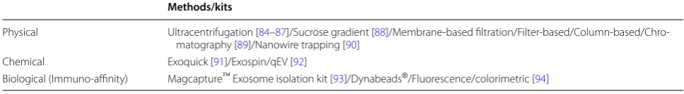
TDE isolation and purification among a mixture of EVs
are technically unavailable at the moment. Therefore,
novel isolation methods are crucial to enrich the specific
subtypes [76]. Three general approaches for exosome
iso-lation were summarized in Table 2 based on: (1) Physical
characters including size and gradient density
centrifu-gation (DGC) and ultracentrifucentrifu-gation (UC)
(increas-ing centrifugal force
≥
100,000
g
) apply to progressively
eradicate unwanted smaller debris and bigger
subpopu-lations of vesicles as a gold standard [77]. Furthermore,
filtration and size exclusion chromatography (SEC) were
considered as an important approach in this category.
UC is a labor intensive and time-consuming procedure
that requires specialist laboratory equipment that can be
combined with the other modalities such as sucrose
gra-dient and poly ethylene glycol (PEG) to increase the yield
[78].
(2) Chemical properties, samples incubated with a PEG
based on their solubility and exosomes separate
centrifu-gation or filtration [79]. Currently, several exosome
pre-cipitation kits such as ExoQuick
™, Exospin and the other
kits are commercially available [80].
(3) Immunoplate- and immunobead-based affinity
isolation can be accompanied by performing molecular
labeling of the exosome, including
CD81
,
CD9
,
CD63
,
TSG101
,
HSP 70
and
Alix.
Magcapture
™exosome
isola-tion kit PS and
CD63
dynabeads
®beads work based on
this approach. An ELISA-based method was also
devel-oped for exosome detection, in support of
functional-ized approach via specific antibodies. Characterization
of exosomes based on morphology via scanning electron
microscope (SEM) and transmission electron microscopy
(TEM) can be determined. Then nanoparticle tracking
assay (NTA) and dynamic light scattering (DLS) verify
wanted vesicle size samples. Finally, their molecular
pro-filing can be defined through conventional ELISA, PCR
and western blotting [81, 82].
Alternatively, microfluidic based exochips and poly
dimethyl siloxane (PDMS) innovative sorting platform
devices by electromagnetic and electrophoretic
manipu-lations have been developed to isolate exosomes. This
technology has many advantages such as being user
friendly, with quantitative readouts, high sensitivity, is
economic, fast and requires minimal sample handling
[83].
Molecular markers
Colorectal cancer has two types including sporadic
and hereditary, the first of the two (65%) [95] is directly
impressed by personal life-style and the second one
con-sists of familial adenomatous polyposis (
FAP
), due to
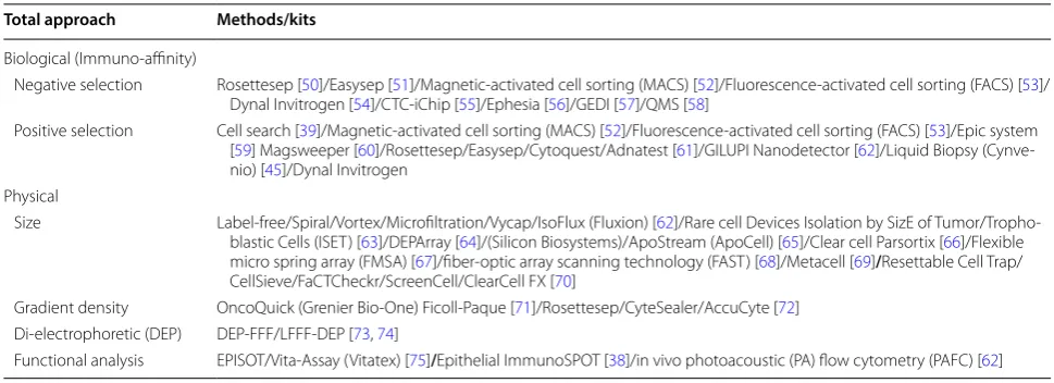
Table 1 Enrichment/isolation approaches of CTCs based on the inherent characteristics
Total approach Methods/kits
Biological (Immuno-affinity)
Negative selection Rosettesep [50]/Easysep [51]/Magnetic-activated cell sorting (MACS) [52]/Fluorescence-activated cell sorting (FACS) [53]/
Dynal Invitrogen [54]/CTC-iChip [55]/Ephesia [56]/GEDI [57]/QMS [58]
Positive selection Cell search [39]/Magnetic-activated cell sorting (MACS) [52]/Fluorescence-activated cell sorting (FACS) [53]/Epic system
[59] Magsweeper [60]/Rosettesep/Easysep/Cytoquest/Adnatest [61]/GILUPI Nanodetector [62]/Liquid Biopsy
(Cynve-nio) [45]/Dynal Invitrogen
Physical
Size Label-free/Spiral/Vortex/Microfiltration/Vycap/IsoFlux (Fluxion) [62]/Rare cell Devices Isolation by SizE of
Tumor/Tropho-blastic Cells (ISET) [63]/DEPArray [64]/(Silicon Biosystems)/ApoStream (ApoCell) [65]/Clear cell Parsortix [66]/Flexible
micro spring array (FMSA) [67]/fiber-optic array scanning technology (FAST) [68]/Metacell [69]/Resettable Cell Trap/
CellSieve/FaCTCheckr/ScreenCell/ClearCell FX [70]
Gradient density OncoQuick (Grenier Bio-One) Ficoll-Paque [71]/Rosettesep/CyteSealer/AccuCyte [72]
Di-electrophoretic (DEP) DEP-FFF/LFFF-DEP [73, 74]
Adenomatous polyposis coli (
APC
) gene mutations, and
HNPCC
/lynch syndrome, that is caused by
MMR
genes
[96].
Colorectal CTC markers included carcinoembryonic
antigen (
CEA/CEACAM5
,
7
),
EpCAM
,
CK19
and
CK20
[97,
98]. Colon stem-like cells express
CD44
,
CD166
(
ALCAM
),
CD133
(Prominin-1),
CD29
,
CD24
,
EPCAM
,
doublecortin like kinase 1 (
DCLK1
), Leucine-rich
repeat-containing G protein-coupled receptor 5 (
Lgr5
) [99, 100].
Additionally, there are some known markers in targeted
therapy which have been discussed clinically including
EGFR
,
VEGF, IGF
-
IR
the insulin-like growth factor 1
receptor (IGF-1R), interleukin-4 (
IL
-
4
) and bone
mor-phogenetic protein 4 (
BMP
-
4
) [101].
Analysis of exosome composition indicated that they
express tetraspanins, a class of membrane proteins
including
CD9
,
CD63
and
CD81
[102]. The other
fre-quent exosomal proteins are
EpCAM
,
Alix
, and
TSG101
[103],
GTPases
, cytoskeletal proteins,
annexines
, the heat
shock proteins (
Hsp70
and
Hsp90
) [104] and integrins
[105, 106], of which all of the valuable biomarkers were
drawn in Fig. 2.
Clinical applications to manage patients
CTCs were captured via all the aforementioned
approaches that have been discussed and cultured
in vivo/vitro named patient-derived xenografts (PDXs)
and CTC-derived xenografts (CDXs) although the
estab-lishment of permanent CTC lines is very challengeable
[21, 107].
In this section, clinical studies concerned with the
colo-rectal CTCs will be mentioned; 63 trials were registered
in
https ://clini caltr ials.gov of which 22 of them were
completed and summarized in Table
3. Meta-analyses
and large-scale clinical trials declare that patients with
CTC number
≥
5 (per 7.5 ml) were classified as being
in the aggressive stage IV and would develop distant
metastasis. Meanwhile, CTC level < 3 cells can also be
correlated with unfavorable prognostic factor [108] with
shorter median OS and PFS [109]. Thus, it can be a vital
factor in cancer progression risk assessment and patients
must be stratified to be treated promptly based on
molecular subtypes [110,
111]. Therefore, higher
bers of CTCs are seen in patients with a greater
num-ber of metastatic sites [112]. Regardless of the metastatic
site, CTC enumeration (cell-based assays) are sufficient
enough as a proper cancer monitoring index whenever
CEA
and other markers levels are not measurable [113].
It is worthy to mention that an elevated CTC number
was not necessarily associated with apoptotic CTCs
or CTC debris and could be used to interrogate
meta-static in patients and contribute to run tumor-associated
events [114, 115].
In another site, only five clinical trials using the key
word ‘colorectal exosome’ were registered that none of
them completed. Recently, TDEs have been introduced
as promising drug delivery vehicles in targeting different
organs and their selective cargo must be determined to
increase therapy effectiveness. Thus, scientists are
focus-ing on TDEs components [116] even in inducfocus-ing
anti-tumor immune responses as cancer vaccine candidates
[117]. The plasma TDE cargo is enriched in
immuno-suppressive and immunostimulatory receptor/ligands,
MHC molecules and various tumor-associated antigens
(TAAs). Their content depends on cellular origin variety
and carries oncogenic DNA, microRNAs, proteins and
mRNAs [118] such as GPC1
+, tumor
suppressor-acti-vated pathway 6 (
TSAP6
) [119],
ΔNp73
[120], metastatic
factors (
TNC
,
MET
,
S100A9
,
S100A8
), signal
transduc-tion molecules (
EFNB2
,
JAG1
,
SRC
,
TNIK
), and lipid raft
associated components (
PROM1
,
CAV1
,
FLOT1
and
2
). Ji
et al. reported
Let
-
7a
-
3p
,
let
-
7f
-
1
-
3p
,
miR
-
574
-
5p
,
miR
-451a
,
miR
-
7641
, and
miR
-
4454
are common to all EV
subtypes [121]. In addition to the detection and
co-local-ization of protein complexes in CRC exosomes,
regula-tion of signaling pathways such as Wnt and
EGFR
ligand,
besides autocrine, paracrine, and juxtacrine, contribute
in priming of the metastatic niche [122]. Furthermore,
inhibition of exosome secretion, besides targeting CSCs,
as a new therapeutic strategy, can block tumor associated
secretion before chemotherapy [123,
124] and facilitate
cross talk between stromal cells and tumors in cancer
microenvironment [125].
Table 2 Enrichment/isolation approaches of exosomes based on the inherent characteristics
Methods/kits
Physical Ultracentrifugation [84–87]/Sucrose gradient [88]/Membrane-based
filtration/Filter-based/Column-based/Chro-matography [89]/Nanowire trapping [90]
Chemical Exoquick [91]/Exospin/qEV [92]
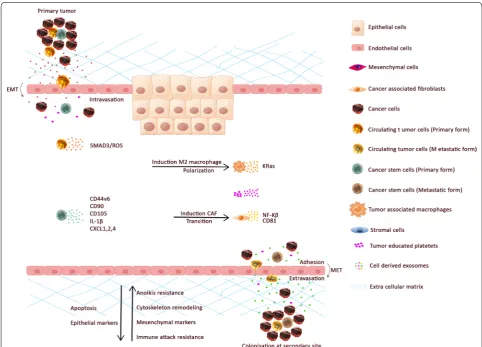
Crosstalk in tumor microenvironment (TME)
Metabolic cells reprogramming, loss of cell
connec-tion with overexpression of matrix metalloproteinases
(
MMP
), cancer cells diapedesis and its integration to
define target sites contribute in metastasis cascade.
Tumor microenvironment (TME) consists of CAFs,
extracellular matrix (ECM), cancer- tumor-associated
vasculature and inflammatory immune cells. Mediating
the crosstalk between tumor and tumor-associated cells
identify as a viable step in cancer development (Fig.
3)
[126, 127].
Primary TDE conveys messages to the other cells which
exist in TME, as well as modifying the microenvironment
through their cargo. Not only does TDE play a pivotal
role, but also the exosomes secreted by cancer-associated
factors including CAFs, tumor-associated macrophages
(TAMs), endothelium, leukocytes and progenitor cells
should be considered as significant characteristics in
cancer progression [128]. TDE is also important in the
regulation of macrophage polarization and CAF
transi-tion [129].
The data related to the TDE roles in CRC are limited
but it was approved that TDE in other cancers promotes
invasiveness by regulating signaling pathway, for
exam-ple, primary TDEs enhance
SMAD3/ROS
signaling and
induce CTC survival and cell adhesion. Furthermore, the
levels of TDEs markers which participated in EMT
pro-cess cellular movement and cell–cell signaling in
can-cer patients’ blood correlated with the disease stage [3].
MiRNAs encapsulated in EVs play a significant role in
metastasis such as circulating exosomal
microRNA
-
203
via inducing TAM in CRC [130], [130]. Cha et al. showed
that the
KRAS
status of CRC have a direct influence on
the type of miRNAs enriched in exosomes [131].
Con-ditioned media harvested from M2 macrophages which
consist of derived exosomes promote CRC motility and
invasion throughout
IL6
,
Wnt5a
,
TNFα
and
EGF
mole-cules [132].
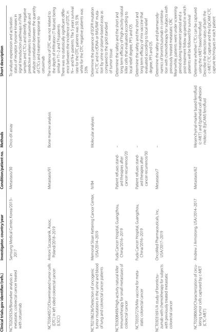
Table
3
T
he c
omplet
e clinic
al trials of cir
cula
ting tumor c
ells (
CT
Cs) in c
olor
ec
tal c
anc
er
Clinical trials .go v iden tifier/(r ef s.) In vestiga tor , c oun tr y/y ear Condition/pa tien t no . M ethods Shor t description NC T02450422/T he det ec tion of cir cu-lating tumor cells
(C
TCs) in patients
with CR C under going cr yosur ger y
combined with
DC-CIK tr eatment W ang , China/2013–2015 II–IV/60 Flo w c yt ometr y RT -PCR Test C TCs fr
om patients r
eceiv
ed cr
yosur
-ger
y and/or
DC-CIK tr eatment, 1 da y bef or
e and 2
da
ys af
ter
NC
T01640444/I
nfluence of BRAF and
PIK3K status in patients with RAS wild- type metastatic color
ec tal car cinoma and < 3 C TC (VISNU-2) Díaz-Rubio
, Aranda, Sastr
e, Spain/2012– 2018 M etastatic/240 C TC count
Influence of BRAF and PIK3K status on the efficac
y of FOLFIRI
+ Be vacizumab or Cetuximab NC T01163305/PE T-C
T and C
TCs in CR
C
Br
igett
e, Hong K
ong/2010–2017
M
etastatic/84
PE
TS
can, RECIST Cr
iter ia A ssessing Chemotherap y ( oxaliplatin or irinot ecan) r esponse (measur ing tumor metabolic) NC T01943500/C ollec
tion of blood
specimens f or C TC analysis Sanz-Altamira, USA/2012–2017 II–IV/14 C TC count
Test the sensitivit
y of a pr
opr ietar y filtration de vice desig ned t o captur
e and concen
-trat e C TCs NC T03337347/Clinical sig nificance of det ec
ting CEA and CK20 mRNA-positiv
e
cells in CR
C patients Duda, Cz ech R epublic/2004–2017 I–IV/256 C TC count RT -PCR D et er
mine the cor
relations of C
TC in the
blood and bone mar
ro
w of CR
C patients
with CEA and CK20 mRNA-positiv
e cells
as a negativ
e pr og nostic fac tor NC T01628328/C olonic st ent and tumor cell dissemination
Poon, Hong k
ong/2010–2012 II–IV/40 FA C S A ssess impac
t of metallic st
ent inser
tion f
or
obstruc
ting measur
ing the le
vel of C
TCs
bef
or
e and af
ter colonoscopic st
enting vs colonoscop y NC T01722903/D et ec
tion of C
TCs in patients under going sur ger y f or stage IV CR C Kaifi, USA/2012–2015 M etastatic/26 FMSA de vice Cell sear ch D et ec
tion of C
TCs dur
ing CR
C syn- and
metachr
onous liv
er and lung metastases
NC
T01212510/Study of cir
culating mar
k-ers in serum of patients tr
eat ed f or metastatic CR C ( Coca-Colon) M
ichel and R
ouen, F
rance/2010–2016
M
etastasis/200
C
TC count Real-time R
T-PCR
M
easur
e of tumor mar
kers (blood rat
e of
A
CE, CA19-9, C
TC, c
tDNA)
NC
T00351572/F
requenc
y of C
TCs in
stage II and stage III colon cancer patients
Sa w yer , C anada/2006–2006 II–III/30 Cell sear ch D et ec
t of C
TC in patients who ha
ve had sur ger y f or CR C pr
esence and r
ecur
rence
NC
T01640405/Study of first line tr
eat
-ment of patients with metastatic CR
C
not pr
eviously tr
eat
ed and with thr
ee or
mor
e C
TC (
VISNU-1)
Díaz-Rubi and Aranda and Sastr
e, Spain/2012–2018 M etastasis/350 C TC count To e valuat e FOLFO X + be vacizumab versus FOLFO XIRI + be
vacizumab as first line
tr
eatment of patients with metastatic CRC not pr
eviously tr
eat
ed and with
thr
ee or mor
e C
TCs
D
et
er
mine the C
or
relation of RAS, BRAF
and PI3K mutations and clinical anti- tumor
ac
tivit
y out
Table
3
(c
on
tinued)
Clinical trials .go v iden tifier/(r ef s.) In vestiga tor , c oun tr y/y ear Condition/pa tien t no . M ethods Shor t description NC T02029326/Biomarker analysis in
metastatic color
ec
tal cancer tr
eat ed with cetuximab Samsung M edical C ent er , K or ea/2013– 2017 M etastasis/30
Onco dX assa
y
To analyz
e expr
ession and ac
tivation
status of r
ecept or t yr osine k inases in sig nal transduc tion path wa
ys in FNA
samples and
C
TCs and identify negativ
e pr edic tiv e mar kers t
o cetuximab and
analyz
e cor
relation bet
w
een the quantit
y of C TCs and tr eatment r esponse t o cetuximab NC T03640572/Disseminat ed tumor cells (D TC
) in lef
t sided color
ec tal cancer (LSC C ) Ant oni Sz czapanik , A ssoc , Poland/2018–2019 M etastasis/91 Bone mar ro w analysis
The incidence of D
TC was not r
elat
ed t
o
the depth of infiltration (
T f eatur e) being similar in T1–2 and T4 patients Ther
e was no statistically sig
nificant diff
er
-ence bet
w
een the incidence of D
TC in N − and N + patients . T he 5 y ears sur vival rat e f
or the D
TC patients was 59, 5%
while f
or the D
TC negativ
e patients was
53%
NC
T02186236/D
et
ec
tion of oncogenic
tumor mutations in the ur ine and blood
of lung and color
ec
tal cancer patients
M
emor
ial Sloan K
ett er ing C ancer C ent er , USA/2014–2019 IV/84 M olecular analyses D et er
mine the pr
esence of EGFR mutation
in C
TC and in cfDNA or RAS/RAF muta
-tion
b
y ur
ine or plasma-based
assa
y
as
compar
ed t
o the gold standar
d of tumor tissue NC T03008499/H igh-ac tivit
y natural k
iller
immunotherap
y f
or small metastases of
color ec tal cancer Fuda C ancer Hospital , Guangzhou , China/2016–2019 Patient r efuses stand -ar
d therapies af
ter cancer r ecur rence/20 – D et er
mine the saf
et
y and the shor
t and
long t
er
m efficac
y of high-ac
tivit
y natural
killer cells that e
valuat ed accor ding t o local r elief deg ree
, PFS and OS
NC
T03357276/M
ix vaccine f
or meta -static color ec tal cancer Fuda C ancer Hospital , Guangzhou , China/2016–2019 Patient r efuses stand -ar
d therapies af
ter cancer r ecur rence/30 – D et er
mine the saf
et
y and the shor
t and
long t
er
m efficac
y of mix vaccine that
evaluat
ed accor
ding t
o local r
elief
deg
ree
, PFS and OS
NC
T03031691/A study of br
ontic
tu
-zumab with chemotherap
y f or subjec ts with pr eviously tr eat ed metastatic color ec tal cancer OncoM ed P har maceuticals , I nc , USA/2017–2019 M etastasis/7 – D et er
mine the saf
et
y and phar
macody
-namics of br
ontic
tuzumab in combina
-tion with chemotherap
y f or subjec ts with pr eviously tr eat
ed metastatic CR
C
M
ean
while
, patients w
ent under scr
eening
per
iod dur
ing tr
eatment per
iod and a
post
-tr
eatment f
ollo
w up per
iod in which
patients will be f
ollo w ed f or sur vival NC T02080650/Charac ter
ization of cir
cu
-lating tumor cells captur
ed b y c-ME T (C TC-ME T) Andr
ew J Ar
mstr ong , USA/2014–2017 M etastasis/62 M esench ymal-mar
ker based f
er
rofluid
(c-ME
T) and Epithelial
cell adhesion molecule (EpCA M) f er rofluid D et er
mine whether C
TCs can be captur
ed
using the cME
T based f
er
rofluid
D
escr
ibe the det
ec
tion rat
es of both the
c-ME
T C
TC captur
e and the EpCA
M C
TC
captur
e t
Table
3
(c
on
tinued)
Clinical trials .go v iden tifier/(r ef s.) In vestiga tor , c oun tr y/y ear Condition/pa tien t no . M ethods Shor t description NCT00924092/An open label phase I Study t
o e
val the saf
et
y and t
olerabilit
y
of a vaccine (
GI-6207) consisting of
whole , heat -k illed r ecombinant saccha -rom yces cer evisiae (y east) genetically modified t o expr
ess cea pr
ot
ein in
adults with metastatic
CEA-expr
essing
Ra
vi A M
adan, M.D . USA/2009–2019 M etastasis/25 M olecular analyses D et er
mine the saf
et
y and t
olerabilit
y of
escalating doses of a heat
ed-k
illed y
east
-based vaccine that tar
gets tumors that expr ess CEA Evaluat
e CD4 and CD8 immunolog
ic response t o y east antigen. To e valuat e
evidence of clinical benefit such as PFS, OR and C
TCs decr
easing via assessment
of tumor
mar
kers
NC
T00560560/Study using CP
-751,871 in
patients with stage iv color
ec
tal cancer
that has not r
esponded t o pr evious anti-cancer tr eatments Pfiz er C T.go v C all C ent er Pfiz er , USA,
Spain and Unit
ed K
ingdom/2007–2013
IV/168
C
TC count
This study will t
est if ther
e is an
y sur
vival
benefit in patients with r
efrac
tor
y
metastatic color
ec
tal cancer that r
eceiv
e
CP
-751,871
NC
T00483080/Study of NGR-h
TNF as
single agent in patients aff
ec
ted b
y
color
ec
tal cancer (
CR C ) M olM ed S.p .A. I taly/2006–2013 M etastasis/46 –
Evaluation of the saf
etly of NGR-h
TNF on
patients who pr
eviously tr eat ed with fluor op yr imidine , o
xaliplatin and ir
inot
e-can based r
eg
imens and cor
relation with
sur
vival
NC
T00335595/Study of be
vacizumab
alone or combined with capecitabine and o
xaliplatin as suppor
t therap
y in
metastatic color
ec
tal cancer patients
Enr
ique Aranda, M.D
.; ph.D ., E duar do Díaz-Rubio , M.D .; ph.D
. and Spanish
Cooperativ e Gr oup f or G astr oint estinal Tumor Therap y ( TTD), Spain/2006–2013 M etastasis/480 C TC count Compar
e the fr
ee time t
o disease
pr
og
ression of combination therap
y
with capecitabine
, o
xaliplatin and
be
vacizumab until disease pr
og ression versus capecitabine , o xaliplatin and be vacizumab f
or 6 c
ycles f ollo w ed b y be
vacizumab until disease pr
og
ression or
a pr
ematur
e dr
op out of the study
NC
T02020291/P
hase I study t
o e valuat e saf et y, t olerabilit
y, anti-tumour ac
tivit
y
and pk pr
ofiles of f
ox
y-5 in metastatic
br
east, colon or pr
ostat e cancer W ntR esear ch AB , D enmar k/2013–2016 M etastasis/31 C TC count D ev elop F ox
y-5 as a first in class
anti-metastatic
cancer
drug via inhibition the
de
velopment of metastasis b
y r educing the motilit y of cancer cells and incr easing the sur vival rat
es of patients
Interestingly, an acidic and hypoxic microenvironment
stimulates the release of TDE and is involved in epithelial
adheres junctions and cytoskeleton remodeling pathways
[133]. In addition, TDEs may potentially collaborate in
the dynamic regulation of the tumor fate and is
consid-ered as a valuable diagnostic non-invasive approach [34,
134].
Cancer stem cells regulate tumor
microenvironment via exosomes
CSCs or “tumor-initiating cells”, a rare subpopulation
are capable of self-renewal and differentiate into
special-ized cells through symmetric division and therapeutic
resistance drive tumor growth [135]. Nowadays, CSCs
are investigated in various ranges of solid tumors. CSCs
derived EVs contribute in tumor initiation, progression,
angiogenesis, invasion and metastasis formation [136].
Tumor exosome RNAs induce the expression of
interleukin-1β through NF-κB signaling leading to the
survival of neutrophil sustain. Colorectal CSCs secreted
CXCL1
and
2
and attracted neutrophils primed via IL-1β
to promote CRC cells tumorigenesis [137]. Moreover,
exosomes may transfer mutant
KRAS
to recipient cells
and trigger increases in
IL
-
8
production, neutrophil
recruitment as well as the formation of the neutrophil
extracellular trap (NET), leading to the deterioration of
CRC [138].
CD44v6
CSC-derived exosomes contribute
to cancer development by non-cancer initiating cells to
acquire the CSC phenotype [139].
EVs-derived CSCs with variable patterns of miRNA
can convey their oncogenic features in order to affect
cancer proliferation, progression, invasion, metastasis
[140], activate angiogenesis and stimulate tumor immune
escape mechanisms [141, 142] (Fig. 3).
Conclusion
Tumor metastasis is still the main principle of cancer
death, highlighting the importance of investigating an
updating approach to control it. Cross talks among tumor
cells and derived-exosomes play a significant role in a
dynamic network of cancer microenvironment.
There-fore, their recognition and characterization are a crucial
step in accurate comprehension of molecular and
cel-lular oncology. Tracking cancer related markers in body
fluid could be helpful to measure residual disease
pres-ence, recurrpres-ence, relapse and resistance and address the
needs of clinicians and patients. Liquid biopsy,
includ-ing CTCs and TDEs as a noninvasive tool in the field of
precision medicine, provides substantially helpful
infor-mation regarding diagnosis, prognosis, predictive and
pharmacodynamics.
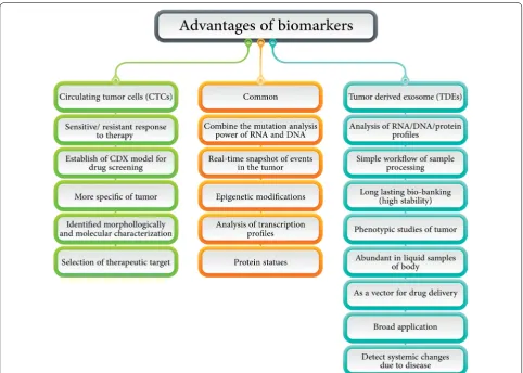
In spite of numerous merits that can be counted for
CTCs and TDEs separately or simultaneously (Fig.
4), it
should be noted that the most challengeable and
disad-vantageous of them concern isolation and purification
due to methodological restrictions (sensitivity and
speci-ficity) and standardization because heterogeneity must be
resolved. For example, by inducing the apoptosis of CTCs
by intervening ROS-mediated DNA damage can inhibit
the CTCs metastasis along the the EGF pathway which is
cleared by ingenuity exosome pathway analysis [143]. In
another study, it was proved that TDEs have equivalent
prognostic values to CTCs in the investigated metastatic
cancers. Patients with favorable CTC counts can have
further prognostic stratification using TDEs [144].
Lab on chip (LOC) technology, in order to grow
aware-ness about the point-of-care testing in cancer was
devel-oped and because of low consumption of a sample and
high compatibility with the liquid biopsy concept and
personalized medicine it has been welcomed [145,
146].
This precious dream can come true with the analysis of
patient-activated social networks and systems medicine.
P4 medicine that is predictive, personalized,
preven-tive, and participatory can be helpful in this field, next
to gene-panel testing due to next-generation sequencing
(NGS) technology [147] and plays a critical role in
cover-ing the current shortcomcover-ings of liquid biopsy regardcover-ing
practicality, standardization, and the result comparisons.
Despite many techniques regarding CTC exosome
capturing and subgrouping are available in clinics; the
need for optimization of downstream analysis is tangible.
Additionally, distinguishing between CTCs with high and
low metastatic status as well as between TDEs and
nor-mal status is absolutely vital. In conclusion, liquid biopsy
is an expanding field in the management of CRC patient
in different stages. It is highly recommended that further
research be done on CTCs and TDEs alone or
simulta-neously until both can serve as valuable biomarkers in
clinics.
Abbreviations
CRC : Colorectal cancer; CTCs: Circulating tumor cells; TDEs: Tumor-derived exosomes; CT: Computed tomography; MRI: Magnetic resonance imaging; TEPs: Tumor-educated platelets; LOC: Lab-on-a-chip; NGS: Next-generation sequencing; CSCs: Cancer stem cells; ECM: Extracellular matrix; EMT: Epithelial mesenchymal transition; MET: Mesenchymal epithelial transition; OS: Overall survival; PFS: Progression-free survival; EVs: Extracellular vesicles; MVBs: Mul-tivesicular bodies; CK: Cytokeratin; EPCAM: Epithelial cell adhesion molecule; DGC: Gradient density centrifugation; UC: Ultracentrifugation; SEC: Size exclusion chromatography; PEG: Poly ethylene glycol; SEM: Scanning electron microscope; TEM: Transmission electron microscopy; NTA: Nanoparticle track-ing assay; DLS: Dynamic light scattertrack-ing; PDMS: Poly dimethyl siloxane; FAP: Familial adenomatous polyposis; APC: Adenomatous polyposis coli; DCLK1: Doublecortin like kinase 1; LGR5: Leucine-rich repeat-containing G protein-coupled receptor 5; IGF-IR: the insulin-like growth factor 1 receptor; IL-4: Inter-leukin-4; BMP-4: Bone morphogenetic protein 4; Hsp70: Heat shock proteins; PDXs: Patient-derived xenografts; CDXs: CTC-derived xenografts; TSAP6: Tumor suppressor-activated pathway 6; CAFs: Carcinoma-associated fibroblasts; DKK4: Dickkopf-related protein 4; TAAs: tumor-associated antigens; MMP: Matrix metalloproteinases; TME: Tumor microenvironment; Mef2c: Myocyte enhancer factor 2c; HCC: Hepatocellular carcinoma; HDGF: Hepatoma-derived growth factor; GSCs: Glioma stem cells; CLIC1: contain functionally active Cl-intracellular channel 1; NET: Neutrophil extracellular trap; EGFR: Epidermal growth factor receptor; DEP-FFF: Dielectrophoretic field-flow fractionation; VEGF: Vascular endothelial growth factor.
Acknowledgements
We are grateful of our colleague at Iran University of Medical Sciences and Royan Stem Cell Technology Company who provided insight and expertise that greatly assisted the research. Apart from, it must be declared that the authors received no specific funding for this work.
Authors’ contributions
SV and ZM conceived of the presented idea. SV collected, interpreted and analyzed data and wrote the drafting of the article. ZM and ME developed, revised and approved the theory. RR and AA performed the critical revision and verified the whole concept. SV and ZM encouraged the other author to investigate and supervised the findings of this work. All authors discussed the results. All authors read and approved the final manuscript.
Funding Not applicable.
Availability of data and materials
Data sharing is not applicable to this article as no new data were created or analyzed in this study and openly available in [repository name at http://doi. org/[doi] and reference number.
Ethics approval and consent to participate Not applicable.
Consent for publication
All of the current study data were searched systematic and were used by refer-ence citation and all of the authors consent to publication.
Competing interests
The authors whose names are listed certify that they have NO affiliations in any organization or entity with any financial interest and non-financial interest in the subject matter or materials discussed in this manuscript.
Author details
1 Oncopathology Research Center, Iran University of Medical Sciences (IUMS),
Hemmat Street (Highway), Next to Milad Tower, Tehran, Iran. 2 Department
of Molecular Medicine, Faculty of Advanced Technologies in Medicine, Iran University of Medical Sciences, Tehran, Iran. 3 Department of Stem Cells
and Developmental Biology, Cell Science Research Center, Royan Institute for Stem Cell Biology and Technology, ACECR, Tehran, Iran. 4 Department
of Medical Oncology, Dana-Farber Cancer Institute, Harvard Medical School, Boston, USA.
Received: 17 April 2020 Accepted: 27 June 2020
References
1. Bray F, Ferlay J, Soerjomataram I, Siegel RL, Torre LA, Jemal A. Global cancer statistics 2018: GLOBOCAN estimates of incidence and mortality worldwide for 36 cancers in 185 countries. CA Cancer J Clin. 2018;68:394–424.
2. Van Cutsem E, Cervantes A, Nordlinger B, Arnold D. Metastatic colorec-tal cancer: ESMO clinical practice guidelines for diagnosis, treatment and follow-up. Ann Oncol. 2014;25(Suppl 3):iii1–9.
3. Fu Q, Zhang Q, Lou Y, Yang J, Nie G, Chen Q, et al. Primary tumor-derived exosomes facilitate metastasis by regulating adhesion of circulating tumor cells via SMAD3 in liver cancer. Oncogene. 2018;37(47):6105–18. 4. Diamantis A, Magiorkinis E, Koutselini H. Fine-needle aspiration (FNA)
biopsy: historical aspects. Folia Histochem Cytobiol. 2009;47(2):191–7. 5. Mousavi S, Moallem R, Hassanian SM, Sadeghzade M, Mardani R,
Ferns GA, et al. Tumor-derived exosomes: potential biomarkers and therapeutic target in the treatment of colorectal cancer. J Cell Physiol. 2019;234(8):12422–32.
6. Punt CJ, Koopman M, Vermeulen L. From tumour heterogeneity to advances in precision treatment of colorectal cancer. Nat Rev Clin Oncol. 2017;14(4):235.
7. Zhai Z, Yu X, Yang B, Zhang Y, Zhang L, Li X, et al. Colorectal cancer het-erogeneity and targeted therapy: clinical implications, challenges and solutions for treatment resistance. Semin Cell Dev Biol. 2017;64:107–15. 8. Karachaliou N, de Las Mayo Casas C, Molina-Vila MA, Rosell R. Real-time liquid biopsies become a reality in cancer treatment. Ann Transl Med. 2015;3(3):36.
9. Brock G, Castellanos-Rizaldos E, Hu L, Coticchia C, Skog J. Liquid biopsy for cancer screening, patient stratification and monitoring. Transl Can-cer Res. 2015;4(3):280–9.
10. Issa IA, Noureddine M. Colorectal cancer screening: an updated review of the available options. World J Gastroenterol. 2017;23(28):5086. 11. Lopez A, Harada K, Mizrak Kaya D, Dong X, Song S, Ajani JA. Liquid
biopsies in gastrointestinal malignancies: when is the big day? Expert Rev Anticancer Ther. 2018;18(1):19–38.
12. Crowley E, Di Nicolantonio F, Loupakis F, Bardelli A. Liquid biopsy: monitoring cancer-genetics in the blood. Nat Rev Clin Oncol. 2013;10(8):472–84.
13. Alix-Panabières C, Pantel K. Circulating tumor cells: liquid biopsy of cancer. Clin Chem. 2013;59(1):110–8.
14. Soler A, Cayrefourcq L, Mazard T, Babayan A, Lamy PJ, Assou S, et al. Autologous cell lines from circulating colon cancer cells captured from sequential liquid biopsies as model to study therapy-driven tumor changes. Sci Rep. 2018;8(1):15931.
15. van Zijl F, Krupitza G, Mikulits W. Initial steps of metastasis: cell invasion and endothelial transmigration. Mutat Res. 2011;728(1–2):23–34. 16. Ashworth TR. A case of cancer in which cells similar to those in the
17. Steinert G, Scholch S, Niemietz T, Iwata N, Garcia SA, Behrens B, et al. Immune escape and survival mechanisms in circulating tumor cells of colorectal cancer. Cancer Res. 2014;74(6):1694–704.
18. Kowalik A, Kowalewska M, Gozdz S. Current approaches for avoiding the limitations of circulating tumor cells detection methods-implica-tions for diagnosis and treatment of patients with solid tumors. Transl Res. 2017;185(58–84):e15.
19. Shibue T, Weinberg RA. EMT, CSCs, and drug resistance: the mechanistic link and clinical implications. Nat Rev Clin Oncol. 2017;14(10):611–29. 20. Xu L, Shamash J, Lu Y-J. Circulating Tumor Cells: a window to
under-stand cancer metastasis, monitor and fight against cancers. J Cancer Res Updates. 2015;4(1):13–29.
21. Zhou L, Dicker DT, Matthew E, El-Deiry WS, Alpaugh RK. Circulating tumor cells: silent predictorsof metastasis. F1000Res. 2017;6(F10000 Faculty Rev):1445.
22. Satelli A, Mitra A, Brownlee Z, Xia X, Bellister S, Overman MJ, et al. Epithelial–mesenchymal transitioned circulating tumor cells capture for detecting tumor progression. Clin Cancer Res. 2015;21(4):899–906. 23. Najafi M, Goradel NH, Farhood B, Salehi E, Solhjoo S, Toolee H, et al. Tumor microenvironment: interactions and therapy. J Cell Physiol. 2019;234(5):5700–21.
24. Zhang T, Boominathan R, Foulk B, Rao C, Kemeny G, Strickler JH, et al. Development of a novel c-MET-based CTC detection platform. Mol Cancer Res. 2016;14(6):539–47.
25. Hardingham JE, Grover P, Winter M, Hewett PJ, Price TJ, Thierry B. Detec-tion and clinical significance of circulating tumor cells in colorectal cancer—20 years of progress. Mol Med. 2015;21(Suppl 1):S25–31. 26. Zhang W, Xia W, Lv Z, Ni C, Xin Y, Yang L. Liquid biopsy for cancer:
circulating tumor cells, circulating free DNA or exosomes? Cell Physiol Biochem. 2017;41(2):755–68.
27. Wang W, Wan L, Wu S, Yang J, Zhou Y, Liu F, et al. Mesenchymal marker and LGR5 expression levels in circulating tumor cells correlate with colorectal cancer prognosis. Cell Oncol. 2018;41:495–504.
28. Li P, Kaslan M, Lee SH, Yao J, Gao Z. Progress in Exosome Isolation Tech-niques. Theranostics. 2017;7(3):789–804.
29. Pan BT, Johnstone RM. Fate of the transferrin receptor during matura-tion of sheep reticulocytes in vitro: selective externalizamatura-tion of the receptor. Cell. 1983;33(3):967–78.
30. Harding C, Stahl P. Transferrin recycling in reticulocytes: pH and iron are important determinants of ligand binding and processing. Biochem Biophys Res Commun. 1983;113(2):650–8.
31. Hessvik NP, Llorente A. Current knowledge on exosome biogenesis and release. Cell Mol Life Sci. 2018;75(2):193–208.
32. He C, Zheng S, Luo Y, Wang B. Exosome theranostics: biology and translational medicine. Theranostics. 2018;8(1):237–55.
33. Wang Z, Chen JQ, Liu JL, Tian L. Exosomes in tumor microenvironment: novel transporters and biomarkers. J Transl Med. 2016;14(1):297. 34. Jia Y, Chen Y, Wang Q, Jayasinghe U, Luo X, Wei Q, et al. Exosome:
emerging biomarker in breast cancer. Oncotarget. 2017;8(25):41717–33. 35. Micalizzi DS, Haber DA, Maheswaran S. Cancer metastasis through the
prism of epithelial-to-mesenchymal transition in circulating tumor cells. Mol Oncol. 2017;11:770–80.
36. Goeminne JC, Guillaume T, Symann M. Pitfalls in the detec-tion of disseminated non-hematological tumor cells. Ann Oncol. 2000;11(7):785–92.
37. Shen Z, Wu A, Chen X. Current detection technologies for circulating tumor cells. Chem Soc Rev. 2017;46(8):2038–56.
38. Zhang J, Chen K, Fan ZH. Circulating tumor cell isolation and analysis. Adv Clin Chem. 2016;75:1–31.
39. Allard WJ, Matera J, Miller MC, Repollet M, Connelly MC, Rao C, Tibbe AG, Uhr JW, Terstappen LW. Tumor cells circulate in the peripheral blood of all major carcinomas but not in healthy subjects or patients with nonmalignant diseases. Clin Cancer Res. 2004;10(20):6897–904. 40. de Wit S, van Dalum G, Lenferink AT, Tibbe AG, Hiltermann TJ, Groen
HJ, et al. The detection of EpCAM(+) and EpCAM(−) circulating tumor cells. Sci Rep. 2015;5:12270.
41. Murtaza M, Dawson SJ, Tsui DW, Gale D, Forshew T, Piskorz AM, et al. Non-invasive analysis of acquired resistance to cancer therapy by sequencing of plasma DNA. Nature. 2013;497(7447):108–12.
42. Hao SJ, Wan Y, Xia YQ, Zou X, Zheng SY. Size-based separation methods of circulating tumor cells. Adv Drug Deliv Rev. 2018;125:3–20.
43. Cayrefourcq L, De Roeck A, Garcia C, Stoebner PE, Fichel F, Garima F, et al. S100-EPISPOT: a new tool to detect viable circulating melanoma cells. Cells. 2019;8(7):755.
44. Goedecke N, Bollhalder M, Bernet R, Silvan U, Snedeker J. Easy and accu-rate mechano-profiling on micropost arrays. J Vis Exp. 2015;105:e53350. 45. Winer-Jones JP, Vahidi B, Arquilevich N, Fang C, Ferguson S, Harkins D,
et al. Circulating tumor cells: clinically relevant molecular access based on a novel CTC flow cell. PLoS ONE. 2014;9(1):e86717.
46. Ueno Y, Furukawa K, Matsuo K, Inoue S, Hayashi K, Hibino H. On-chip graphene oxide aptasensor for multiple protein detection. Anal Chim Acta. 2015;866:1–9.
47. Stott SL, Hsu CH, Tsukrov DI, Yu M, Miyamoto DT, Waltman BA, et al. Isolation of circulating tumor cells using a microvortex-generating herringbone-chip. Proc Natl Acad Sci USA. 2010;107(43):18392–7. 48. Sheng W, Ogunwobi OO, Chen T, Zhang J, George TJ, Liu C, et al.
Capture, release and culture of circulating tumor cells from pan-creatic cancer patients using an enhanced mixing chip. Lab Chip. 2014;14(1):89–98.
49. Huang T, Xu C, Xiao J, Wang Q, Wang Y, Zhang Y, et al. Determination of the optimal detection time of circulating tumor cells for the postopera-tive monitoring of colorectal cancer. Oncol Lett. 2020;19(4):2996–3002. 50. Munugalavadla V, Mariathasan S, Slaga D, Du C, Berry L, Del Rosario
G, et al. The PI3K inhibitor GDC-0941 combines with existing clini-cal regimens for superior activity in multiple myeloma. Oncogene. 2014;33(3):316–25.
51. Lapin M, Tjensvoll K, Oltedal S, Buhl T, Gilje B, Smaaland R, et al. MIN-DEC—an enhanced negative depletion strategy for circulating tumour cell enrichment. Sci Rep. 2016;6:28929.
52. Wang X, Sun L, Zhang H, Wei L, Qu W, Zeng Z, et al. Microfluidic chip combined with magnetic-activated cell sorting technology for tumor antigen-independent sorting of circulating hepatocellular carcinoma cells. PeerJ. 2019;7:e6681.
53. Vishnoi M, Peddibhotla S, Yin W, Scamardo AT, George GC, Hong DS, et al. The isolation and characterization of CTC subsets related to breast cancer dormancy. Sci Rep. 2015;5:17533.
54. Kallergi G, Politaki E, Alkahtani S, Stournaras C, Georgoulias V. Evaluation of isolation methods for circulating tumor cells (CTCs). Cell Physiol Biochem. 2016;40(3–4):411–9.
55. Karabacak NM, Spuhler PS, Fachin F, Lim EJ, Pai V, Ozkumur E, et al. Microfluidic, marker-free isolation of circulating tumor cells from blood samples. Nat Protoc. 2014;9(3):694–710.
56. Sharma S, Zhuang R, Long M, Pavlovic M, Kang Y, Ilyas A, et al. Circulat-ing tumor cell isolation, culture, and downstream molecular analysis. Biotechnol Adv. 2018;36(4):1063–78.
57. Galletti G, Sung MS, Vahdat LT, Shah MA, Santana SM, Altavilla G, et al. Isolation of breast cancer and gastric cancer circulating tumor cells by use of an anti HER2-based microfluidic device. Lab Chip. 2014;14(1):147–56.
58. Wu Y, Deighan CJ, Miller BL, Balasubramanian P, Lustberg MB, Zborowski M, et al. Isolation and analysis of rare cells in the blood of cancer patients using a negative depletion methodology. Methods. 2013;64(2):169–82.
59. Werner SL, Graf RP, Landers M, Valenta DT, Schroeder M, Greene SB, et al. Analytical validation and capabilities of the Epic CTC Platform: enrichment-free circulating tumour cell detection and characterization. J Circ Biomark. 2015;4:3.
60. Dawson SJ, Tsui DW, Murtaza M, Biggs H, Rueda OM, Chin SF, et al. Analysis of circulating tumor DNA to monitor metastatic breast cancer. N Engl J Med. 2013;368(13):1199–209.
61. Andreopoulou E, Yang LY, Rangel KM, Reuben JM, Hsu L, Krishnamur-thy S, et al. Comparison of assay methods for detection of circulating tumor cells in metastatic breast cancer: adnaGen AdnaTest Breast-Cancer Select/Detect versus Veridex Cell Search system. Int J Breast-Cancer. 2012;130(7):1590–7.
62. Ferreira MM, Ramani VC, Jeffrey SS. Circulating tumor cell technologies. Mol Oncol. 2016;10(3):374–94.
64. Hiltermann TJ, Pore MM, van den Berg A, Timens W, Boezen HM, Liesker JJ, et al. Circulating tumor cells in small-cell lung cancer: a predictive and prognostic factor. Ann Oncol. 2012;23(11):2937–42.
65. Krebs MG, Sloane R, Priest L, Lancashire L, Hou JM, Greystoke A, et al. Evaluation and prognostic significance of circulating tumor cells in patients with non-small-cell lung cancer. J Clin Oncol. 2011;29(12):1556–63.
66. Hvichia GE, Parveen Z, Wagner C, Janning M, Quidde J, Stein A, et al. A novel microfluidic platform for size and deformability based separation and the subsequent molecular characterization of viable circulating tumor cells. Int J Cancer. 2016;138(12):2894–904.
67. Harouaka RA, Zhou MD, Yeh YT, Khan WJ, Das A, Liu X, et al. Flexible micro spring array device for high-throughput enrichment of viable circulating tumor cells. Clin Chem. 2014;60(2):323–33.
68. Ao Z, Liu X. Fiber-Optic Array Scanning Technology (FAST) for detection and molecular characterization of circulating tumor cells. Methods Mol Biol. 2017;1634:235–46.
69. Eliasova P, Pinkas M, Kolostova K, Gurlich R, Bobek V. Circulating tumor cells in different stages of colorectal cancer. Folia Histochem Cytobiol. 2017;55(1):1–5.
70. Ribeiro-Samy S, Oliveira MI, Pereira-Veiga T, Muinelo-Romay L, Carvalho S, Gaspar J, et al. Fast and efficient microfluidic cell filter for isolation of circulating tumor cells from unprocessed whole blood of colorectal cancer patients. Sci Rep. 2019;9(1):8032.
71. Harouaka RA, Nisic M, Zheng SY. Circulating tumor cell enrichment based on physical properties. J Lab Autom. 2013;18(6):455–68. 72. Banko P, Lee SY, Nagygyorgy V, Zrinyi M, Chae CH, Cho DH, et al.
Technologies for circulating tumor cell separation from whole blood. J Hematol Oncol. 2019;12(1):48.
73. Gascoyne PR, Shim S. Isolation of circulating tumor cells by dielectro-phoresis. Cancers. 2014;6(1):545–79.
74. Waheed W, Alazzam A, Mathew B, Christoforou N, Abu-Nada E. Lateral fluid flow fractionation using dielectrophoresis (LFFF-DEP) for size-inde-pendent, label-free isolation of circulating tumor cells. J Chromatogr B. 2018;1087–1088:133–7.
75. Alix-Panabieres C, Vendrell JP, Pelle O, Rebillard X, Riethdorf S, Muller V, et al. Detection and characterization of putative metastatic precursor cells in cancer patients. Clin Chem. 2007;53(3):537–9.
76. Lim J, Choi M, Lee H, Kim YH, Han JY, Lee ES, et al. Direct isolation and characterization of circulating exosomes from biological samples using magnetic nanowires. J Nanobiotechnol. 2019;17(1):1.
77. Mol EA, Goumans MJ, Doevendans PA, Sluijter JPG, Vader P. Higher functionality of extracellular vesicles isolated using size-exclusion chro-matography compared to ultracentrifugation. Nanomed Nanotechnol Biol Med. 2017;13(6):2061–5.
78. Li A, Zhang T, Zheng M, Liu Y, Chen Z. Exosomal proteins as potential markers of tumor diagnosis. J Hematol Oncol. 2017;10(1):175. 79. Yang D, Zhang W, Zhang H, Zhang F, Chen L, Ma L, et al. Progress,
opportunity, and perspective on exosome isolation—efforts for effi-cient exosome-based theranostics. Theranostics. 2020;10(8):3684–707. 80. Soares Martins T, Catita J, Martins Rosa I, da AB Cruz e Silva O, Henriques
AG. Exosome isolation from distinct biofluids using precipitation and column-based approaches. PLoS ONE. 2018;13(6):e0198820. 81. Zarovni N, Corrado A, Guazzi P, Zocco D, Lari E, Radano G, et al.
Inte-grated isolation and quantitative analysis of exosome shuttled proteins and nucleic acids using immunocapture approaches. Methods. 2015;87:46–58.
82. Nakai W, Yoshida T, Diez D, Miyatake Y, Nishibu T, Imawaka N, et al. A novel affinity-based method for the isolation of highly purified extracel-lular vesicles. Sci Rep. 2016;6:33935.
83. Shrirao AB, Fritz Z, Novik EM, Yarmush GM, Schloss RS, Zahn JD, et al. Microfluidic flow cytometry: the role of microfabrication meth-odologies, performance and functional specification. Technology. 2018;6(1):1–23.
84. Zhang Z, Wang C, Li T, Liu Z, Li L. Comparison of ultracentrifugation and density gradient separation methods for isolating Tca8113 human tongue cancer cell line-derived exosomes. Oncol Lett. 2014;8(4):1701–6. 85. Skottvoll FS, Berg HE, Bjorseth K, Lund K, Roos N, Bekhradnia S, et al.
Ultracentrifugation versus kit exosome isolation: nanoLC-MS and other
tools reveal similar performance biomarkers, but also contaminations. Future Sci OA. 2019;5(1):FSO359.
86. Greening DW, Xu R, Ji H, Tauro BJ, Simpson RJ. A protocol for exosome isolation and characterization: evaluation of ultracentrifugation, density-gradient separation, and immunoaffinity capture methods. Methods Mol Biol. 2015;1295:179–209.
87. Thery C, Amigorena S, Raposo G, Clayton A. Isolation and characteriza-tion of exosomes from cell culture supernatants and biological fluids. Curr Protoc Cell Biol. 2006;30:3–22.
88. Gupta S, Rawat S, Arora V, Kottarath SK, Dinda AK, Vaishnav PK, et al. An improvised one-step sucrose cushion ultracentrifugation method for exosome isolation from culture supernatants of mesenchymal stem cells. Stem Cell Res Therapy. 2018;9(1):180.
89. Lobb R, Moller A. Size exclusion chromatography: a simple and reliable method for exosome purification. Methods Mol Biol. 2017;1660:105–10. 90. Lim J, Choi M, Lee H, Han JY, Cho Y. A novel multifunctional nanowire
platform for highly efficient isolation and analysis of circulating tumor-specific markers. Front Chem. 2018;6:664.
91. Tang YT, Huang YY, Zheng L, Qin SH, Xu XP, An TX, et al. Comparison of isolation methods of exosomes and exosomal RNA from cell culture medium and serum. Int J Mol Med. 2017;40(3):834–44.
92. Ding M, Wang C, Lu X, Zhang C, Zhou Z, Chen X, et al. Comparison of commercial exosome isolation kits for circulating exosomal microRNA profiling. Anal Bioanal Chem. 2018;410(16):3805–14.
93. Patel GK, Khan MA, Zubair H, Srivastava SK, Khushman M, Singh S, et al. Comparative analysis of exosome isolation methods using culture supernatant for optimum yield, purity and downstream applications. Sci Rep. 2019;9(1):5335.
94. Jiang Y, Shi M, Liu Y, Wan S, Cui C, Zhang L, et al. Aptamer/AuNP biosen-sor for colorimetric profiling of exosomal proteins. Angew Chem. 2017;56(39):11916–20.
95. Burt R. Inheritance of Colorectal Cancer. Drug Discov Today Dis Mech. 2007;4(4):293–300.
96. Barnetson RA, Tenesa A, Farrington SM, Nicholl ID, Cetnarskyj R, Porteous ME, et al. Identification and survival of carriers of muta-tions in DNA mismatch-repair genes in colon cancer. N Engl J Med. 2006;354(26):2751–63.
97. Burz C, Pop VV, Buiga R, Daniel S, Samasca G, Aldea C, et al. Circulating tumor cells in clinical research and monitoring patients with colorectal cancer. Oncotarget. 2018;9(36):24561–71.
98. Bork U, Rahbari NN, Scholch S, Reissfelder C, Kahlert C, Buchler MW, et al. Circulating tumour cells and outcome in non-metastatic colorec-tal cancer: a prospective study. Br J Cancer. 2015;112(8):1306–13. 99. Sanders MA, Majumdar AP. Colon cancer stem cells: implications in
carcinogenesis. Front Biosci. 2011;16:1651–62.
100. Fanali C, Lucchetti D, Farina M, Corbi M, Cufino V, Cittadini A, et al. Can-cer stem cells in colorectal canCan-cer from pathogenesis to therapy: con-troversies and perspectives. World J Gastroenterol. 2014;20(4):923–42. 101. Li CJ, Zhang X, Fan GW. Updates in colorectal cancer stem cell research.
J Cancer Res Therap. 2014;10(Suppl):233–9.
102. Matsumura T, Sugimachi K, Iinuma H, Takahashi Y, Kurashige J, Sawada G, et al. Exosomal microRNA in serum is a novel biomarker of recur-rence in human colorectal cancer. Br J Cancer. 2015;113(2):275–81. 103. Manri C, Yokoi T, Nishida H. Size-selective harvesting of extracellular
vesicles for strategic analyses towards tumor diagnoses. Appl Biochem Biotechnol. 2017;182(2):609–23.
104. Rappa G, Mercapide J, Anzanello F, Pope RM, Lorico A. Biochemical and biological characterization of exosomes containing prominin-1/CD133. Mol Cancer. 2013;12:62.
105. Demory Beckler M, Higginbotham JN, Franklin JL, Ham AJ, Halvey PJ, Imasuen IE, et al. Proteomic analysis of exosomes from mutant KRAS colon cancer cells identifies intercellular transfer of mutant KRAS. Mol Cell Proteomics. 2013;12(2):343–55.
106. Chiba M, Kimura M, Asari S. Exosomes secreted from human colorectal cancer cell lines contain mRNAs, microRNAs and natural antisense RNAs, that can transfer into the human hepatoma HepG2 and lung cancer A549 cell lines. Oncol Rep. 2012;28(5):1551–8.
107. Norcic G. Liquid biopsy in colorectal cancer-current status and potential clinical applications. Micromachines. 2018;9(6):300.
cancer-a prospective evaluation of circulating tumor cells, 18F-fluoro-deoxyglucose positron-emission tomography and the RECIST criteria. Ann Oncol. 2017;28(7):1576–81.
109. Cohen SJ, Punt CJ, Iannotti N, Saidman BH, Sabbath KD, Gabrail NY, et al. Relationship of circulating tumor cells to tumor response, progression-free survival, and overall survival in patients with metastatic colorectal cancer. J Clin Oncol. 2008;26(19):3213–21.
110. Cristofanilli M, Pierga JY, Reuben J, Rademaker A, Davis AA, Peeters DJ, et al. The clinical use of circulating tumor cells (CTCs) enumeration for staging of metastatic breast cancer (MBC): international expert consen-sus paper. Crit Rev Oncol Hematol. 2019;134:39–45.
111. Li J, Fu W, Zhang W, Li P. High number of circulating tumor cells predicts poor survival of cutaneous melanoma patients in China. Med Sci Monit. 2018;24:324–31.
112. Gallagher DJ, Milowsky MI, Ishill N, Trout A, Boyle MG, Riches J, et al. Detection of circulating tumor cells in patients with urothelial cancer. Ann Oncol. 2009;20(2):305–8.
113. Jia S, Zhang R, Li Z, Li J. Clinical and biological significance of circulating tumor cells, circulating tumor DNA, and exosomes as biomarkers in colorectal cancer. Oncotarget. 2017;8(33):55632–45.
114. Allen JE, Saroya BS, Kunkel M, Dicker DT, Das A, Peters KL, et al. Apop-totic circulating tumor cells (CTCs) in the peripheral blood of metastatic colorectal cancer patients are associated with liver metastasis but not CTCs. Oncotarget. 2014;5(7):1753–60.
115. Hou JM, Krebs MG, Lancashire L, Sloane R, Backen A, Swain RK, et al. Clinical significance and molecular characteristics of circulating tumor cells and circulating tumor microemboli in patients with small-cell lung cancer. J Clin Oncol. 2012;30(5):525–32.
116. Shahabipour F, Barati N, Johnston TP, Derosa G, Maffioli P, Sahebkar A. Exosomes: nanoparticulate tools for RNA interference and drug deliv-ery. J Cell Physiol. 2017;232(7):1660–8.
117. Fan WTX, Huang E, Zhang JJ. Exosomes from CIITA-transfected CT26 cells enhance antitumor effects. Asian Pac J Cancer Prev. 2013;14(2):987–91.
118. Harada T, Yamamoto H, Kishida S, Kishida M, Awada C, Takao T, et al. Wnt5b-associated exosomes promote cancer cell migration and prolif-eration. Cancer Sci. 2017;108(1):42–52.
119. Silva J, Garcia V, Rodriguez M, Compte M, Cisneros E, Veguillas P, et al. Analysis of exosome release and its prognostic value in human colorec-tal cancer. Genes Chromosomes Cancer. 2012;51(4):409–18.
120. Soldevilla B, Rodriguez M, San Millan C, Garcia V, Fernandez-Perianez R, Gil-Calderon B, et al. Tumor-derived exosomes are enriched in DeltaNp73, which promotes oncogenic potential in acceptor cells and correlates with patient survival. Hum Mol Genet. 2014;23(2):467–78. 121. Ji H, Chen M, Greening DW, He W, Rai A, Zhang W, et al. Deep
sequenc-ing of RNA from three different extracellular vesicle (EV) subtypes released from the human LIM1863 colon cancer cell line uncovers distinct miRNA-enrichment signatures. PLoS ONE. 2014;9(10):e110314. 122. Lim JW, Mathias RA, Kapp EA, Layton MJ, Faux MC, Burgess AW, et al.
Restoration of full-length APC protein in SW480 colon cancer cells induces exosome-mediated secretion of DKK-4. Electrophoresis. 2012;33(12):1873–80.
123. Hu Y, Yan C, Mu L, Huang K, Li X, Tao D, et al. Fibroblast-derived exosomes contribute to chemoresistance through priming cancer stem cells in colorectal cancer. PLoS ONE. 2015;10(5):e0125625.
124. Wang MSZ, Amoah Barnie P. Crosstalk among colon cancer-derived exosomes, fibroblast-derived exosomes, and macrophage phenotypes in colon cancer metastasis. Int Immunopharmacol. 2020;81:106298. 125. Ji H, Greening DW, Barnes TW, Lim JW, Tauro BJ, Rai A, et al. Proteome
profiling of exosomes derived from human primary and meta-static colorectal cancer cells reveal differential expression of key metastatic factors and signal transduction components. Proteomics. 2013;13(10–11):1672–86.
126. Huang H, Zheng X, Cai C, Yao Z, Lu S, Meng X, et al. Exosomes derived from breast cancer lung metastasis subpopulations promote tumor self-seeding. Biochem Biophys Res Commun. 2018;503(1):242–8. 127. Maia J, Caja S, Strano Moraes MC, Couto N, Costa-Silva B.
Exosome-based cell-cell communication in the tumor microenvironment. Front Cell Dev Biol. 2018;6:18.
128. Luga V, Zhang L, Viloria-Petit AM, Ogunjimi AA, Inanlou MR, Chiu E, et al. Exosomes mediate stromal mobilization of autocrine Wnt-PCP signal-ing in breast cancer cell migration. Cell. 2012;151(7):1542–56. 129. Baig MSRA, Rajpoot S, Liu D, Savai R, Banerjee S, Kawada M, Faisal SM,
Saluja R, Saqib U, Ohishi T, Wary KK. Tumor-derived exosomes in the regulation of macrophage polarization. Inflamm Res. 2020;69(5):435–51. 130. Takano Y, Masuda T, Iinuma H, Yamaguchi R, Sato K, Tobo T, et al.
Circulating exosomal microRNA-203 is associated with metastasis pos-sibly via inducing tumor-associated macrophages in colorectal cancer. Oncotarget. 2017;8(45):78598–613.
131. Cha DJ, Franklin JL, Dou Y, Liu Q, Higginbotham JN, Demory Beck-ler M, et al. KRAS-dependent sorting of miRNA to exosomes. eLife. 2015;4:e07197.
132. Lan J, Sun L, Xu F, Liu L, Hu F, Song D, et al. M2 macrophage-derived exosomes promote cell migration and invasion in colon cancer. Cancer Res. 2019;79(1):146–58.
133. Parolini I, Federici C, Raggi C, Lugini L, Palleschi S, De Milito A, et al. Microenvironmental pH is a key factor for exosome traffic in tumor cells. J Biol Chem. 2009;284(49):34211–22.
134. Cossetti C, Iraci N, Mercer TR, Leonardi T, Alpi E, Drago D, et al. Extracel-lular vesicles from neural stem cells transfer IFN-gamma via Ifngr1 to activate Stat1 signaling in target cells. Mol Cell. 2014;56(2):193–204. 135. Shackleton M, Quintana E, Fearon ER, Morrison SJ.
Heterogene-ity in cancer: cancer stem cells versus clonal evolution. Cell. 2009;138(5):822–9.
136. Ciardiello C, Cavallini L, Spinelli C, Yang J, Reis-Sobreiro M, de Candia P, et al. Focus on extra