Open Access
Research article
Decrease in oxidative phosphorylation yield in presence of butyrate
in perfused liver isolated from fed rats
Jean-Louis Gallis
1, Pierre Tissier
1, Henri Gin
2and
Marie-Christine Beauvieux*
1,2Address: 1Centre de Résonance Magnétique des Systèmes Biologiques, UMR 5536 CNRS-UB2, 146 rue Léo Saignat, 33076 F-Bordeaux Cedex France and 2Service de Nutrition et Diabétologie, Hôpital Haut-Lévêque, Avenue de Magellan, F-33604 Pessac France
Email: Jean-Louis Gallis - [email protected]; Pierre Tissier - [email protected]; Henri Gin - [email protected]; Marie-Christine Beauvieux* - [email protected]
* Corresponding author
Abstract
Background: Butyrate is the main nutrient for the colonocytes but the effect of the fraction reaching the liver is not totally known. A decrease in tissue ATP content and increase in respiration was previously demonstrated when livers were perfused with short-chain fatty acids (SCFA) such as butyrate, or octanoate.
In fed rats the oxidative phosphorylation yield was determined on the whole isolated liver perfused with butyrate in comparison with acetate and octoanoate (3 mmol/L). The rate of ATP synthesis was determined in the steady state by monitoring the rate of ATP loss after inhibition of (i) cytochrome oxidase (oxidative phosphorylation) with KCN (2.5 mmol/L) and (ii) glyceraldehyde 3-phosphate dehydrogenase (glycolysis) with IAA (0.5 mmol/L). The ATP flux, estimated by 31P
Nuclear Magnetic Resonance, and the measured liver respiration allowed the ATP/O ratio to be determined.
Results: ATP turnover was significantly lower in the presence of butyrate (0.40 ± 0.10 µmoles/ min.g, p = 0.001, n = 7) and octanoate (0.56 ± 0.10 µmoles/min.g, p = 0.01, n = 5) than in control (1.09 ± 0.13 µmoles/min.g, n = 7), whereas perfusion with acetate induced no significant decrease (0.76 ± 0.10 µmoles/min.g, n = 7). Mitochondrial oxygen consumption was unchanged in the presence of acetate (1.92 ± 0.16 vs 1.86 ± 0.16 for control) and significantly increased in the presence of butyrate (p = 0.02) and octanoate (p = 0.0004) (2.54 ± 0.18 and 3.04 ± 0.15 µmoles/ min.g, respectively). The oxidative phosphorylation yield (ATP/O ratio) calculated in the whole liver was significantly lower with butyrate (0.07 ± 0.02, p = 0.0006) and octanoate (0.09 ± 0.02, p = 0.005) than in control (0.30 ± 0.05), whereas there was no significant change with acetate (0.20 ± 0.02).
Conclusion: Butyrate or octanoate decrease rather than increase the rate of ATP synthesis, resulting in a decrease in the apparent ATP/O ratio. Butyrate as a nutrient has the same effect as longer chain FA. An effect on the hepatic metabolism should be taken into account when large quantities of SCFA are directly used or obtained during therapeutic or nutritional strategies.
Published: 28 August 2007
BMC Physiology 2007, 7:8 doi:10.1186/1472-6793-7-8
Received: 26 April 2007 Accepted: 28 August 2007
This article is available from: http://www.biomedcentral.com/1472-6793/7/8 © 2007 Gallis et al; licensee BioMed Central Ltd.
Background
We have previously reported that unlike acetate, the short-chain fatty acid (SCFA) butyrate enhances the rate of net ATP consumption in isolated perfused liver of rat [1]. However, the contribution of oxidative phosphorylation remains to be demonstrated. SCFAs are physiologically produced in the colon of mammals as a result of micro-bial fermentation of resistant starch and other dietary fib-ers. It has been reported in humans [2] that fermentation of 80 g of mainly soluble fibers can theoretically yield 300 to 800 mmol SCFA and human nutritional recommenda-tions are at least 30 g of fiber/day [3]. Three SCFA (acetate, propionate and n-butyrate) account for 83% of all SCFAs produced and they are distributed in a fairly constant ratio of 60:25:15, butyrate accounting for about 13% (from 40 to 100 mmol) [4].
A part of the absorbed SCFAs plays a role in maintaining the functional integrity of colonocytes. Butyrate is the main substrate for the aerobic energy metabolism and a trophic factor of the colonocytes [5,6]. Provision of butyrate alone has been shown to increase mucosal growth and epithelial proliferation in the intestine [7]. In humans with intestinal bowel inflammation or colic resection, local trophicity can be improved by irrigation with SCFA [8] or butyrate alone [9], suggesting their ther-apeutic interest in humans. Moreover, the properties of butyrate on cell growth and the cell cycle are not strictly restricted to the colonic cells, as butyrate has been used to restore differentiated hepatocyte-specific functions in a human liver cell line [10].
Besides this trophic effect of SCFAs, another part of them reaches the liver via the portal vein and is metabolized. A removal by the liver close to 100% of butyrate has been thus evidenced in Wistar rats adapted to a high fiber diet [11] and butyrate was also taken up by the liver at a high rate after intracecal loads in the rat [12].
Acetate and butyrate are oxidized and propionate is a glu-coneogenic substrate. However, even fatty acids can indi-rectly produce a stimulation of gluconeogenesis in livers perfused with gluconeogenic substrates, such as lactate [13]. The liver poorly uses lactate in fed rats, but oleate and mainly octanoate are known to reduce the threshold of the lactate hepatic use in gluconeogenesis [14]. A high production of acetylCoA from octanoate (which is inde-pendent of the carnitine acyltransferase system) has been proposed in isolated hepatocytes to explain the drastic increase in the utilization of lactate, compared to longer FFA [14], whereas acetate was practically ineffective in stimulating lactate utilization. The first step in the meta-bolic pathways of acetate and butyrate is activation in the acyl-CoA derivatives to be used in the cell. All these ATP-consuming steps (activation, neoglucogenesis) need to be
compensated under physiological conditions by an increase in ATP synthesis in order to maintain an energetic steady state in the cell. Moreover, acyl-CoA are subjected to the mitochondrial β-oxidation that produces acetyl-CoA and reduced cofactors (NADH+H+, FADH
2). The
entry of acetyl-CoA into the Krebs cycle also generates some reduced cofactors. The latter have to be reoxidized by the mitochondrial respiratory chain, which consumes oxygen [15,16]. This reoxidation induces a proton flux occurring across the mitochondrial inner membrane that leads in part to a coupled ATP synthesis by means of oxi-dative phosphorylation.
Among medium-length FFA, octanoate is usually used to investigate the effect of FFA on energy metabolism and it has been shown to decrease the mitochondrial ATP/O ratio, thus reflecting the oxidative phosphorylation yield [17]. However, few studies have been devoted to butyrate which, unlike acetate, enhances the rate of net ATP con-sumption in perfused isolated rat liver [1].
Evaluation of the ATP/O ratio in the whole organ requires the determination of both oxygen consumption and the rate of ATP synthesis. The in vivo ATP turnover can be measured by using NMR magnetization-transfer tech-niques [18,19], although this method is controversial [20]. From studies combining the estimation of ATP turn-over by NMR spectroscopy and respiration by polarogra-phy in the whole isolated rat liver, it is possible to calculate the ATP/O ratio [21,22].
Our aim was to investigate the effect of butyrate on the oxidative phosphorylation yield in the whole isolated rat liver. For this purpose, the chemical inhibition of the ATP synthesis pathways (glycolysis and oxidative phosphor-ylation) made it possible to evaluate the contribution of each pathway to the rate of liver ATP synthesis by moni-toring the ATP content using 31P NMR [21].
Results
Effect of substrate on ATP content
A typical 31P NMR spectrum of an isolated rat liver
per-Typical 31P Nuclear Magnetic Resonance spectra of an isolated rat liver Figure 1
Typical 31P Nuclear Magnetic Resonance spectra of an isolated rat liver. A: After 30-min control KHB normothermic
fusion. Livers were also perfused with acetate, butyrate or octanoate. The addition of 3 mmol/L of acetate induced a decrease in the ATP content and led to a new steady state of ATP content stabilized at 59 ± 4.6% after 20–30 min-utes (p = 0.03 vs KHB). The addition of 3 mmol/L of butyrate or octanoate induced a more rapid decrease in ATP content and led to a new steady state of ATP content at a level of 42 ± 4.9% for butyrate and 43 ± 5% for octanoate after 20–30 minutes (both p < 0.001 vs KHB). The observed decrease in the ATP content was not reversi-ble after removal of the substrates [1].
This result confirmed a different effect on ATP content of two groups of fatty acids [1]: (i) acetate, which is not con-cerned by the β-oxidation pathway, did not significantly affect the net ATP consumption whereas the ATP content was significantly decreased after 20–30 min perfusion,
and (ii) butyrate and octanoate, which are oxidized in the β-oxidation pathway, induced a large decrease in ATP con-tent due to a significant increase in the net ATP consump-tion.
Effect of substrate on ATP turnover: calculation of ATP synthesis rates
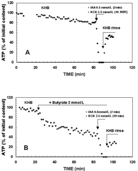
KHB group (Figure 2A)
After the ATP steady state was reached, the inhibition of both oxidative phosphorylation and glycolysis (IAA + KCN addition) induced a drastic fall in ATP content reach-ing zero within 2–3 min at a rate of 1.09 ± 0.13 µmole/ min.g (n = 7) (Table 1) in the whole organ. This rate was 100-fold faster than the rate of basal ATP consumption (considered as negligible).
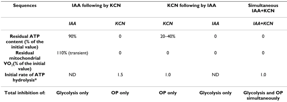
Fatty acid group (Figures 1B and 2B)
When the steady state of ATP content was reached in the presence of each fatty acid (20–30 min perfusion), the total ATP synthesis rate was calculated when IAA + KCN were added. The values gave an ATP synthesis rates of 0.76 ± 0.10, 0.40 ± 0.10 and 0.56 ± 0.10 µmole/min.g for ace-tate (n = 7), butyrate (n = 7) and octanoate (n = 5), respec-tively. Therefore, there was a significantly lower ATP turnover in the presence of butyrate (p = 0.001) and octanoate (p = 0.01) compared to the KHB group (Table 1).
O2 consumption (VO2)
During KHB perfusion (basal condition), mitochondrial liver consumption was 1.86 ± 0.16 µmol oxygen/min.g ww (n = 7). The addition of 3 mmol/L of each substrate (acetate, butyrate and octanoate) induced an increase in oxygen consumption and reached a new steady state for mitochondrial VO2 after 15 min of perfusion (Table 1) increasing with chain length, thus confirming previous results [1]. Addition of IAA + KCN induced the total and immediat inhibition of mitochondrial respiration, lead-ing to a decrease in a few seconds in effluent oxygen to a concentration around 24% (0.59 µmol O2/min.g ww) of the initial concentration, thus reflecting KCN-insensitive oxygen consumption in agreement with recent results [24]. Moreover, it has been previously demonstrated that only ≈30% of the mitochondrial respiration was devoted to ATP synthesis [24,25].
Validation of the method of inhibition of ATP synthesis (Table 2)
When KCN was used alone, a rapid and partial decrease in ATP signal was observed: the ATP content was not totally decreased and remained at a stable level around 20 to 40% of the initial value. However, the total inhibition of mitochondrial respiration in a few seconds indicated that the ATP plateau, observed after addition of KCN alone, probably originated from glycolysis. A subsequent addi-Time course of liver ATP content throughout the entire
pro-tocol perfusion Figure 2
tion of IAA slowly induced total removal of this residual ATP content, confirming this hypothesis.
The comparison of the ATP decline between the inhibi-tion condiinhibi-tions with KCN alone or simultaneous KCN+IAA, showed that the rate of ATP decrease was not changed by the presence of IAA at the onset of inhibition. At first sight, this should mean only a slight contribution of glycolysis to ATP synthesis in the liver.
On the other hand, when glycolysis was previously inhib-ited by IAA (0.5 mmol/L; 2 min), we observed a slight decrease in ATP content to a new steady state reached within 10 min. This decrease accounted for a maximum of 10% of the initial ATP content, indicating a slight contri-bution of glycolysis to total ATP synthesis. Moreover,
there was a transient (5 min) VO2 increase (< 10 ± 2%)
which returned to baseline. The further addition of KCN in a delay of 15 min after the addition of IAA induced an immediate and drastic fall in ATP content (to zero) and VO2, thereby showing that mitochondrial respiration and ATP synthesis were totally inhibited. In this sequential inhibition, the apparent rate of ATP decrease was at least 1.5-fold faster than that observed during simultaneous KCN+IAA inhibition, indicating a compensatory stimula-tion of oxidative phosphorylastimula-tion after the inhibistimula-tion of glycolysis alone. Therefore the sequential inhibition was not suitable for the purpose of our study.
Finally, in order to avoid overestimation of ATP synthesis flux, the use of this method of inhibition to determine ATP synthesis implies no increase in the activity of mito-Table 1: Calculation of mitochondrial ATP synthesis yield in isolated liver perfused with Krebs-Henseleit Buffer or substrates
ATP synthesis rate VO2 ATP/O
KHB 1.09 ± 0.13 1.86 ± 0.16 0.30 ± 0.05
(n = 7) - -
-Acetate 0.76 ± 0.10 1.92 ± 0.16 0.20 ± 0.02
(n = 7) NS NS NS
Butyrate 0.40 ± 0.10* 2.54 ± 0.18* 0.07 ± 0.02**
(n = 7) p = 0.001 p = 0.02 p = 0.0006
Octanoate 0.56 ± 0.10*** 3.04 ± 0.15*** 0.09 ± 0.018***
(n = 5) p = 0.01 p = 0.0004 p = 0.005
ATP synthesis rate and mitochondrial VO2 are expressed in µmole/min.g liver ww. ATP/O = ATP (µmol ATP)/2 VO2 (µatoms oxygen). The control group was perfused with Krebs-Henseleit buffer (KHB); the fatty acids were perfused at 3 mmol/L. Values are given as mean ± SEM (Significant ANOVA and t-test).
* p < 0.03 versus acetate, ** p = 0.0006 versus acetate, *** NS versus butyrate.
Table 2: Effects of various sequences of ATP synthesis inhibition in KHB group
Sequences IAA following by KCN KCN following by IAA Simultaneous IAA+KCN
IAA KCN KCN IAA IAA+KCN
Residual ATP content (% of the
initial value)
90% 0 20–40% 0 0
Residual mitochondrial VO2(% of the initial
value)
110% (transient) 0 0 0 0
Initial rate of ATP hydrolysis*
ND 1.5 1.0 ND 1.0
Total inhibition of: Glycolysis only OP only OP only Glycolysis only Glycolysis and OP simultaneously
chondrial ATP-consuming systems consecutively to the inhibitor addition. In the isolated mitochondria, it has been demonstrated that a rapid reduction of the proton motive force following CN- addition induced the change
of F0–F1 ATP synthase activity to ATPase activity, hydrolyz-ing ATP originated from cytosol via the ATP/ADP translo-case [26,27]. Hence, we performed complementary kinetic studies of change of whole liver ATP content in presence or in absence of carboxyatractyloside (200 µmol/ L), the specific, non competitive and irreversible inhibitor of translocase [28], when KCN and IAA were used. There was no significant difference of the rate of ATP decrease, demonstrating that in response to the large KCN-induced decrease of proton motive force, liver cytosolic ATP was not hydrolysed in situ by mitochondria.
Yield of ATP synthesis: ATP/O ratio (Table 1)
The ATP/O ratios (µmol ATP/µatom oxygen) were calcu-lated from the previous values of ATP synthesis rate and mitochondrial oxygen consumption. Perfusion with butyrate led to an ATP/O ratio significantly lower (p = 0.0006) than that of the control group and similar to those obtained in the presence of octanoate. No signifi-cant variation was observed in the presence of acetate compared to control.
These results demonstrated that fatty acids entering the β-oxidation pathway decreased the oxidative phosphoryla-tion yield by 77% and 70% for butyrate and octanoate, respectively, whereas acetate, which enters the TCA cycle directly, had no significant effect.
Discussion
Butyrate is one of the SCFAs (acetate, butyrate and propi-onate) produced by the intestinal bacterial fermentation of unabsorbed soluble carbohydrate. After their uptake by the colon, they are metabolized by the colonocytes with an action on cell growth and cell cycle, while the remain-ing fraction reaches the liver via the portal vein where it is metabolized. Moreover, SCFAs (butyrate) are used to sup-plement total parenteral nutrition in patients with short bowel syndrome or intestinal malabsorption syndromes. Besides their trophic effects on hepatocytes [10], it is of interest to know the influence of these substrates on oxi-dative phosphorylation since this may reflect the supply of ATP required for the anabolic pathways. To be free of the known reciprocal influences on SCFA on their absorp-tion and on their liver metabolism [29,30], our study con-cerned only the inherent effect of separate substrates.
Among medium-length FFA, octanoate is usually used to investigate the effect of FFA on energy metabolism and it has been shown to decrease the mitochondrial ATP/O ratio, thus reflecting the oxidative phosphorylation yield [17]. However, few studies have been devoted to butyrate
which, unlike acetate, enhances dramatically the rate of net ATP consumption in perfused isolated rat liver [1].
The present work mainly sought to estimate the effects of butyrate on the yield of in situ mitochondrial oxidative phosphorylation in perfused rat liver. NRM spectroscopy made it possible to monitor continuously the ATP content and its turnover in real time in a whole organ or organism. The in vivo ATP turnover can be measured by using NMR magnetization-transfer techniques [18,19], although this method is controversial [20] leading to a very large over-estimation of ATP fluxes in the liver [18]. Indeed, its basis is the use of exchange reactions between terminal phos-phate of ATP and inorganic phosphos-phate (Pi) at the level of (i) mitochondrial ATP synthase or (ii) glycolysis (glyceral-dehyde-3-phosphate dehydrogenase/phospho-glycerate kinase couple); the phosphate exchange can be increased during the NMR magnetization-transfer experiment with-out any net ATP synthesis. Nevertheless, from studies combining the estimation of ATP turnover by NMR spec-troscopy and respiration by polarography in the whole isolated rat liver, it is possible to calculate the ATP/O ratio [21,22]. The kinetic method used here to evaluate ATP turnover was based on the complete and rapid inhibition of the processes involved in cellular ATP synthesis, namely, oxidative phosphorylation (inhibited by KCN) and glycolysis (inhibited by IAA). The rates of ATP synthe-sis and consumption were equal at the steady state. Dur-ing blockage of the ATP synthesis pathways, the ATP concentration in the organ diminished on the basis of the cellular ATP consumption rate. In fact, the transient increase in oxygen uptake upon previous addition of IAA during sequential inhibition (as shown in the comple-mentary experiment) suggests that there is some degree of adjustment to the impaired metabolic state, in the pres-ence or abspres-ence of SCFA. Indeed, the apparent ATP syn-thesis flux was consistently higher when oxidative phosphorylation was inhibited after glycolysis than when their inhibitors were added together or when KCN was used alone (more than 50% increase). This result suggests that when glycolysis was inhibited before oxidative phos-phorylation, the mitochondria compensated for the loss of glycolytic ATP. Therefore, the sequential inhibition pro-tocol is not suitable for our purpose and both oxidative phosphorylation and glycolysis must be simultaneously inhibited in order to avoid compensatory and reciprocal stimulation. Even if the IAA inhibition was not immedi-ate, the contribution of glycolysis to ATP content and ATP flux remained less than 10% at the onset of KCN inhibi-tion.
acceptors. In addition, the activity of the respiratory com-plexes affects the respiration rate. Nevertheless, the contri-bution of the various components to the flux control of mitochondrial respiration has been a matter of endless debate for several decades. The production of acyl-CoA via short-chain acyl-CoA synthase consumes one ATP/mole FFA whatever the chain length. Butyryl-CoA and octanoyl-CoA are then β-oxidized providing acetyl-octanoyl-CoA and reduced cofactors. Acetyl-CoA finally enters the citric cycle. Among various factors, the rate of respiration depends in part on the amount of reduced cofactors (FADH2 and NADH+H+) produced during β-oxidation
and the citric cycle.
The present work confirms that the respiration rate increased with chain length at the concentration used, according to the process of the β-oxidation pathway [31,1], the lowest respiration rate being observed in the presence of acetate. Because respiration was greatly stimu-lated in the presence of butyrate and octanoate, an increase in coupled ATP synthesis was expected. However, the ATP content (resulting theoretically from both con-sumption and synthesis) was unexpectedly decreased at the same rate when butyrate and octanoate were added, reaching a plateau after at least 20 minutes, as previously demonstrated [1]. In this condition, glycolysis was proba-bly inhibited since these fatty acids have been shown to inhibit this pathway in isolated hepatocytes [32]. Moreo-ver, fatty acids have been identified as potent activators of hepatic gluconeogenesis in presence of neoglucogenic substrates such as lactate and pyruvate [13,14]. Therefore, we attempted to estimate in situ the flux of ATP synthesis originating from mitochondria, counterbalancing all ATP-consuming processes.
The ratio of synthetic flux of ATP to the mitochondrial liver respiration allowed the phosphorylative oxidation yield to be calculated. Butyrate or octanoate reduced the oxidative phosphorylation rate and increased the respira-tion rate, leading to the decrease in mitochondrial ATP/O ratio. Octonoate and longer-chain FA have been demon-strated to impair mitochondrial activity due to (i) an intrinsic uncoupling of the respiratory chain (direct effect), hence reducing ATP synthesis [17,33] and/or (ii) a change in the proportion of electron supply to the cou-pling site due to the increase in FADH2/NADH+H+ ratio
resulting from β-oxidation [17]. Since the respiration rate depends partly and directly on the amount of reduced cofactors, the latter process could explain in part the dependence from the chain length of the decrease in ATP/ O ratio. The low values of hepatic ATP/O ratio calculated in the control fed rats demonstrated that the in situ mito-chondrial activity in the whole liver was slight and phe-nomenologically near to state IV, as also observed in the mitochondria freshly isolated from the liver of fed rats
[34]. These low ATP/O ratios were far from the P/O ratio of 2.48 indirectly estimated in the perfused liver isolated from 24-hr fasted rat [35] and far from the maximal phos-phorylation capacity. In our study the addition of butyrate as well as octanoate induced a large decrease in the yield of phosphorylation, implying that substrate oxidation leads to a large enthalpy supply.
Conclusion
The difference in the hepatic oxidation pathway of the physiological substrates acetate and butyrate could explain their different action on energy metabolism. Butyrate is the main nutrient for the colonocytes but the large remaining fraction reaching the liver [11] has the same effect as longer-chain FFA. An effect on hepatic metabolism has to be taken into account when large quantities of SCFA are found in therapeutic purposes, especially in long-term acarbose treatment of patients with metabolic syndromes such as type 2 diabetes [36] or NASH [37], thereby reducing the risk of cardiovascular events [38]. Acarbose, an alpha-glucosidase inhibitor, reduces postprandial plasma glucose excursions by delay-ing the absorption of carbohydrate from the small intes-tine. It increases (i) the amount of starch entering the colon and (ii) butyrate production, as shown in fer-mented samples collected in humans undergoing this treatment [39,40]. Moreover, long-chain FFA have been shown to play a large part in the pathogenesis of insulin resistance in human obesity [41], in skeletal muscle and liver [42]. The specific question of the role of butyrate in the metabolic syndrome remains to be elucidated.
Methods
ChemicalsHigh-grade chemicals were purchased from Sigma Chem-ical (St. Louis, Missouri, USA) except where otherwise specified.
Animals and liver perfusion
Media were diluted daily from concentrated stock solu-tions. Standard Krebs-Heinseleit buffer (KHB) was com-posed of (in mmol/L) 120 NaCl, 4.7 KCl, 1.2 MgSO4, 25 NaHCO3, 1.2 KH2PO4-K2HPO4, 1.3 CaCl2, 0.3 Na-pyru-vate and 2.1 Na-lactate (pH = 7.35 at 37°C). Acetate, butyrate or octanoate were added to the KHB (3 mmol/L for each substrate). This concentration was chosen as we have previously demonstrated that it leads to maximal oxygen consumption and allows the ATP content steady state to be reached more quickly [1].
(kcal/kg) 4.05% lipids, 3.95% fiber, 18.70% protein, 5.35% minerals and 55.85% carbohydrates [1]. Lipid sup-ply consisted of a mixture of saturated and unsaturated long-chain fatty acids (C16 and C18). Rats were anesthe-tized by intraperitoneal injection of pentobarbital sodium (50 mg/kg of rat), and the technique of liver antegrade perfusion through the portal vein has been described pre-viously [43]. Briefly, the liver (4–6 g) was perfused in nor-mothermic and well oxygenated conditions with KHB at 37°C regulated by a thermostatic bath. The perfused liver was then excised from the rat abdomen and transferred to a 20 mm-diameter NMR cell. All experiments were carried out after a 20–30 min metabolic equilibration period in KHB.
The perfusate was pumped through a Silastic© home-made
oxygenator, gassed with 95% O2 and 5% CO2 (1 bar pres-sure). Perfusion flow was kept constant (5 ml/min.g liver wet weight) and was sufficient to ensure good oxygena-tion of the liver. Perfusate temperature and pH were mon-itored both before entering and after leaving the liver by continuous-flow pH electrodes and temperature elec-trodes.
Determination of ATP/O ratio
In order to suppress cytosolic and ATP mitochondrial pro-duction, perfusion was performed using both IAA (0.5 mmol/L) to inhibit glyceraldehyde 3-phosphate dehydro-genase (glycolysis) and KCN (2.5 mmol/L) to inhibit cytochrome oxidase (oxidative phosphorylation). In com-plementary experiments we showed that the addition of 0.5 mmol/L IAA for two minutes is sufficient to prevent glycolysis totally and irreversibly, without preventing the ATP mitochondrial metabolism (data not shown).
Three groups of animals were used:
(i) NMR measurement group: A first group was submitted to continuous NMR measurements in order to study the kinetic of the ATP content: the sequence was as follows: (i) initial KHB metabolic equilibration period (20–30 min), (ii) addition (or not = control) of substrate (3 mmol/L, 40–60 min) and (iii) in presence (or not = control) of sub-strate, addition of both IAA (0.5 mmol/L, 2 min) and KCN (2.5 mmol/L, 10 min), followed by (iv) a KHB rinse phase.
(ii) Respiratory measurement group: A second group was devoted to respiratory measurements through a similar sequence.
(iii) Both NMR and respiratory measurement group: A third group corresponded to experiments with sequential NMR and respiratory measurements (requiring removal of NMR cell from magnet).
Validation of the method of ATP synthesis inhibition
Complementary experiments were performed in order to evaluate the effect of the inhibition of the glycolytic path-way on mitochondrial ATP synthesis. For this purpose, three groups of livers were used:
First group: only KCN (2.5 mmol/L, 40 min) was added to inhibit oxidative phosphorylation.
Second group: a sequential inhibition was performed with KCN added firstly (2.5 mmol/L, 40 min) and fol-lowed by a delayed (15 min after the onset of KCN addi-tion) IAA addition (0.5 mmol/L, 2 min).
Third group: a sequential inhibition was performed with IAA (0.5 mmol/L, 2 min) added firstly and followed by a delayed (15 min) KCN addition (2.5 mmol/L).
The study complied with the Guide for the Care and Use of Laboratory Animals, U.S. Department of Health and Human Services, N.I.H. Publication No. 85-23, revised (1985).
31P NMR measurements
NMR measurements on isolated liver were performed on a Bruker DPX-400 wide-bore spectrometer operating at 9.4 T. 31P and 13C NMR spectra were recorded at 161.9
and 100.6 MHz respectively, with a 31P/13C double-tuned
20 mm probe. The magnetic field was adjusted to the water proton signal. 31P-NMR spectra were obtained
with-out proton decoupling every 1 min and 30 seconds (148 free induction decays, FIDs) throughout the sequence pro-tocol, except during inhibitor addition times, every 20 sec-onds (40 FIDs) in order to increase the determination accuracy. Radiofrequency pulses of 25 µs duration (70° flip angle) with 0.2 s acquisition time, 0.4 s total recycling time, 10000 Hz spectral width and 4 K data points were used for data acquisition. A Lorentzian line broadening of 15 Hz was applied prior to Fourier transformation for 31P
NMR spectra. An external reference of 13 µmoles methyl-ene diphosphonic acid (MDPA set at 18.40 ppm) was used to quantify the phosphorylated metabolites.
Hepatic ATP levels were estimated from peak areas and expressed as a percentage of the initial value obtained at the beginning of the 30-min equilibration period in KHB.
Evaluation of ATP turnover
addi-tion of IAA+KCN. Vs was expressed in µmoles ATP/min.g wet weight (ww).
Oxygen consumption measurements
Isolated perfused livers were placed in an NMR cell similar to that used for the NMR experiments. The total oxygen consumption of liver was continuously monitored using O2 Clark electrodes, placed in the perfusion circuit just before and after the liver, using an oxygenmeter model 781 (Strathkelvin Instrument, Glasgow, Scotland). Oxy-gen consumption (µmol O2/min.g liver ww) VO2 = (per-fusate O2 content - effluent O2 content) × liver flow rate
(ml/mn.g ww) was calculated, where perfusate O2 content was expressed in µmol/ml. The mitochondrial respiration component of tissue respiration was calculated by sub-tracting the KCN-insensitive oxygen consumption from the total O2 consumption.
Statistics
All results were expressed as means ± SEM. Statistical anal-ysis was performed with Microsoft® Excel 2000 (Microsoft
Corporation). One-way analysis of variance (ANOVA) was performed for all data analyses. A t test was performed following the analysis of variance (p value lower than 0.05 was considered to be significant).
Abbreviations
ATP- Adenosine triphosphate.
ATP/O- Oxidative phosphorylation yield.
FFA- Free fatty acids.
IAA- Iodacetic acid.
KHB- Krebs Henseleit buffer.
NMR- Nuclear magnetic resonance.
OP- Oxidative phosphorylation.
SCFA- Short-chain free fatty acids.
Authors' contributions
MCB participated in the design and coordination of the study and was involved in drafting the manuscript.
PT carried out all the experimental procedures.
HG was highly involved in revising the manuscript.
JLG designed the study, carried out the analysis and data interpretation and was involved in revising the manu-script.
All authors read and approved the final manuscript.
Acknowledgements
The authors thank Ray Cooke for revising the manuscript. This work was supported by grants from the Conseil Régional d'Aquitaine, Centre National de la Recherche Scientifique and Institut de Recherche sur les Boissons (contrat 2000/05)
References
1. Beauvieux MC, Tissier P, Gin H, Canioni P, Gallis JL: Butyrate impairs energy metabolism in isolated perfused liver of fed rats. J Nutr 2001, 131:1986-1992.
2. Royall D, Wolever TM, Jeejeebhoy KN: Clinical significance of colonic fermentation. Am J Gastroenterol 1990, 85:1307-1312. 3. Rapport Glucides et Santé: Etat des lieux, évaluation et
recom-mandations. Agence Française de Sécurité Sanitaire des Aliments, Maisons-Alfort: AFSSA; 2004.
4. Nyman M, Asp NG, Cummings J, Wiggins H: Fermentation of die-tary fiber in the intestinal tract: comparison between man and rat. Br J Nutr 1986, 55:487-496.
5. Jacobash G, Schmiedl D, Kruschewsli M, Schmehl K: Dietary starch and chronic inflammatory bowel diseases. Int J Colorectal Dis
1999, 14:201-211.
6. Bartholome AL: Supplementation of total parenteral nutrition with butyrate acutely increases structural aspects of intesti-nal adaptation after 80% jejunoileal resection in neonatal pig-lets. J Par Ent Nutr 2004, 28(4):210-222.
7. Sakata T: Influence of short chain fatty acids on intestinal growth and functions. Adv Exp Med 1997, 427:191-199. 8. Cummings JH: Short-chain fatty acid enemas effective in the
treatment of distal ulcerative colitis. Gastroenterol Hepatol 1997,
9:149-153.
9. Scheppach W, Sommer H, Kirchner T, Paganelli GM, Bartram P, Christl S, Richter F, Dusel G, Kasper H: Effect of butyrate enemas on the colonic mucosa in distal ulcerative colitis. Gastroenterol
1992, 103(1):51-56.
10. Yoon JH, Lee HV, Lee JS, Park JB, Kim CY: Development on a non-transformed human liver cell line with differentiated-hepa-tocyte and urea-synthetic functions: applicable for artificial liver. Int J Artif Organs 1999, 22:769-777.
11. Demigné C, Yacoub C, Rémésy C: Effects of absorption of large amounts of volatile fatty acids on rat liver metabolism. J Nutr
1986, 116:77-86.
12. Rémésy C, Demigné C, Chartier F: Origin and utilization of vola-tile fatty acids in the rat. Reprod Nutr Dev 1980, 20:1339-1349. 13. Gonzalez-Manchon C, Martin-Requiro A, Ayuso MS, Parrilla R: Role
of endogenous fatty acids in the control of hepatic gluconeo-genesis. Arch Biochem Biophys 1992, 292:95-101.
14. Morand C, Rémésy C, Demigné C: Fatty acids are potent modu-lators of lactate utilization in isolated hepatocytes from fed rats. Am J Physiol 1993, 264:E816-823.
15. Mollica MP, Iossa S, Liverini G, Soboll S: Stimulation of oxygen consumption following addition of lipid substrates in liver and skeletal muscle from rats fed a high-fat diet. Metabolism
1999, 48:1230-1235.
16. Scholtz R, Schwabe U, Soboll S: Influence of fatty acids on energy metabolism. 1. Stimulation of oxygen consumption, ketogenesis and CO2 production following addition of
octanoate and oleate in perfused rat liver. Eur J Biochem 1984,
141:223-230.
17. Leverve X, Sibille B, Devin A, Piquet MA, Espié P, Rigoulet M: Oxida-tive phosphorylation in intact hepatocytes: quantitaOxida-tive characterization of the mechanisms of change in efficiency and cellular consequences. Mol Cell Biochem 1998, 184:53-65. 18. Thoma WJ, Ugurbil K: Saturation-transfer studies of ATP-Pi
exchange in isolated perfused rat liver. Biochim Biophys Acta
1987, 893:225-231.
19. Sheldon JG, Williams SP, Fulton AM, Brindle KM: 31P NMR mag-netization transfer study of the control of ATP turnover in Saccharomyces cerevisiae. Proc Natl Acad Sci USA 1996,
93:6399-6404.
Publish with BioMed Central and every scientist can read your work free of charge
"BioMed Central will be the most significant development for disseminating the results of biomedical researc h in our lifetime."
Sir Paul Nurse, Cancer Research UK
Your research papers will be:
available free of charge to the entire biomedical community
peer reviewed and published immediately upon acceptance
cited in PubMed and archived on PubMed Central
yours — you keep the copyright
Submit your manuscript here:
http://www.biomedcentral.com/info/publishing_adv.asp
BioMedcentral
21. Beauvieux MC, Tissier P, Couzigou P, Gin H, Canioni P, Gallis JL: Eth-anol perfusion increases the yield of oxidative phosphoryla-tion in isolated liver of fed rats. Biochim Biophys Acta 2002,
1570:135-140.
22. Dufour S, Thiaudière E, Gallis JL, Canioni P, Diolez P: Yield of oxi-dative phosphorylation in resting perfused liver: saturation ad biochemical analyses [abstract]. Magn Reson Mater Phys Biol Med 1996, 4:329.
23. Baillet-Blanco L, Beauvieux MC, Gin H, Rigalleau V, Gallis JL: Insulin induces a positive relationship between the rates of ATP and glycogen changes in isolated rat liver in presence of glucose; a 31P and 13C NMR study. Nutr Metab 2005, 31:32.
24. Dewar BJ, Bradford BU, Thurman RG: Nicotine increases hepatic oxygen uptake in the isolated perfused rat liver by inhibiting glycolysis. J Pharmacol Exp Therapeutics 2002, 301:930-937. 25. Taylor WM, Van de Pol E, Bygrave FL: On the stimulation of
res-piration by α-adrenergic agonists in perfused rat liver. Eur J Biochem 1986, 155:319-322.
26. Schwerzmann K, Pedersen PL: Regulation of the mitochondrial ATP synthase/ATPase complex. Arch Biochem Biophys 1986,
250:1-18.
27. Gaballo A, Zanotti F, Papa S: Structures and interactions of pro-teins involved in the coupling function of the protonmotive F(0)F(1)-ATP synthase. Curr Protein Pept Sci 2002, 3:451-460.
28. Dahout-Gonzalez C, Nury H, Trezeguet V, Lauquin GJ, Pebay-Pey-roula E, Brandolin G: Molecular, functional, and pathological aspects of the mitochondrial ADP/ATP carrier. Physiology
2006, 21:242-249.
29. Gordon MJ, Crabtree B: The effects of propionate and butyrate on acetate metabolism in rat hepatocytes. Int J Biochem 1992,
24:1029-1031.
30. Martin L, Dumon H, Lecannu G, Nguyen P, Champ M: Apparition dans la veine porte des acides gras à chaîne courte perfusés in situ dans le colon proximal du porc sain [abstract]. Nutr Clin Metab 1999, 13:52s.
31. Plomp PJAM, van Roermund CWT, Groen AK, Meijer AJ, Tager JM:
Mechanism of the stimulation of respiration by fatty acids in rat liver. Febs Lett 1985, 193:243-246.
32. Morand C, Besson C, Demigné C, Rémésy C: Importance of the modulation of glycolysis in the control of lactate metabolism by fatty acids in isolated hepatocytes from fed rats. Arch Bio-chem Biophys 1994, 309:254-260.
33. Soboll S, Stucki J: Regulation of the degree of coupling of oxida-tive phosphorylation in intact rat liver. Biochim Biophys Acta
1985, 807:245-254.
34. Delmas-Beauvieux MC, Leducq N, Thiaudière E, Diolez P, Gin H, Canioni P, Gallis JL: Liposomes as fatty acids carriers in isolated rat liver: effect on energy metabolism and on isolated mito-chondria activity. Magn Reson Mat Phys Biol Med 2000,
10(1):43-51.
35. Soboll S, Oh MH, Brown GC: Control of oxidative phosphoryla-tion, gluconeogenesis, ureagenesis, and ATP turnover in iso-lated perfused rat liver by top-down metabolic control analysis. Eur J Biochem 1998, 254:194-201.
36. Abuissa H, Bel DS, O'Keefe JR Jr: Strategies to prevent type 2 diabetes. Curr Med Res Opin 2005, 21:1107-1114.
37. Yamagishi S, Nakamura K, Inoue H: Acarbose is a promising ther-apeutic strategy for the treatment of patients with non alco-holic steatohepatitis (NASH). Med Hypotheses 2005,
65:377-379.
38. Zeymer U: Cardiovascular benefits of acarbose in impaired glucose tolerance and type 2 diabetes. Int J Cardiol 2006,
107:11-20.
39. Weaver GA, Tangel CT, Krause JA, Parfitt MM, Jenkins PL, Rader JM, Lewis BA, Miller TL, Wolin MJ: Acarbose enhances human colonic butyrate production. J Nutr 1997, 127:717-723. 40. Wolever MS, Chiasson JJ: Acarbose raises serum butyrate in
human subjects with impaired glucose tolerance. Brit J Nutr
2000, 84:57-61.
41. Boden G: Free fatty acids – the link between obesity and insu-lin resistance. Endocr Pract 2001, 7:44-51.
42. Boden G: Effects of free fatty acids (FFA) on glucose metabo-lism for insulin resistance and type 2 diabetes. Exp Clin Endo-crinol Diabetes 2003, 111:121-124.
43. Delmas-Beauvieux MC, Gallis JL, Rousse N, Clerc M, Canioni P:
Phosphorus-31 nuclear magnetic resonance of isolated rat