Open Access
Preliminary communication
Study on the effects of nitrilotriproprionic acid and
4,5-dihydroxy-1,3-benzene disulphonate on the fractionation of
beryllium in human serum using graphite furnace atomic
absorption spectrometry
Chadi H Stephan
1, Michel Fournier
2, Pauline Brousseau
2and
Sébastien Sauvé*
1Address: 1Department of Chemistry, Université de Montréal, PO 6128, Station Centre-Ville, Montréal, QC, H3C 3J7, Canada and 2INRS-Institut Armand-Frappier, 245 Hymus, Pointe-Claire, QC, H9R 3G6, Canada
Email: Chadi H Stephan - [email protected]; Michel Fournier - [email protected]; Pauline Brousseau - [email protected]; Sébastien Sauvé* - [email protected] * Corresponding author
Abstract
Background: Occupational exposure to beryllium may cause Chronic Beryllium Disease (CBD), a lung disorder initiated by an electrostatic interaction with the MHC class II human leukocyte antigen (HLA). Molecular studies have found a significant correlation between the electrostatic potential at the HLA-DP surface and disease susceptibility. CBD can therefore be treated by chelation therapy. In this work, we studied the effect of two complexing agents, nitrilotriproprionic acid (NTP) and 4,5-dihydroxy-1,3-benzene disulphonate (Tiron), on the fractionation of beryllium in human serum analysed by graphite furnace atomic absorption spectrometry (GFAAS).
Results: We found the average serum beryllium concentration of fourteen non-exposed individuals to be 0.53 (± 0.14) µg l-1, with 21 (± 3)% of the beryllium mass bound to the low
molecular weight fraction (LMW), and 79 (± 3)% bound to the high molecular weight fraction (HMW). The addition of Tiron increased the beryllium mass in the HMW fraction, while NTP was not seen to have any influence on the fractionation of beryllium between the two fractions. NTP was, however, shown to complex 94.5% of the Be mass in the LMW fraction. The beryllium GFAAS detection limit, calculated as three times the standard deviation of 10 replicates of the lowest standard (0.05 µg L-1), was 6.0 (± 0.2) ng L-1.
Conclusion: The concentration of beryllium or its fractionation in human serum was not affected by sex or smoking habit. On average, three quarters of the beryllium in serum were found in the HMW fraction. Of the two ligands tested, only Tiron was effective in mobilising beryllium under physiological conditions, thus increasing the Be content in the HMW fraction.
Published: 14 May 2008
Chemistry Central Journal 2008, 2:10 doi:10.1186/1752-153X-2-10
Received: 13 February 2008 Accepted: 14 May 2008
This article is available from: http://journal.chemistrycentral.com/content/2/1/10
© 2008 Stephan et al
Background
Beryllium, the first of the alkaline earth metals, is natu-rally found in mineral rocks, coal, soil, and volcanic dust [1]. Beryllium ore is mined and purified for its use in nuclear reactors, weapons, aircraft and space vehicle struc-tures, x-ray machines, as well as telecommunication and high-tech devices [2]. Occupational exposure to beryllium may cause Chronic Beryllium Disease (CBD), a lung dis-order characterised by a granulomatous inflammation ini-tiated by an electrostatic interaction with the MHC class II human leukocyte antigen (HLA) [3,4]. Molecular epide-miological studies have shown that interaction between beryllium and specific HLA-DP alleles is a factor in disease susceptibility [5]. Furthermore, molecular modelling has been used to investigate a potential mechanistic basis for these observations. A significant correlation has been found between the risk of chronic beryllium disease asso-ciated with specific alleles, and the predicted electrostatic surface potential, suggesting that the alleles associated with the most negatively charged proteins carry the great-est risk of beryllium sensitisation and disease [6].
At present, CBD can be treated but not cured. Adrenocor-tical steroids such as prednisone, prednisolone and dex-amethasone act to reduce the inflammation and immune response to beryllium but cannot eliminate beryllium from organs or tissues, a key factor in stopping CBD [7]. Chelation therapy [1,8,9] or metal encapsulation [10] can eventually provide an alternative or adjunctive treatment to accelerate beryllium clearance from organs and tissues. In this work, we looked at the natural distribution of beryllium in serum and investigated the effect of two sequestering agents, nitrilotriproprionic acid (NTP) and 4,5-dihydroxy-1,3-benzene disulphonate (Tiron) (Figure 1), on the distribution of beryllium. We used a mobilisa-tion index (MI) [11-13] to represent the relative ability of a complexing agent to compete for the metal of interest and mobilise it under physiological conditions. The MI is defined as:
Nitrilotripropionic acid was selected because of its phys-ico-chemical properties: it complexes beryllium in a tetra-hedral complex [Be(NTP)]- where the beryllium cation
lies at the centre of a slightly distorted tetrahedron of C3υ symmetry, with a longer Be-N bond and three equal Be-O bonds [14]. Tiron is a hydrophilic chelator and was selected because of its reported efficiency in mobilising beryllium [15,16], restoring the altered biochemical parameters [17] and improving the altered hepatorenal biochemistry and ultramorphology in different rat tissues and organs [18].
Beryllium, like any other metal ions in serum, can be frac-tionated into four distinct groups: rigidly bound to metal-loproteins; loosely bound to other types of proteins (labile equilibrium); complexed by the so-called low-molecular-weight fraction (LMW); and occurring as free (or hydrated) metal ions [12].
In this study, we looked first at the natural distribution of beryllium in serum and the influence of NTP and Tiron on this distribution. We focused on two operationally-defined fractions; the first fraction combines the Be rigidly and loosely bound to proteins, and is called the high molecular weight fraction (HMW); whilst the second frac-tion, that is the low molecular weight fraction (LMW), combines the Be complexed to the low molecular-weight fraction or that which occurs in its free ionic form. We sep-arated the HMW and LMW fractions by ultrafiltration with Centricon centrifugal filter devices at a 10,000 nom-inal molecular weight limit (NMWL) cut-off. Beryllium quantification in serum and serum fractions was carried out using an optimised graphite furnace atomic absorp-tion spectrometer (GF-AAS). The accuracy of the analytical method was tested using Seronorm samples (Seronorm trace elements whole blood level 2, STEWB; Ref # 201605; Lot # 0503109).
Results and discussion
Beryllium in serum
The average beryllium concentration in fourteen non-exposed individuals (9 females and 5 males; 9 non smok-ers and 5 smoksmok-ers) found to be 0.53 (± 0.14) µg L-1.
Nei-ther sex nor smoking habit was shown to have a significant influence on the concentration of beryllium in serum (independent sample T-test, p > 0.05). The method detection limit, calculated as three times the standard deviation of 10 replicates of the lowest standard (0.05 µg L-1), was 6.0 (± 0.2) ng L-1. The accuracy of the method
MI
total concentration of low molecular weight beryllium
in the pre
= ssence of the complexing agent
total concentration of low mol
eecular weight beryllium
in the absence of the complexing agent
Chemical structures of nitrilotripropionic acid (NTP) and 4,5-dihydroxy-1,3-benzene disulphonate (Tiron)
Figure 1
varied from 99 to 104% and was verified by analysing a control sample (STEWB level 2 certified blood material with a concentration of 5.9 (± 0.5) µg Be L-1).
Beryllium distribution in serum
On average, 21 (± 3)% of the beryllium mass was bound to the LMW fraction, with 79 (± 3)% bound to the HMW fraction (Table 1). Neither sex nor smoking habit was shown to have a significant influence on the distribution of beryllium between the LMW and the HMW fractions (independent sample T-test, p > 0.05). These findings agree with the work of Stiefel et al. [19] conducted on human and guinea pig blood. They reported that the cor-puscular part of all samples contained from 2 to 10% of the total Be, the LMW fraction contained between 17 and 33%, with 60 to 73% of the total Be bound to the HMW fraction – or more specifically bound to the pre-albumins and the γ-globulins fractions. They also reported that the Be distribution between the two protein regions depends on the absolute concentration of Be in blood. At concen-trations lower than 1 µg L-1, most of the Be mass (> 90%)
was bound to the γ-globulins fraction, while at higher concentrations most of the Be mass was found bound to the pre-albumins fraction. This is very similar to the chem-ical speciation of aluminium in human serum, were almost all the Al mass in the HMW fractions was bound to transferrin belonging to the γ-globulins protein fractions [20-22]. Beryllium and aluminium have very similar chemical properties [1]. In an attempt to better under-stand beryllium chemistry in human body fluids, Sutton and Burastero [23] have simulated the speciation of beryl-lium in plasma fluid (LMW fraction) and reported most of the beryllium at pH 7.4 to be aqueous BeCO3 (49.4%) and BeOH+ (39.9%). A reproduction of this speciation is
presented in Figure 2, where in using the chemical equi-librium software Mineql+ [24] we show that adding cit-rate and oxalate, should complex most of the Al in the LMW human serum [20,22,25-27]. We obtained similar results and found that most of the beryllium at pH 7.4 is shown to exist as aqueous Be(OH)+ (60.5%), BeCO
3
(27.4%) and Be(OH)2 (10.2%). Neither citrate nor oxalate influenced the speciation of beryllium at serum pH, although both ligands complexed beryllium at acidic pH with maxima of 16.2% (at pH = 3) and 9% (at pH = 5.5) for Be(oxalate) and Be(citrate) respectively.
In an attempt to investigate the chelation effect of Tiron and NTP, as well as study their effect on the repartition of beryllium in human serum, dose-response experiments were conducted showing the influence of both complex-ing agents on the repartition of beryllium between the two serum fractions. We tested different equilibration times (2, 4 and 6 h), but found no differences for both ligands, suggesting that the exchange reactions rates are almost instantaneous and that equilibrium is reached within the
2 hour equilibration timeframe employed. This is similar to the Al-transferrin exchange reaction rate with desferri-oxamine B (DFO) where the addition of DFO increased the amount of ultrafiltrable Al sharply up to 90% of the total plasma Al content [28,29]. Due to the similarity between Be and Al "hard ions", and DFO, NTP and Tiron "complexing agents with soft oxygen donors", we should expect almost instant exchange reaction rates for the Be-Tiron-HMW and Be-NTP-HMW systems, thus confirming that the 2-h equilibration time is more than enough for the exchange to occur.
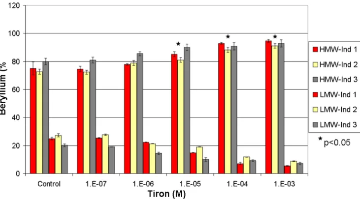
In spiked and non-spiked samples (Figures 3 and 4), Tiron showed an increasing pattern of the beryllium mass in the HMW fraction. Such results contradict the general behav-iour of complexing agents, which usually tend to increase the metal concentration in the LMW fraction [12,13]. In the spiked samples, the addition of Tiron increased the mass fraction of beryllium in the HMW from an average of 76 (± 4)%, reaching a maximum of 93 (± 2)% at 10-3
M. Conversely, the average concentration of beryllium in the LMW fraction decreased from 24 (± 4)% to 7 (± 2)%. This increase in the Be-HMW fraction becomes significant at 10-5 M (One-Way Anova, Fisher's LSD, with P < 0.05).
We observed the same pattern in non-spiked samples. The HMW beryllium content increased from an average of 79 (± 3)% to 90 (± 4)% at 10-3 M, while the LMW beryllium
content decreased from 20 (± 3)% to 10 (± 4)%. This change in the Be-HMW fraction is only significant at 10-3
M. These observations confirm that Tiron complexes Be under serum conditions and increases the beryllium con-tent in the HMW fraction by generating a mobilisation index that decreases with increasing Tiron concentrations (Figure 5). Tiron complexes Be in a 2:1 ratio [30,31], most probably generating a less charged complex that tends to be more soluble in the HMW fraction than in the hydrophilic LMW fraction. We suspect that such behav-iour might increase the toxicity of beryllium, but several recent studies have shown that Tiron (with or without the aid of adjuvants) restored the altered biochemical physio-logical parameters and oxidative stress response in rats exposed to beryllium [15,18,31].
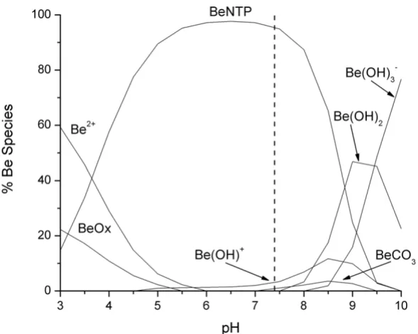
HMW and LMW fractions, but showed a capacity to com-plex most of the beryllium in the LMW, as shown by the chemical speciation simulations (Figure 8). At a concen-tration of 10-7 M, NTP was shown to complex 94.5% of the Be in the LWM followed by 3.4% and 1.1% of the Be occurring as Be(OH)+ and BeCO3, respectively. These results support earlier findings which suggest that NTP would be an ideal complexing agent for Be [14].
Conclusion
Neither sex nor smoking habit was shown to influence beryllium concentration or its fractionation in human serum. On average, three quarters of the beryllium in serum were found to be bind to the HMW fraction. Tiron, unlike NTP, showed a significant interaction with beryl-lium under physiological conditions by increasing the Be content in the HMW fraction – in contrast to that which is usually observed for standard complexing agents used in chelation therapy. The addition of NTP did not affect the MW distribution between the two fractions but was shown to complex most of the Be in the LMW fraction. Further work could employ anion exchange fast protein liquid chromatography (FPLC) coupled with electrospray tandem mass spectrometry (ES-MS-MS) or inductively coupled plasma mass spectrometry to confirm the nature of the ligands or proteins that complex Be in both serum fractions [20-22,27].
Experimental
Reagents
All of the reagents used were of analytical grade or better. Antifoam B silicone emulsion (J.T. Baker, NJ, USA), ammonium hydroxide (certified A.C.S. Plus, Fisher scien-tific, NJ, USA), ethylenediaminetetraacetic acid disodium salt dihydrate (EDTA) (Fluka chemika, Switzerland), Tri-ton X-100 (Acros, NJ, USA), nitric acid (trace metal grade, Fisher Scientific, Ontario, Canada), beryllium plasma standard solution (Specpure, Alfa Aesar, MA, USA), nitrilotripropionic acid (NTP) (MP Biomedicals, Ohio, USA) and 4,5-dihydroxy-1,3-benzene disulphonate (Tiron) (Acros Organics, New Jersey, USA).
Solutions
Serum samples were diluted with a Nash reagent (NR) solution prepared weekly and containing 5% (v/v) nitric acid, 5% (v/v) of ammonium hydroxide, 0.2% (v/v) Tri-ton X-100, 0.2% (v/v) antifoam B and 0.5% (w/v) of EDTA in distilled-deionised water. Solutions of NTP and Tiron were prepared at 10-1, 10-2, 10-3, 10-4 and 10-5 M by
dissolving the appropriate mass in 2% (v/v) HNO3 aque-ous solution. A 50 µg L-1 working Be (II) solution was
pre-pared by dilution of the beryllium stock solution (1000
µg L-1) in 2% (v/v) HNO
3. The Be standard solution was
prepared daily by dilution of the working Be (II) solution
Speciation of Be in the LMW of serum fluid ([Be] 5.8*10-5 mM; [Ca] 1.3 mM; [Cl] 108 mM; [CO
3] 24 mM; [K] 4.2 mM; [Mg] 0.8
mM; [Na] 142 mM; [PO4] 2 mM; [SO4] 0.5 mM; [Cit] 0.1 mM; [Ox] 0.01 mM)
Figure 2
Speciation of Be in the LMW of serum fluid ([Be] 5.8*10-5 mM; [Ca] 1.3 mM; [Cl] 108 mM; [CO
3] 24 mM; [K] 4.2 mM; [Mg] 0.8
with the Nash reagent to give a final concentration of 0.5
µg L-1.
Sample preparation
Blood from unexposed individuals was collected in BD vacutaine SST tubes (BD Franklin lakes, New Jersey, USA). Tubes were left at room temperature for 30 minutes to allow clot formation and then centrifuged at 500 g for 10
minutes. Supernatants of each individual were collected and recombined to ensure sample homogenisation. Each sample was divided into twelve 2.5 mL sub-samples in borosilicate disposable culture tubes, two served as con-trols, five were spiked with NTP and five were spiked with Tiron at concentrations ranging from 10-7 to 10-3 M.
Sub-samples were left to equilibrate for two hours. A 2 mL aliquot of each sub-sample was transferred to a Centricon centrifugal device with ultracel YM-10 (10,000 MWCO) (Millipore Corporation, Ireland) and centrifuged at 4500 g for 90 minutes. The LMW and HMW fractions obtained were analysed for beryllium by graphite furnace atomic absorption spectrometry (GFAAS) after respective 2-fold and 5-fold dilutions with NR. The same experimental pro-cedure was repeated on serum samples that were spiked with 1 µg L-1 of beryllium. We also measured the
beryl-lium concentration in fourteen individuals and looked at the natural distribution of beryllium between the HMW and LMW fractions. Serum was diluted 5-fold with NR before GFAAS analysis.
Graphite furnace atomic absorption spectrometer
A Varian AA280Z Zeeman atomic absorption spectrome-ter, equipped with a Zeeman background correction, GTA 120 graphite tube atomiser and PSD 120 programmable sample dispenser was used for the atomic absorption measurement of beryllium at 234.9 nm with a spectral bandwidth of 1.0 nm. A beryllium hollow cathode lamp (Varian, Part No. 5610100500) was used as a light source operated at 5 mA. Pyrolytic graphite coated partitioned tubes (Varian partition tubes, Part No. 63-100012-00)
Table 1: Concentration and natural distribution of beryllium in the human serum of non-exposed individuals.
Sample Sex Smoking Serum (µg L-1) LMW (%) HMW (%)
1 F S 0.54 (0.08) 22 (2) 78 (3) 2 F NS 0.46 (0.04) 24 (3) 76 (3) 3 F S 0.47 (0.09) 19 (1) 81 (1) 4 F NS 0.42 (0.05) 24 (2) 76 (2) 5 F NS 0.49 (0.07) 21 (2) 79 (2) 6 M NS 0.44 (0.02) 25 (3) 75 (3) 7 F NS 0.45 (0.01) 25 (3) 75 (3) 8 F NS 0.44 (0.01) 19 (1) 81 (1) 9 F NS 0.46 (0.01) 24 (3) 76 (3) 10 M NS 0.45 (0.01) 16 (3) 84 (3) 11 M S 0.78 (0.03) 23 (4) 77 (4) 12 F NS 0.69 (0.03) 21 (5) 79 (5) 13 M S 0.45 (0.04) 20 (3 80 (3) 14 M S 0.86 (0.01) 21 (3) 79 (3)
Mean 0.53 (0.14) 21 (3) 79 (3)
F = female; M = male; S = smoking; NS = non smoking
Influence of Tiron on the repartition of beryllium between the HMW and the LMW fractions in three spiked individuals Figure 3
Influence of Tiron on the repartition of beryllium between the HMW and the LMW fractions in three non-spiked individuals Figure 4
Influence of Tiron on the repartition of beryllium between the HMW and the LMW fractions in three non-spiked individuals. * Significant difference calculated by One-Way Anova, Fisher's LSD.
Mobilisation Index (MI) of Be as a function of Tiron concentration Figure 5
Influence of NTP on the repartition of beryllium between the HMW and the LMW fractions in three spiked individuals Figure 6
Influence of NTP on the repartition of beryllium between the HMW and the LMW fractions in three spiked individuals. * Signif-icant difference calculated by One-Way Anova, Fisher's LSD.
Influence of NTP on the repartition of beryllium between the HMW and the LMW fractions in three non-spiked individuals Figure 7
were used for all experiments. High purity Argon (99.99%) was used as the carrier gas. A beryllium hollow cathode lamp (Varian, Part No. 5610100500) was used as a light source. Instrument control, sample results, signal graphics and data collection are controlled by the Spec-trAA Worksheet software for the Windows® XP operating
system. Peak area values were used for beryllium signal and background (BG) measurements. The instrumental conditions and the furnace program are listed in Table 2.
Chemical Speciation
For the chemical speciation calculations, we used MINEQL+ (version 4.5 for Windows – Environmental
Research Software, Hallowell, ME), a chemical equilib-rium modelling system that can be used to perform calcu-lations at low temperatures (0–50°C) and low to moderate ionic strength (< 0.5 M). MINEQL+ operates over three steps: creation of a system by selecting chemical components from a menu with the possibility of adding new ligands, then scanning the thermodynamic database and finally running the calculations with actual measured concentrations included. The output data module yields the activity for each species of each component. Our chemical speciation calculations were made assuming the following parameters (unless specified otherwise): T = 37°C (fixed), ionic strength: I = 0.01 M (fixed), Log pCO2 = -3.5 (open atmosphere). Different chemical equilibrium software should yield similar results when the same stabil-ity constants are used. The following input data were added to the MINEQL+ thermodynamic database; Log KBe-Ox = 3.47, Log KBe-(Ox)2 = 5.24, Log KBe-NTP = 13.94, Log KH3-NTP = 11.54, Log KH2-NTP -1 = 10.31 and Log K
H-NTP -2 =
7.59.
Statistical analysis
Statistical analysis was performed with SPSS (version 13 for Windows, SPSS Inc, Chicago, IL) using independent sample T-tests to monitor significant differences among groups. We also used One-Way ANOVA tests with Fisher's
Table 2: The furnace programme for the determination of beryllium in serum.
Step Temp (°C) Time (s) Argon flow rate (L min-1)
Drying 85 5 0.3
Drying 95 30 0.3
Drying 120 20 0.3
Pre-pyrolysis 450 22 0.3 Pyrolysis 1000 17 0.3 Atomizing 2900 3 0
Cleaning 2900 2 0.3
Influence of NTP on the speciation of Be in the LMW of serum fluid ([Be] 5.8*10-5 mM; [NTP] 10-4 mM; [Ca] 1.3 mM; [Cl] 108
mM; [CO3] 24 mM; [K] 4.2 mM; [Mg] 0.8 mM; [Na] 142 mM; [PO4] 2 mM; [SO4] 0.5 mM; [Cit] 0.1 mM; [Ox] 0.01 mM) Figure 8
Influence of NTP on the speciation of Be in the LMW of serum fluid ([Be] 5.8*10-5 mM; [NTP] 10-4 mM; [Ca] 1.3 mM; [Cl] 108
Open access provides opportunities to our colleagues in other parts of the globe, by allowing
anyone to view the content free of charge.
Publish with
Chemistry
Central and every
scientist can read your work free of charge
W. Jeffery Hurst, The Hershey Company.
available free of charge to the entire scientific community peer reviewed and published immediately upon acceptance cited in PubMed and archived on PubMed Central yours you keep the copyright
Submit your manuscript here:
http://www.chemistrycentral.com/manuscript/
LSD (least significant difference) to make pair wise com-parisons as a way of monitoring significant changes within a group.
Authors' contributions
All authors contributed to the experimental design. CHS carried out nearly all of the laboratory work, the initial interpretation of the data and the initial write-up. PB and MF contributed their expertise especially with the biolog-ical work, and SS focused on the analytbiolog-ical chemistry.
Acknowledgements
We thank the Natural Sciences and Engineering Research Council of Can-ada and the Institut de Recherche Robert-Sauvé en Santé et en Sécurité du Travail for financial support.
References
1. Wong CY, Woollins JD: Beryllium Coordination Chemistry.
Coordination Chemistry Reviews 1994, 130:243-273.
2. Jameson CW: Introduction to the Conference on Beryllium-Related Diseases. Environmental Health Perspectives 1996,
104:935-936.
3. Fontenot AP, Falta MT, Freed BM, Newman LS, Kotzin BL: Identifi-cation of pathogenic T cells in patients with beryllium-induced lung disease. Journal of Immunology 1999, 163:1019-1026. 4. Amicosante M, Sanarico N, Berretta F, Arroyo J, Lombardi G, Lechler R, Colizzi V, Saltini C: Beryllium binding to HLA-DP molecule carrying the marker of susceptibility to berylliosis glutamate beta 69. Human Immunology 2001, 62:686-693.
5. Berretta F, Butler RH, Diaz G, Sanarico N, Arroyo J, Fraziano M, Aichinger G, Wucherpfennig KW, Colizzi V, Saltini C, Amicosante M:
Detailed analysis of the effects of Glu/Lys beta 69 human leu-kocyte antigen-DP polymorphism on peptide-binding specif-icity. Tissue Antigens 2003, 62:459-471.
6. McCanlies EC, Kreiss K, Andrew M, Weston A: HLA-DPB1 and chronic beryllium disease: A HuGE review. Am J Epidemiol
2003, 157:388-398.
7. Rossman MD: Chronic beryllium disease: Diagnosis and man-agement. Environmental Health Perspectives 1996, 104:945-947. 8. Flora SJS, Mathur S, Mathur R: Effects of
Meso-2,3-Dimercapto-succinic Acid Or 2,3-Dimercaptopropane 1-Sulfonate on Beryllium-Induced Biochemical-Alterations and Metal Con-centration in Male-Rats. Toxicology 1995, 95:167-175.
9. Seiler HG, Sigel H, Sigel A: Handbook on toxicity of inorganic compounds
New York, Marcel Dekker; 1988:753-758.
10. Kumberger O, Riede J, Schmidbaur H: Metal-Binding by Amino-Acids - Preparation and Crystal-Structures of Lithium, Sodium, and Potassium Hydrogen Bis-L-Pyroglutamate.
Chemische Berichte-Recueil 1992, 125:1829-1834.
11. Jarvis NV, Wagener JM, Jackson GE: Metal-Ion Speciation in Blood-Plasma As A Tool for Elucidating the In-Vivo Behavior of Radiopharmaceuticals Containing Sm-153 and Ho-166.
Journal of the Chemical Society-Dalton Transactions 1995:1411-1415. 12. May PM, Williams DR: Computer simulation of chelation
ther-apy Plasma mobilizing index as a replacement for effective stability constant. FEBS Letters 1977, 78:134-138.
13. Peter MM, Michael JW, David RW, Andrew MC: Metal binding by pharmaceuticals. Part 5. Interaction of Cd(II), Ni(II) and Pb(II) with the intracellular hydrolysis products of the anti-tumour agent ICRF 159 and its inactive homologue ICRF 192. Inflammation Research 1984, V15:448-453.
14. Chinea E, Dominguez S, Mederos A, Brito F, Arrieta JM, Sanchez A, Germain G: Nitrilotripropionic Acid (Ntp) and Other Polyamino Carboxylic-Acids As Sequestering Agents for Beryllium(Ii) - X-Ray Crystal-Structure of Sodium (Nitrilot-ripropionato)Beryllate(Ii) Trihydrate, Na[Be(Ntp)]Center-Dot-3H(2)O. Inorg Chem 1995, 34:1579-1587.
15. Nirala S, Bhadauria M, Mathur R, Mathur A: Influence of tocophe-rol, propolis and piperine on therapeutic potential of tiferron against beryllium induced toxic manifestations. Journal of Applied Toxicology 2008, 28:44-54.
16. Sharma P, Johri S, Shukla S: Beryllium-induced toxicity and its prevention by treatment with chelating agents. Journal of Applied Toxicology 2000, 20:313-318.
17. Mathur S, Flora SJS, Mathur R, Dasgupta S: Mobilization and Dis-tribution of Beryllium Over the Course of Chelation-Ther-apy with Some Polyaminocarboxylic Acids in the Rat. Human & Experimental Toxicology 1993, 12:19-24.
18. Nirala S, Bhadauria M, Mathur R, Mathur A: Amelioration of beryl-lium induced alterations in hepatorenal biochemistry and ultramorphology by co-administration of tiferron and adju-vants. Journal of Biomedical Science 2007, 14:331-345.
19. Steifel T, Schulze K, Tolg G: Analysis of trace elements distrib-uted in blood. Fresenius' Journal of Analytical Chemistry 1980,
300:189-196.
20. Sanz-Medel A, Soldado Cabezuelo AB, Milacic R, Polak TB: The chemical speciation of aluminium in human serum. Coordina-tion Chemistry Reviews 2002, 228:373-383.
21. Soldado Cabezuelo AB, Gonzalez EB, Sanz-Medel A: Quantitative studies of aluminium binding species in human uremic serum by fast protein liquid chromatography coupled with electrothermal atomic absorption spectrometry. Analyst
1997, 122:573-577.
22. Soldado Cabezuelo AB, Bayon MM, Gonzalez EB, Alonso JI, Sanz-Medel A: Speciation of basal aluminium in human serum by fast protein liquid chromatography with inductively coupled plasma mass spectrometric detection. Analyst 1998,
123:865-869.
23. Sutton M, Burastero SR: Beryllium Chemical Speciation in Ele-mental Human Biological Fluids. Chem Res Toxicol 2003,
16:1145-1154.
24. Schecher WD, McAvoy DC: MINEQL+. Hallowell, ME., Environ-mental Research Software; 2003.
25. Ohman LO, Martin RB: Citrate As the Main Small-Molecule Binding Al3+ in Serum. Clinical Chemistry 1994, 40:598-601. 26. Bantan T, Milacic R, Pihlar B: Possibilities for speciation of
Al-cit-rate and other negatively charged Al complexes by anion-exchange FPLC-ICP-AES. Talanta 1998, 46:227-235.
27. Bantan T, Milacic R, Pihlar B: Quantitative determination of trace amounts of Al-citrate by anion-exchange FPLC-ETAAS. Talanta 1998, 47:929-941.
28. Marques HM: Kinetics of the Release of Aluminum from Human Serum Dialuminum Transferrin to Citrate. Journal of Inorganic Biochemistry 1991, 41:187-193.
29. Harris WR, Berthon G, Day JP, Exley C, Flaten TP, Forbes WF, Kiss T, Orvig C, Zatta PF: Speciation of aluminum in biological sys-tems. Journal of Toxicology and Environmental Health 1996,
48:543-568.
30. Shukla S, Sharma P, Johri S, Mathur R: Influence of chelating agents on the toxicity and distribution of beryllium in rats.
Journal of Applied Toxicology 1998, 18:331-335.