R E S E A R C H
Open Access
Prostaglandin E2 alters Wnt-dependent migration
and proliferation in neuroectodermal stem cells:
implications for autism spectrum disorders
Christine T Wong
1,2, Eizaaz Ahmad
3, Hongyan Li
1and Dorota A Crawford
1,2,3*Abstract
Prostaglandin E2 (PGE2) is a natural lipid-derived molecule that is involved in important physiological functions. Abnormal PGE2signalling has been associated with pathologies of the nervous system. Previous studies provide evidence for the interaction of PGE2and canonical Wnt signalling pathways in non-neuronal cells. Since the Wnt pathway is crucial in the development and organization of the brain, the main goal of this study is to determine whether collaboration between these pathways exists in neuronal cell types. We report that PGE2interacts with canonical Wnt signalling through PKA and PI-3K in neuroectodermal (NE-4C) stem cells. We used time-lapse microscopy to determine that PGE2increases the final distance from origin, path length travelled, and the average speed of migration in Wnt-activated cells. Furthermore, PGE2alters distinct cellular phenotypes that are characteristic of Wnt-induced NE-4C cells, which corresponds to the modified splitting behaviour of the cells. We also found that in Wnt-induced cells the level ofβ-catenin protein was increased and the expression levels of Wnt-target genes (Ctnnb1,Ptgs2,Ccnd1,Mmp9) was significantly upregulated in response to PGE2treatment. This confirms that PGE2 activated the canonical Wnt signalling pathway. Furthermore, the upregulated genes have been previously associated with ASD. Our findings show, for the first time, evidence for cross-talk between PGE2and Wnt signalling in neuronal cells, where PKA and PI-3K might act as mediators between the two pathways. Given the importance of PGE2and Wnt signalling in prenatal development of the nervous system, our study provides insight into how interaction between these two pathways may influence neurodevelopment.
Keywords:Prostaglandin E2, Wnt signalling, Neuroectodermal stem cells, Cell motility, Proliferation, Autism
Background
The plasma membrane phospholipids play a fundamental role in the nervous system and act as a reservoir for sec-ond messenger molecules important for the development and normal functioning of the brain. Prostaglandin E2 (PGE2) is a bioactive fatty acid that is derived from
ara-chidonic acid, a major structural component of plasma membrane phospholipids, through the enzymatic me-tabolism of cyclooxygenases−1 and−2 (COX-1,-2) and then prostaglandin synthases [1]. Extracellular stimuli such as immunological and infectious agents [2-4], en-vironmental toxins such as mercury and lead [5], and
exposure to drugs including misoprostol and valproic acid [6] can trigger the local production of PGE2 via
specific biosynthetic pathways, resulting in altered cell signal transmission that modulates biological functions such as sleep, fever, inflammation, and pain [7].
The diverse action of PGE2 is achieved through the
activation of 4 different G-protein coupled E-prostanoid receptors (EP1 through 4) [8,9]. The divergent role of PGE2 is amplified by the variety of different
kinase-mediated signalling cascades that can be activated through its EP receptors, such as the protein kinase A (PKA), phosphatidylinositide 3-kinases (PI-3K), and protein kinase C (PKC) pathways [10].
During the early stages of pregnancy, there are elevated levels of COX-2 and PGE synthases, enzymes responsible for the production of PGE2, which is indicative of the
involvement of PGE2in prenatal development [11]. We * Correspondence:[email protected]
1
School of Kinesiology and Health Science, York University, 4700 Keele Street, Toronto, Ontario M3J 1P3, Canada
2
Neuroscience Graduate Diploma Program, York University, 4700 Keele Street, Toronto, Ontario M3J 1P3, Canada
Full list of author information is available at the end of the article
have previously shown that the expression profiles of EP receptors during mouse embryonic development changes depending on the embryonic stage, with EP receptor ex-pression highest during E7 (Embryonic day 7) and E15, which corresponds to peak periods of neurogenesis [12]. It has been shown that PGE2plays a regulatory role in
mem-brane excitability and synaptic transmission in neurons [13]. PGE2 increases the dendritic length and complexity
of Purkinje neurons, and can also alter neuronal firing activity in the developing brain [14]. PGE2is involved in
synaptic plasticity and neuroprotection [15], and can also be involved in neuronal cell death and apoptosis [16,17]. Prostaglandins have also been reported to induce the differentiation of neuronal cells [18]. Moreover, the inhib-ition of COX-2, can suppress neurogenesis and prolifera-tion of neural progenitor cells [19]. These studies show the important role PGE2can play during normal
develop-ment of the nervous system. Furthermore, previous re-search found that PGE2can exert various effects on cell
development, proliferation, and migration in a diversity of cell lines. It has been shown that PGE2 stimulates cell
growth and motility in osteoblasts [20], prostate cancer cells [21], and pancreatic stellate cells [22]. The migration of vascular smooth muscle cells [23], intestinal subepithe-lial myofibroblasts [24], dendritic cells [25], hepatocellular carcinoma cells [26], and mesangial cells [27] can all be regulated by PGE2. However, the effects of PGE2on neural
stem cell behaviour and movement are not well character-ized. Our previous studies provide some insight into the molecular mechanisms of abnormal PGE2signalling on
neuronal cells. We have found that exposure to PGE2
results in the retraction of neurites and the elevation of calcium amplitude fluctuations in growth cones of dif-ferentiated Neuro-2A cells [12,28].
Abnormal fatty acid metabolism through the PGE2
pathway may contribute to the pathology of neurodeve-lopmental disorders such as Autism Spectrum Disorders (ASD) [29]. Abnormal levels of PGE2 and other fatty
acid metabolites have been identified as potential bio-markers for ASD [30]. PGE2 can act as an endogenous
modulator for cerebellar development in the rat brain affecting social interaction and sensory behaviour, which are characteristic behaviours altered in ASD [31]. A clin-ical study showed that maternal exposure to the drug misoprostol (prostaglandin E analogue), has been associ-ated with the development of Moebius syndrome and autistic-like symptoms [32-34].
Current literature also provides evidence that PGE2
signalling interacts with another crucial developmental pathway, the canonical Wnt (wingless-related MMTV integration site) signalling pathway in various non-neuronal cells [35] such as osteocytes [36], prostate and colon cancer cells [37], hematopoietic stem cells [38], and mesenchymal stem cells [39]. Wnt signalling is
tightly regulated in early development and is required for the formation of the nervous system [40]. The canonical Wnt signalling pathway is composed of a network of pro-teins that modify cell communication and interactions with other cells. Wnt proteins bind to cell surface Frizzled (FZD) receptors, where the signal is then transduced to β-catenin, activating the transcription of Wnt target genes. The Wnt molecules are vital to embryonic devel-opment since they can moderate cell proliferation and differentiation by participating in the determination of cell fates [41]. Previous literature shows that conver-gence of PGE2-dependent signalling with the canonical
Wnt pathway can occur at the level ofβ-catenin through EP1-4 receptors, including the association of the Gαs
sub-unit with Axin, the stimulation of the cAMP/PKA path-way, or the phosphorylation of GSK-3β by PI-3K [42]. However, the interaction of PGE2 and Wnt signalling is
not well-characterized in the nervous system. To activate and study canonical Wnt signalling in an in vitro model system, Wnt Agonist (WntA), 2-amino-4-[3,4-(methylene-dioxy)benzylamino]-6-(3-methoxyphenyl) pyrimidine, can serve as a useful reagent. WntA is a cell-permeable pyr-imidine compound that mimics the effects of Wnt by functioning through the canonical pathway via upregulat-ing TCF-activity without inhibitupregulat-ing the activity of GSK-3β [43]. This is important because GSK-3βplays a regulatory role in many signalling pathways other than Wnt so an agonist that blocks GSK-3βcould have disparate effects in cellular models.
This study investigates the effects of PGE2interaction
with the Wnt signalling pathway on the behaviour of murine neuroectodermal (NE-4C) stem cells. We dem-onstrate that PGE2modifies canonical Wnt signalling in
NE-4C stem cells by altering components of cell motility such as final distance travelled, path length travelled, average speed of migration, as well as cell splitting be-haviour. We also reveal that PGE2 can alter the protein
expression of non-phospho (active) β-catenin (Ser33/37/ Thr41), as well as the expression of specific Wnt-target genes. Interestingly, the genes implicated in our study have been previously associated with ASD. To our knowledge, we show for the first time, that PGE2signalling interacts
with the Wnt pathway in neural stem cells to affect cell behaviour and gene transcription. Our study furthers our understanding of the possible mechanisms by which intracellular cross-talk between PGE2and Wnt signalling
may contribute to cell migration and proliferation during prenatal development of the nervous system.
Results
Expression of EP1-4 receptors in NE-4C cells
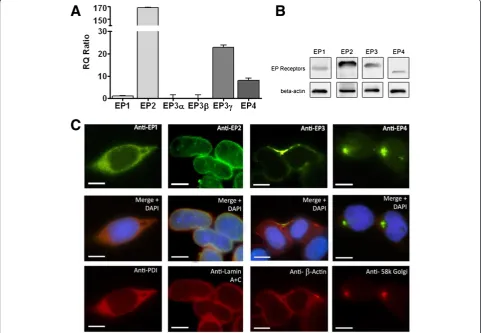
To determine whether NE-4C cells endogenously ex-press the receptors of PGE2, we performed real-time
immunocytochemistry. Our results show that in NE-4C cells, EP2 had the highest mRNA expression followed by EP3γand EP4 receptors. Endogenous EP1 and EP3β receptor expression was considerably low in NE-4C cells, while the EP3αtranscript level was nearly absent and may be considered negligible. The relative quantity (RQ = 1) values of EP1, EP2, EP3α, EP3β, EP3γ, and EP4 transcripts expression were 3, 542, 0, 1, 391, and 15, respectively (Figure 1A). Western blot results confirm the expression of all four EP receptors in NE-4C cells (Figure 1B). The localization of the EP receptors in NE-4C cells was also detected with immunocytochemistry using EP1-4 specific antibodies along with antibodies against various cellular organelles including the nuclear envelope, Golgi apparatus, the endoplasmic reticulum, andβ-Actin (Figure 1C). Our results show that EP1 re-ceptors were localized in the ER membrane, EP2 recep-tors were uniformly expressed around the nucleus and co-localized with the nuclear envelope marker, EP3 re-ceptors were located at the plasma membrane, and EP4
receptors at the Golgi apparatus. Hence, NE-4C cells can act as an appropriate experimental model to study PGE2signalling.
Prostaglandin E2increases the cell motility of
Wnt-induced NE-4C cell migration
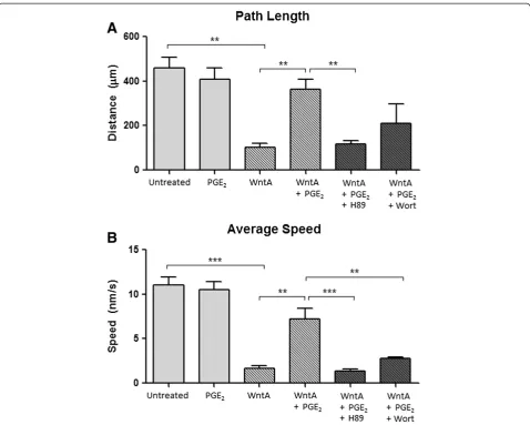
The effect of PGE2 on Wnt-dependent migration of
NE-4C cells was determined using Nikon Eclipse Ti-E microscope with NIS Elements time-lapse tracking soft-ware over a 24 hour period. Final distance, path length, and average speed were quantified after exposure to 1μM PGE2, 2 μM Wnt Agonist (WntA), or 2 μM WntA with
the addition of 1μM PGE2. Thefinal distancewas defined
as the distance between the initial and final positions of the cell, represented as a straight line distance. Thepath lengthwas the total distance travelled from the initial to the final cell position. Theaverage speedof a cell was cal-culated by dividing the total distance travelled by the time it took to travel between the two positions.
The results show that untreated NE-4C cells moved an average final distance of 65.6μm following a 24 hour period (Figure 2A). The addition of PGE2 to the cells
resulted in a final distance of 56.2 μm which was not significantly different from the untreated control (65.6μm). WntA only treatment resulted in a significant decrease in final distance of 21.3μm (p= 0.00242) when compared to the control. The addition of PGE2 to
WntA-treated cells resulted in a final distance of 45.0μm, which is an increase by 23.6μm (p= 0.04371), as compared to WntA only-treated cells. It represents a 211% increase from the WntA-regulated movement. Visualization of final distance through dispersion XY position plots clearly illustrates that PGE2 signalling
restores the Wnt-regulated suppression of cell move-ment (Figure 2B, WntA + PGE2).
The quantification of path length (Figure 3A) revealed the same pattern. The path length of untreated cells was 458.9 μm. As compared to untreated cells, PGE2 only
treatment did not result in a significant change (408.6μm), but WntA treatment significantly decreased the path length to 103.3 μm (p= 0.00189). Addition of PGE2to
WntA-treated cells led to a path length of 362.1 μm. This is a 350% increase (p= 0.00928) compared to WntA only-treated cells.
Quantification of average speed showed that PGE2
treated cells travelled at a speed of 10.5 nm/s, which was not significantly different from untreated NE-4C cells that moved at a speed of 11.0 nm/s (Figure 3B). WntA only treatment resulted in a decreased average cell speed of 1.65 nm/s (p= 0.00065). Addition of PGE2to
WntA-treated cells resulted in an average speed of 7.34 nm/s.
Figure 2PGE2-dependent effect on final distance travelled from origin. (A)Final distance from origin was 65.6, 56.2, 21.3, 45.0μm,
respectively. The error bars represent + SEM and values were considered significant at *p< 0.05, **p< 0.01.(B)The Dispersion XY position plots illustrate the effect of PGE2on Wnt-induced behaviour, where addition of PGE2to Wnt-activated cells increased the final distance. Addition of
H89 (PKA blocker) and Wort (PI-3K) blocker reduces the effect PGE2. Measurements represent an average of 150 cells from three independent
This suggests that addition of PGE2elevated the average
speed by 439%; an increase of 5.59 nm/s (p= 0.00946) when compared to WntA only-treatment.
In summary, administration of PGE2 treatment leads
to significant changes in WntA-regulated cell behaviours such as final distance, path length, and average speed. PGE2 treatment significantly restored the cell kinematic
measures which were suppressed by WntA treatment.
Prostaglandin E2modulates Wnt-induced cell behaviour
through PKA and PI-3K kinases
Previous studies in embryonic kidney and colon cancer cells determined that the convergence of PGE2signalling
on the Wnt pathway occurred through the activation of PKA or PI-3K [44-46]. To determine whether PGE2
treatment alters Wnt-induced cell migration behaviour via these kinases in NE-4C cells, we used dihydrochloride hydrate (H89) to block PKA and Wortmannin (Wort) to
block PI-3K. Our results show a trend across final dis-tance, path length, and average speed (Figures 2 and 3). With the addition of H89 to WntA + PGE2treated cells,
all cell motility measures significantly decreased compared to the WntA + PGE2treated cells, resulting in movement
profiles that were not statistically different from the WntA-only condition. Specifically, H89-treated cells travelled a final distance of 20.32 μm from the origin (p= 0.02477), path length of 116.01 μm (p= 0.00567), and at an average speed of 1.37 nm/s (p= 0.00073) (Figure 2A and 2B).
With the addition of Wort to WntA + PGE2 treated
cells, there was a decreasing trend in final distance and path length but it was not significantly different from PGE2+ WntA treated cells. Only average speed
signifi-cantly decreased to 2.76 nm/s (N= 3; p= 0.00422) com-pared to the WntA + PGE2treatment. Post hoc Dunnett
Wort conditions were not significantly different from the WntA-only treatment, indicating that H89 and Wort significantly diminished the effect of PGE2 on
WntA-treated cells. This indicates that PGE2likely acts through
PKA and PI-3K to elicit effects on the WntA-dependent cell motility. However, it appears that H89 may have had a greater effect, suggesting that PGE2may predominately
act through PKA.
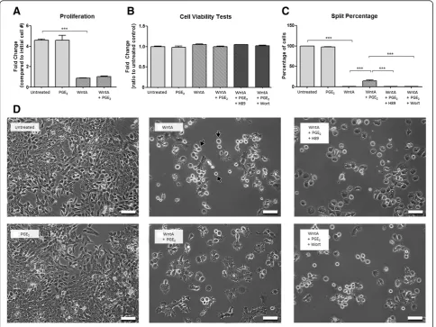
Prostaglandin E2alters cell proliferation behaviour of
NE-4C cells induced by Wnt agonist treatment
Previous literature reveals that PGE2may also affect cell
proliferation via the Wnt signalling pathway in prostate and colon cancer cells [37] and hematopoietic [38] and mesenchymal [39] stem cells. We studied the effects of PGE2 on NE-4C cell proliferation using NIS Elements
software. The cells were exposed to 1 μM PGE2, 2μM
WntA, or 2μM WntA with the addition of 1μM PGE2.
Furthermore, H89 or Wort was added to PGE2+ WntA
treated cells to determine the effective role of these kinases. The initial number of cells was compared to the final number of cells following 24 hours treatment. PGE2 treatment led to an increase in cell number by
4.60-fold, which was not significantly different from the untreated cells that proliferated by a 4.59 fold-increase (Figure 4A). Administration of WntA resulted in a fold-change of 0.86 (p< 0.001) which was significantly lower than untreated cells. Addition of PGE2to WntA-treated
cells (WntA + PGE2) resulted in a fold-change of 1.03,
which was not significant from the WntA only treated condition. Although we observed lower levels of prolif-eration in the WntA, WntA + PGE2and WntA + PGE2+
Blocker conditions, we confirmed no change in cell viabil-ity between the conditions tested (Figure 4B).
Figure 4PGE2-dependent effect on proliferation behaviour. (A)Over the experimental duration of 24 hours, the number of cells changed by
However, we observed distinct differences in cell pheno-type between the WntA, WntA + PGE2and WntA + PGE2
with H89 or Wort treatment. A majority of the cells treated with WntA adopted a shiny circular shape (indi-cated by black arrows, Figure 4D). This was not as preva-lent in the WntA + PGE2 condition. However, the cells
treated with WntA + PGE2and Wort blocker, adopted the
shiny circular phenotype seen in the WntA condition. Cells treated with WntA + PGE2and H89 blocker adopted
a circular appearance as well but a smaller population of these round cells were shiny.
Our experiments showed that cell viability was not affected but a distinct shiny circular cell appearance was observed, which is characteristic of a cell just prior to splitting into two daughter cells. Therefore, we also quantified thesplit percentage, defined as the percentage of cells that successfully divided into two daughter cells during the recorded time period. As expected, the NE-4C untreated cells demonstrated a split percentage of 100% (Figure 4C), indicating that all cells entering a mitotic phase resulted in cell division. A similar pattern was seen in PGE2-treated cells (97.5%). However, treatment of
WntA resulted in a significant decrease of split percentage to 0% (p< 0.001), where mitotic cells appeared to become arrested in a round stage denoted in Figure 4D (WntA Image) with black arrows. The addition of 1μM PGE2to
WntA-treated cells produced a significant increase in split percentage to 14.7% (p< 0.001, Figure 4C) as compared to
WntA only treatment. The cells appear to resume their flat morphology. These results suggest that PGE2
treat-ment can modify Wnt-induced proliferation behaviour such as split percentage. Following treatment with either H89 or Wort, cells returned to a split percentage of 0% as seen with WntA only treatment (Figure 4C, D). This again indicates that PGE2 likely acts on the Wnt pathway
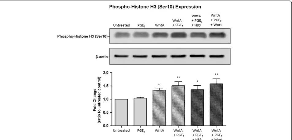
through PKA and PI-3K to modify cell proliferation. To further confirm our results of the cell splitting behav-iour, we measured the level of Phospho-Histone H3 (Ser10) (Figure 5) since phosphorylation at Ser10 is tightly associ-ated with chromosome condensation and segregation that occurs during mitosis [47-49]. Compared to untreated cells, PGE2only-treated cells did not display a significant
differ-ence. However, when compared to untreated NE-4C cells, cells treated with WntA, WntA + PGE2and WntA + PGE2
with H89 or Wort treatment led to a significance increase in Phospho-Histone H3 (Ser10) expression. RQ values were 1.35 (p= 0.033), 1.52 (p= 0.001), 1.36 (p= 0.027), and 1.58 (p= 0.005), respectively. This revealed that although cell numbers were lower under these conditions, the relative expression of Phospho-Histone H3 (Ser10) was significantly higher, indicating that a greater percentage of cells were undergoing mitosis when exposed to these treatments com-pared to untreated cells. This correlates with our finding that a larger proportion of cells under these conditions adopts and seems to be arrested in a round stage charac-teristic of cells undergoing mitosis.
Figure 5PGE2-dependent effect on phospho-histone H3 (Ser10) expression.Western blot analysis was used to determine Phospho-Histone
Prostaglandin E2increases activeβ-catenin expression in
Wnt-induced NE-4C cells
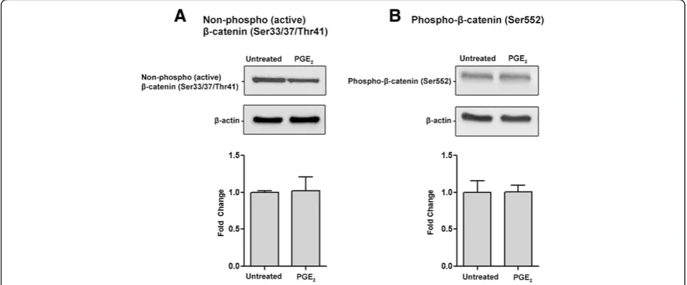
β-catenin is a key effector in the canonical Wnt signalling pathway that regulates downstream gene transcription [50].β-catenin levels can be intricately regulated at mul-tiple phosphorylation sites. Phosphorylation at Ser33, Ser37, and Thr41 leads to its destabilization and primes it for degradation [51], while phosphorylation at Ser552 has been correlated with β-catenin nuclear accumulation [52,53]. We tested the levels of non-phospho-(the active form)β-catenin (Ser33/37/Thr41) and phospho-β-catenin (Ser552). The addition of PGE2 only to NE-4C cells did
not significantly change the levels of either form of β-catenin (Figure 6A and B). However, adding PGE2 to
WntA-induced NE-4C cells lead to a significant 2.1 fold increase in non-phospho-(active) β-catenin (Ser33/37/ Thr41) levels compared to the WntA only treated condi-tion (Figure 7A). There was no significant difference in Phospho-β-catenin (Ser552) levels between the sample conditions (Figure 7B), suggesting that phosphorylation of β-catenin at Ser552 is likely not involved with the behav-ioural differences in NE-4C cells described earlier. These results indicate that PGE2may interact with the canonical
Wnt signalling pathway by regulation of non-phospho-(active)β-catenin (Ser33/37/Thr41) levels.
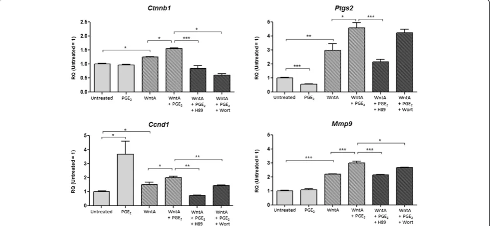
Prostaglandin E2regulates expression of Wnt-target
genes in Wnt-induced NE-4C cells
To investigate whether the addition of PGE2can influence
gene transcription relevant to the canonical Wnt pathway, we screened 29 target genes using Custom TaqMan® Array
Plates. We found that Ctnnb1, Ptgs2, Ccnd1, and Mmp9 were differentially regulated (data not shown). Their expression was confirmed with real-time PCR using RNA derived from the same treatment conditions used for be-havioural analyses, which includes 1μM PGE2, 2μM Wnt
Agonist (WntA), or 2μM WntA with the addition of 1μM PGE2. Kinase blockers (H89 or Wort) were added to PGE2
+ WntA treated cells to determine the potential contribu-tion of PKA and PI3K activity via PGE2 signalling. Our
real-time PCR results indicate that PGE2affects the
expres-sion levels of all Wnt-target genes tested (Figure 8). Ctnnb1 (beta-catenin) levels were not altered with the addition of PGE2 when compared to untreated NE-4C
cells, but cells treated with WntA showed a significant increase of RQ value 1.25 (p= 0.0372). Addition of PGE2
to WntA-induced cells led to a further increase ofCtnnb1 level to an RQ value of 1.55, which was significantly dif-ferent from the WntA-only condition (p= 0.0131). This pattern was consistent with the expression of phospho (active) β-catenin (Ser33/37/Thr41) protein quantified earlier using Western blot analysis. Addition of H89 or Wort to PGE2+ WntA treated cells resulted in RQ values
to 0.83 and 0.60, respectively, compared to untreated cells which was a significant decrease compared to the PGE2+
WntA condition (p> 0.001,p> 0.001). The PKA and PI3K blockers, H89 and Wort, appeared to remove the effect of PGE2on Ctnnb1 expression in WntA-induced cells,
while also reversing the influence onCtnnb1levels from WntA-only treatment. This suggests that PKA and PI3K signalling may modify Ctnnb1 expression through PGE2
signalling.
Figure 6PGE2-dependent effect onβ-catenin expression in NE-4C cells.Western blot analysis was used to determine two forms of active
β-catenin:(A)non-phospho-(active)β-catenin (Ser33/37/Thr41) and(B)phospho-β-catenin (Ser552) (92 kDa). Addition of PGE2to NE-4C cells did
NE-4C cells treated with PGE2alone had a significant
decrease in Ptgs2 (prostaglandin-endoperoxide synthase 2; gene encoding COX-2) mRNA levels compared to un-treated cells (RQ = 0.56, p< 0.001), while cells treated with WntA had a significant increase of RQ value 2.99
(p= 0.00286). In contrast, when PGE2 was added to
WntA-induced NE-4C cells, Ptgs2 expression was fur-ther elevated with an RQ value of 4.59 compared to untreated. This value was significantly different from the PGE2+ WntA condition (p= 0.015). Addition of H89 or
Figure 7PGE2-dependent effect onβ-catenin expression in Wnt-activated NE-4C cells.Western blot analysis was used to determine two
forms of activeβ-catenin: non-phospho-(active)β-catenin (Ser33/37/Thr41) and phospho-β-catenin (Ser552) (92 kDa).(A)The expression of active
β-catenin represented in fold change was 1, 2.09, 1.61, and 1.98, respectively. The error bars represent + SEM and values were considered significantly different from control at *p< 0.05. Only PGE2+ WntA condition was significantly different from WntA only condition.(B)There was no significant
difference in phospho-β-catenin (Ser552) expression between the conditions. Average measurements represent protein from three independ-ent experimindepend-ents (N= 3).β-Actin was used to indicate equal loading.
Figure 8PGE2-dependent effect on Wnt-target genes.Real-time PCR was used to determine the RQ value forCtnnb1,Ptgs2,Ccnd1, and
Mmp9. The expression ofCtnnb1represented in fold change was 1, 0.97, 1.25, 1.55, 0.84, and 0.60, respectively. The fold change expression of
Wort to PGE2+ WntA treated cells resulted in RQ
values of 2.16 and 4.22, but only the H89 treatment was significantly different from the PGE2+ WntA condition
(p< 0.001). This suggests that the effect of PGE2 on
WntA-induced cells may be through PKA.
Expression ofCcnd1(cyclin D1) was also altered. Ad-ministration of PGE2 treatment to NE-4C cells
corre-lated with a significant increase of an RQ value to 3.68 (p= 0.045) compared to untreated cells, while WntA-treated cells had a significant increase of RQ value to 1.50 (p= 0.048). Addition of PGE2to WntA-activated cells was
associated with a further increase of Ccnd1 expression, with an RQ value 1.99 compared to untreated cells, which was significantly different from WntA-only treated cells (p= 0.047). H89 or Wort added to PGE2+ WntA treated
cells had RQ values of 0.74 and 1.42, respectively, which was significantly different from the PGE2+ WntA
condi-tion (p= 0.0054,p= 0.0078). The blockers, H89 and Wort, seemed to attenuate the increase of Ccnd1 levels associ-ated with the addition of PGE2to WntA-induced cells.
In comparison to untreated NE-4C cells, PGE2
treat-ment did not change levels ofMmp9(matrix metallopro-teinase 9). However, when compared to WntA-induced NE-4C cells, addition of PGE2treatment to WntA-treated
cells caused a significant increase in expression level (p< 0.001). Specifically, with WntA treatment, Mmp9 expression was significantly elevated to an RQ value of 2.19 (p < 0.001) compared to untreated cells, but addition of PGE2to WntA-induced cells resulted in a further rise
ofMmp9 expression with an RQ value of 3.00. H89 and Wort were added to PGE2+ WntA treated cells and RQ
values forMmp9were 2.16 and 2.68, respectively, com-pared to the untreated condition. These values were sig-nificantly different from the PGE2+ WntA condition.
This indicates that the use of H89 and Wort diminished the increase in Mmp9 expression as a result of PGE2
treatment on WntA-induced cells.
Overall, these results demonstrate that PGE2can raise
the expression of Wnt-target genes, specifically,Ctnnb1, Ptgs2,Ccnd1, andMmp9, in WntA-induced NE-4C cells. Since H89 and Wort attenuated the changes caused by PGE2, PKA and PI3K likely serve as a molecular link for
the interaction between the PGE2 and canonical Wnt
signalling pathways.
Discussion
Cell migration and proliferation are crucial components of neural development. Previous studies have shown that elevated levels of PGE2can result in increased cell motility
and proliferation in various non-neuronal cells [46,54-56]. Recent evidence indicates that abnormalities in cell be-haviour can result from the interaction between PGE2
with Wnt signalling pathways [44,57]. Our current study provides evidence, for the first time, for the
cross-talk between these two pathways in neural stem cells. We report that PGE2 treatment elicits changes in cell
behaviour such as an increase in components of cell motility and proliferation, as well as expression of Wnt-target genes, in Wnt-activated NE-4C stem cells. More-over, the stimulatory effects of PGE2are subdued through
the inhibition of downstream pathway kinases, PKA and PI-3K, suggesting that PGE2acts through these particular
kinases to converge with the Wnt pathway.
Previous studies have shown that PGE2 can increase
or decrease the activity of canonical Wnt signalling. PGE2activates several components of the canonical Wnt
pathway in colorectal cancer cells (reviewed in [42]). Specifically in these cells, PGE2stimulated a significant
increase in the activity of Wnt transcription factors, T cell factor-4 (Tcf-4), as well as elevated protein levels of Wnt-target genes [58]. PGE2acted through its EP2
recep-tor to modulate β-catenin activity of the Wnt pathway, promoting the growth of colon cancer cells [44]. Wnt activation induced by PGE2also contributed to abnormal
proliferation resulting in enhanced gastric tumorigenesis [57]. Furthermore, PGE2–regulated Wnt signalling had a
hepatoprotective effect, aiding in liver regeneration [59]. In pre-osteoblastic cells, concentration-dependent treat-ment of PGE2 modulated Wnt signalling by altering
protein expression of pathway activators,β-catenin and low-density lipoprotein receptor-related protein 5/6 (LRP 5/6), as well as Wnt inhibitor, dickkopf-1 (DKK-1); low doses of PGE2 promoted the Wnt pathway while high
doses inhibited it [37]. PGE2 also modified Tcf-luciferase
activity of Wnt signalling through the same dose effect [37]. Additionally, in human colorectal adenoma and car-cinoma cells, PGE2treatment up-regulated the protein
ex-pression of the Wnt target gene, leucine-rich G-protein coupled receptor 5 (LGR5), which internalizes FZD co-receptor LRP6 and decreases Wnt activity [60]. Altogether, these studies reveal that the interaction between PGE2and
Wnt signalling can have different effects depending on the dose of PGE2administered and the specific cell type.
We reveal that PGE2 increases the final distance and
path length travelled, as well as the average speed of mi-gration in Wnt-activated neuroectodermal stem NE-4C cells. We also show that PGE2 alters the phenotype of
Our study adds to the current body of research by showing that PGE2interferes with the Wnt pathway by
attenuating Wnt-dependent cell behaviour in NE-4C cells. This is important because Wnt signalling is involved in a myriad of regulatory processes important for the develop-ment and organization of the nervous system [64]. It is thoroughly established that Wnt signalling is instrumental to normal anterior-posterior patterning of the embryo [65]. Wnt proteins are key regulators for the formation of the neural tube, as well as neuronal migration and differ-entiation [40,64]. Wnt signalling also modulates neurite outgrowth [66], axon growth and guidance [67-70], den-dritic development and arborization [71,72], radial migra-tion [73], and synapse formamigra-tion and plasticity [74,75]. Moreover, Wnt signalling is crucial in neuronal fate de-termination, particularly in the specification and differ-entiation of neuronal precursors in the midbrain [76] and forebrain [77,78]. Furthermore, epithelial stem cells require Wnt/β-catenin signalling for proliferation and quiescent division [79] and the balance between re-entry and exit of the cell cycle can be altered by Wnt/β-catenin signalling [80]. Additionally, aberrant cortical progenitor cell proliferation patterns and defective hippocampus de-velopment can result due to abnormal Wnt signalling [81]. Interestingly, recent findings provide evidence that defective Wnt signalling could contribute to the pathogen-esis of psychiatric disorders like schizophrenia and ASD [82-84]. Specifically, Wnt2, located in the putative speech and language region at chromosome 7q31-33, has been identified as a susceptibility gene for autism. [85,86]. Given the importance of Wnt signalling in pre-natal development and the existing interaction between Wnt and PGE2 pathways in NE-4C stem cells,
alter-ations in levels of PGE2via endogenous and exogenous
means may have profound effects on nervous system development.
In addition to quantifying cell behaviour, we also demon-strate that PGE2can affect the expression of non-phospho
(active)β-catenin (Ser33/37/Thr41). Wnt/β-catenin signal-ling occurs through a complex, highly regulated pathway that involves the phosphorylation of multiple sites on β-catenin, which may promote its degradation or activa-tion and subsequent nuclear internalizaactiva-tion. For instance, the phosphorylation of sites Ser33, 37, and Thr41 targets β-catenin for ubiquitination and proteasomal degrada-tion [87,88]. Quantificadegrada-tion of β-catenin that is non-phosphorylated at these sites has become a common measurement for active or stabilizedβ-catenin expression. Phosphorylation ofβ-catenin at the site Ser552 has also been correlated with increased β-catenin/TCF medi-ated transcriptional activity [89,90]. We found that PGE2
treatment administered to Wnt-activated cells increased the expression of non-phospho (active) β-catenin (Ser33/37/Thr41) protein. In contrast, the phospho- β
-catenin (Ser552) levels remained unchanged. It has been established that the regulation of glycogen synthase kin-ase 3 beta (GSK3β) activity may control stabilization of β-catenin and increased levels of non-phospho (active) β-catenin (Ser33/37/Thr41) protein [91]. It is possible that PGE2signalling may modify GSK3βactivity but this
remains to be determined. Nonetheless, the increased levels of non-phospho (active)β-catenin (Ser33/37/Thr41) quantified were in line with our gene expression results that also showed an increase inCtnnb1expression as well as other Wnt-target genes. Ctnnb1 encodes for the β-catenin protein, which can regulate cell growth and ad-hesion and is also a key downstream component of the ca-nonical Wnt pathway. It has also been shown to regulate cortical size; enlarged cortices with increased cortical folds were observed in Ctnnb1 transgenic mice [80]. Interest-ingly, brain overgrowth and abnormal excess in number of neurons was measured in children with autism [92]. Gene expression of Ctnnb1 was altered in both young and adult autistic cases [93]. Furthermore, de novo mu-tations of this gene and its relevant network have been ranked significantly as potential autism candidate genes [94,95]. Within the canonical Wnt pathway, the β-catenin/TCF complex can promote the transcription of target genes including Ptgs2[96],Ccnd1[97,98], and Mmp9 [99,100]. Expression of these genes was in-creased as an effect of elevated PGE2signalling in our
study, and interestingly, previous studies have reported a link between these genes and ASD as described below. Ptgs2, also known as COX-2, is the key enzyme in prostaglandin biosynthesis, including the production of PGE2. COX-2 is a crucial mediator of inflammation and
prostanoid signalling [101,102]. Polymorphism of Ptgs2 has been associated with ASD [103]. A recent clinical study proved the efficacy of a COX-2 inhibitor drug, cele-coxib, as an adjunctive therapy in the treatment of autism: the treatment was superior for treating irritability, social withdrawal, and stereotypy of children with autism [104].
Another gene affected was Ccnd1. This gene encodes for a protein in the cyclin family, which are important regulators in cell cycle progression, transcription, and neur-onal function [105,106]. The increased levels of Ccnd1, as a result of added PGE2, may be involved with the
Diminished expression of 22q11 genes, which disrupts cortical neurogenesis and cell migration, led to alterations inCcnd1levels [109]. The authors explain that a develop-mental disruption, as such, may alter cortical circuitry and establish vulnerability for developmental disorders, includ-ing schizophrenia and autism.
Mmp9is a membrane of the matrix metalloproteinase (MMP) family, which can target many extracellular pro-teins including proteases, growth factors, and adhesion molecules [110] and are involved with the breakdown of the extracellular matrix in normal physiological processes such as embryonic development and tissue remodelling [111]. MMPs are also important in neuronal development, plasticity, and maintenance of neuronal health [112]. Mmp9 has also been shown to regulate the proliferation and migration of embryonic neural stem cells [99] and participate in neuronal differentiation by regulating neur-ite elongation and neuronal cell migration [113-115]. Therefore, altered Mmp9 expression may contribute to the deviant behaviour observed in our study. Mmp9 has also been associated with ASD [116]. Elevated levels of MMP9 protein were found in the amniotic fluid of ASD cases compared to controls [117]. Findings from this study provide evidence that molecular and physiological abnor-malities in ASD may begin prenatally. Mmp9 has also been implicated in Fragile X syndrome (FXS) [118], which is characterized by behaviours at the extreme of the autistic spectrum. Using in a mouse model of fragile x (Fmr1 KO), levels of MMP9 was found to be elevated in the hippocampus of Fmr1 KO mice [119]. Furthermore, Minocycline, a drug that inhibits MMP9 activity, has been shown to promote dendrite spine maturation and improve behavioural performance in Fmr1 KO mice [119]. These researchers continued their work in human trials and found that Minocycline taken as a daily dose for 8 weeks led to behavioural improvements in FXS patients. This was consistent with their fmr1 KO mouse model results, indicating that MMP9 activity alters underlying neural defects that contribute to behavioural abnormalities seen in ASD [120].
Taken altogether, our gene expression results not only show a potential interaction of the PGE2and canonical
Wnt pathway in the nervous system, but also provide further evidence for a link to ASD.
We show that PGE2interacts with canonical Wnt
sig-nalling through PKA and PI-3K to produce the reported behavioural changes in cell motility and proliferation, as well as gene expression. Specifically, we found that inhibit-ing these PGE2 downstream pathway kinases, PKA and
PI-3K with H89 and Wort respectively, reduced the effect of PGE2. This is in line with previous literature, which
found that the convergence of PGE2-dependent effects
and the Wnt pathway can occur through the stimulation of PKA or PI-3K in embryonic kidney cells and colon
cancer cells [44-46]. Moreover, similar stimulatory effects on cell migration induced by PGE2in Wnt-activated
NE-4C cells from our study were also exhibited in prostate cancer cells through the activation of PI-3K [121]. Our re-sults revealed that H89 had a stronger effect than Wort, suggesting that PGE2 may predominately act through
PKA; but future studies are needed to determine which EP receptors are involved. A proposed model is provided in Figure 9.
Increasing evidence for the contribution of environ-mental factors in the etiology of neurodevelopenviron-mental disorders like ASD has prompted urgency to reveal their potential exogenous causes and underlying mechanisms [122]. Environmental factors like exposure to drugs, toxins or infectious agents cause disruptions in PGE2signalling
by increasing the levels of oxidative stress, consequent lipid peroxidation, and the immunological response; these factors and consequences that disturb normal PGE2
sig-nalling have all been linked to ASD [123]. We show that increased PGE2 signalling can modify cell migration,
proliferation behaviour, and gene expression in Wnt-activated NE-4C stem cells. Aberrant cell migration and proliferation are pathophysiologic mechanisms that im-pact the brain broadly, and could be possible factors that contribute to the origination of neurodevelopment disorders. Abnormalities in structure, organization, and connectivity of the brain are all indicators of irregular neural cell migration and proliferation. Local distortions in neural cytoarchitecture, dysplasia, and hypoplasia have been described in brains of autistic subjects [124]. Moreover, structural abnormalities and atypical con-nectivity of the brain in ASD has been identified by a number of research groups [123,125-128]. Noteworthy, areas of the brain that would be most impacted by dysregulation in neuronal migration and proliferation— that is the cerebellum, cerebral cortex, and hippocampus— are also implicated in ASD [124,129-131]. Despite the assumptions that can be made from our in vitro results, in vivo models must be employed to further describe the possible effects of PGE2 and its interaction with
morphogenic signalling pathways, such as Wnt, during prenatal development.
Conclusions
PGE2 is an important bioactive lipid signalling
mol-ecule and its interaction with Wnt signalling pathway could have significant effects on prenatal development. Our study shows for the first time that PGE2can affect
Wnt-dependent cell behaviours and gene expression in neuroectodermal stem cells through PKA and PI-3K. Aberrant PGE2and Wnt signalling have been linked to
Our in vitrostudy provided further evidence that these aberrations may be potential mechanisms in the genesis of neurodevelopment disorders like ASD.
Methods Cell culture
Mouse NE-4C cells were obtained from American Tissue Culture Collection (ATCC) and grown in Minimum Essential Medium (MEM) supplemented with 10% fetal bovine serum, 2 mM glutamine, 1 X penicillin-streptomycin mixture (Invitrogen). Cells were maintained in an incu-bator containing 5% CO2at 95% humidity 37°C. Cells
were plated on 0.01% poly-L-lysine (Sigma) coated 100 mm culture plates (BD Falcon) and were subcul-tured at a 1:10 ratio. Supplemented MEM was changed every 2–3 days.
Cell culture-treatments
NE-4C cells (ATCC) were dissociated with 0.05% trypsin-EDTA, pelleted and resuspended in complete medium as described above. The cells were plated on poly-L-lysine 0.01% (Sigma, MW 70000–150000 kDa) on 35 mm tissue culture dishes (Sarstedt). Plated cells were incubated in 5%
CO2 at 95% humidity 37°C overnight before treatment
with Wnt Agonist (WntA, Calbiochem), prostaglandin E2(PGE2, Sigma) and/or blockers. WntA (2μM), PGE2
(1 μM), H89 dihydrochloride hydrate (H89, 10 μM, Sigma), Wortmannin (WORT, 1 μM, Sigma) or an equivalent volume of vehicle were added to each well. Cells were treated for 24 hours.
Reverse transcription and real-time PCR
Total RNA was extracted from NE-4C cells using the NucleoSpin RNA/Protein Kit (Macherey-Nagel) and was reverse-transcribed into cDNA using MMuLV reverse transcriptase (New England Biolabs) according to manu-facturer’s instructions. Primer3 Input software (v. 0.4.0) was used to design forward and reverse primers for EP receptors and have been previously noted [12]. Selection of Wnt-target genes was determined using Custom TaqMan® Array Plates (Life Technologies) as a screening tool (data not shown). Genes that had a greater than 1.8 fold-change were selected for further validation and forward and reverse primers were designed (Table 1). Real-time PCR was performed using the 7500 Fast Real-time PCR system (Applied Biosystems) and the ΔΔCT method was applied
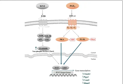
Figure 9A proposed model for PGE2-Wnt interactions in Wnt-induced NE-4C cells.From the compilation of our results (bolded) and other
to calculate the expression of transcripts. Hypoxanthine phosphoribosyl transferase (HPRT) and Phosphoglycerate Kinase 1 (PGK1) served as endogenous controls. The rela-tive quantification (RQ) ratios were determined from the average of three technical replicates from three biological replicates.
Western blot analysis
Total protein was extracted from NE-4C cells using the NucleoSpin RNA/Protein Kit (Macherey-Nagel). Samples were separated by polyacrylamide gel electrophoresis (PAGE). Primary antibodies used for EP expression levels include rabbit polyclonal anti-EP1, −EP2, −EP3, −EP4 (1:200; Santa Cruz Biotechnology). Detection of rabbit monoclonal anti-Phospho-Histone H3 (Ser10) (1:1000; Cell Signaling) was used as a measure of cell splitting be-haviour. Primary antibodies used forβ-catenin expression levels were rabbit monoclonal anti-non-phospho (Active) β-catenin (Ser33/37/Thr41) and rabbit polyclonal anti-phospho-β-catenin (Ser552) (1:1000; Cell Signaling). Blots were reprobed with mouse monoclonal anti-β-Actin (1:10,000; Abcam). Visualization of bound anti-rabbit and anti-mouse horseradish peroxidise-conjugated secondary antibodies was achieved by incubation with ECL Prime Western Blotting Detection Reagent (GE Healthcare) and detection by Geliance 600 Imaging System (Perkin Elmer).
Immunocytochemistry
NE-4C cells were seeded onto 35 mm culture plates containing poly-L-lysine coated coverslips and grown overnight at 37°C. The cells were fixed with 50% acetone and 50% methanol for 20 minutes at−20°C and washed with phosphate buffered saline (4.3 mM Na2HPO4,
137 mM NaCl, 2.7 mM KCl, 1.4 mM KH2PO4). Cells
were then incubated with primary antibodies in PBS
with 0.3% Triton-X 100 and 2% Normal Goat Serum. Cellular localization of the EP receptors was determined by incubation with anti-EP primary antibodies as de-scribed above along with mouse monoclonal anti-Lamin A + C nuclear envelope marker (1:200; Abcam), anti-58 K Golgi marker [anti-58 K-9] (1:100; Abcam), anti-PDI endoplasmic reticulum marker [RL90] (1:100; Abcam) or
β-Actin (1:1000; Abcam) at room temperature for 1 hour. Following primary antibody incubation, cells were washed three times with PBS-T for 15 min and incubated with secondary antibodies in PBS-T and 2% NGS for 1 hour at room temperature in the dark. Secondary antibodies used were anti-rabbit fluorescein isothiocyanate (FITC) (1:100; Jackson ImmunoResearch Laboratories) and anti-mouse Texas Red (1:200; Jackson ImmunoResearch Laboratories). Cells were then washed twice with PBS-T for 10 min, followed by a 20 minute incubation of 4′,6-diamidino-2-phenylindole (DAPI) (1:2000; Molecular Probes) at room temperature. Cells were washed twice with PBS-T for 5 min and coverslips were mounted on glass micro-scope slides with mounting media (Vectashield). The staining was visualized and captured using an Eclipse 80i upright fluorescent microscope with DS-5MC camera (Nikon).
Time-lapse imaging and analysis
Cell behaviour was recorded using Nikon Eclipse Ti-E microscope. Three biological replicates of each treatment condition were performed (N = 3), where an average of 150 cells were tracked. Micrographs were automatically captured every 10 minutes for a 24 hour period from a minimum of three fields. To maintain conditions physiolo-gically suitable for the cells, an enclosed chamber was mounted to the microscope, which was equipped with CO2supply and temperature thermostat. Cells were kept
at 5% CO2, 95% humidity, 37°C. Measurements were
com-pleted using NIS Elements software, including a special-ized tracking module. Final distance from origin, path length, and average speed were tracked and calculated from an average of 150 cells per treatment condition. Ini-tial and final cell counts were used to determine fold change as a measurement of proliferation. Split percentage was quantified as a measurement of proliferation behav-iour. Split percentage was defined as the percentage of cells that fulfilled the complete cell cycle, which was evalu-ated based on whether the parent cell could successfully split into two daughter cells.
Cell viability analysis
Cells were disassociated and diluted with equal volumes of trypan blue dye (4%). Cell count averages were taken from a minimum of four hemacytometer squares to deter-mine cell number and viability.
Table 1 Forward and reverse primer sequences for real-time-PCR
Gene Primer sequences(5′→3′) Amplicon size (base pair)
Hprt F: TCCATTCCTATGACTGTAGATTTTATCAG 75
R: AACTTTTATGTCCCCCGTTGACT
Pgkl F: CAGTTGCTGCTGAACTCAAATCTC 65
R: GCCCACACAATCCTTCAAGAA
Ptgs2 F: CAGCCAGGCAGCAAATCC 81 R: TTATACTGGTCAAATCCTGTGCTCAT
Ctnnbl F: GGACGTTCACAACCGGATTG 71 R: GAGAATAAAGCAACTG CACAAACAA
Ccndl F: GCACTTTCTTTCCAGAGTCATCAA 79 R: CTCCAGAAGGGCTTCAATCTGT
Mmp9 F: TCGCGTGGATAAGGAGTTCTCT 73
Statistical analysis
All numerical data were presented as mean + SEM of three individual experiments. Statistical analysis was performed using student t-test or one-way analysis of variance (ANOVA) followed by Tukey post-hoc compar-isons or Dunnett t-test (2-sided). Differences were con-sidered statistically significant at *p< 0.05, **p< 0.01, or ***p< 0.001.
Abbreviations
ASD:Autism spectrum disorders; COX-1,-2: Cyclooxygenases−1 and−2; Ccnd1: Cyclin D1; Ctnnb1: Beta-catenin; EP1-4: E-prostanoid 1 through 4; H89: Dihydrochloride hydrate; Mmp9: Matrix metalloproteinase 9; NE-4C stem cell: Neuroectrodermal stem cells; PGE2: Prostaglandin E2;
PI-3K: Phosphatidylinositide 3-kinases; PKA: Protein kinase A; PKC: Protein kinase C; Ptgs2: Prostaglandin-endoperoxide synthase 2; Wnt: Wingless-related MMTV integration site; WntA: Wnt agonist; Wort: Wortmannin.
Competing interests
The authors declare that they have no competing interests.
Authors’contributions
CTW designed and performed experiments, acquisition and analysis of data, and wrote the manuscript. EA performed experiments and acquisition of data. HYL assisted with research and discussions. DAC participated in the design and coordination of the study, and was involved in drafting the manuscript. All authors read and approved the final manuscript.
Acknowledgements
This research work was supported by the Natural Sciences and Engineering Research Council of Canada (NSERC).
Author details 1
School of Kinesiology and Health Science, York University, 4700 Keele Street, Toronto, Ontario M3J 1P3, Canada.2Neuroscience Graduate Diploma Program, York University, 4700 Keele Street, Toronto, Ontario M3J 1P3, Canada.3Department of Biology, Faculty of Health, York University, 4700 Keele Street, Toronto, Ontario M3J 1P3, Canada.
Received: 13 September 2013 Accepted: 13 March 2014 Published: 23 March 2014
References
1. Breyer RM, Bagdassarian CK, Myers SA, Breyer MD:Prostanoid receptors: subtypes and signaling.Annu Rev Pharmacol Toxicol2001,41:661–690. 2. Brown AS:Epidemiologic studies of exposure to prenatal infection and
risk of schizophrenia and autism.Dev Neurobiol2012,72(10):1272–1276. 3. Parker-Athill EC, Tan J:Maternal immune activation and autism spectrum
disorder: interleukin-6 signaling as a key mechanistic pathway.
Neurosignals2010,18(2):113–128.
4. Patterson PH:Maternal infection and immune involvement in autism.
Trends Mol Med2011,17(7):389–394.
5. Grandjean P, Landrigan PJ:Developmental neurotoxicity of industrial chemicals.Lancet2006,368(9553):2167–2178.
6. Arndt TL, Stodgell CJ, Rodier PM:The teratology of autism.Int J Dev Neurosci2005,23(2–3):189–199.
7. Sugimoto Y, Narumiya S:Prostaglandin E receptors.J Biol Chem2007,
282(16):11613–11617.
8. Coleman RA, Smith WL, Narumiya S:International Union of Pharmacology classification of prostanoid receptors: properties, distribution, and structure of the receptors and their subtypes.Pharmacol Rev1994,
46(2):205–229.
9. Furuyashiki T, Narumiya S:Stress responses: the contribution of prostaglandin E(2) and its receptors.Nat Rev Endocrinol2011,
7(3):163–175.
10. Andreasson K:Emerging roles of PGE2 receptors in models of neurological disease.Prostaglandins Other Lipid Mediat2010,
91(3–4):104–112.
11. Saint-Dizier M, Guyader-Joly C, Charpigny G, Grimard B, Humblot P, Ponter AA:Expression of enzymes involved in the synthesis of prostaglandin E2 in bovine in vitro-produced embryos.Zygote2011,19(3):277–283. 12. Tamiji J, Crawford DA:Prostaglandin E(2) and misoprostol induce neurite
retraction in Neuro-2a cells.Biochem Biophys Res Commun2010,398(3):450–456. 13. Chen C, Bazan NG:Endogenous PGE2 regulates membrane excitability
and synaptic transmission in hippocampal CA1 pyramidal neurons.
J Neurophysiol2005,93(2):929–941.
14. Dean SL, Wright CL, Hoffman JF, Wang M, Alger BE, McCarthy MM:
Prostaglandin E2 stimulates estradiol synthesis in the cerebellum postnatally with associated effects on Purkinje neuron dendritic arbor and electrophysiological properties.Endocrinology2012,
153(11):5415–5427.
15. Chen C, Bazan NG:Lipid signaling: sleep, synaptic plasticity, and neuroprotection.Prostaglandins Other Lipid Mediat2005,77(1–4):65–76. 16. Shimamura M, Zhou P, Casolla B, Qian L, Capone C, Kurinami H, Iadecola C,
Anrather J:Prostaglandin E type 1 receptors contribute to neuronal apoptosis after transient forebrain ischemia.J Cereb Blood Flow Metab
2013,33(8):1207–1214.
17. Miyagishi H, Kosuge Y, Yoneoka Y, Ozone M, Endo M, Osada N, Ishige K, Kusama-Eguchi K, Ito Y:Prostaglandin E2-induced cell death is mediated by activation of EP2 receptors in motor neuron-like NSC-34 cells.
J Pharmacol Sci2013,121(4):347–350.
18. Choi SY, Choi BH, Suh BC, Chae HD, Kim JS, Shin MJ, Kang SS, Negishi M, Kim KT:Potentiation of PGE(2)-mediated cAMP production during neuronal differentiation of human neuroblastoma SK-N-BE(2)C cells.
J Neurochem2001,79(2):303–310.
19. Goncalves MB, Williams EJ, Yip P, Yanez-Munoz RJ, Williams G, Doherty P:
The COX-2 inhibitors, meloxicam and nimesulide, suppress neurogenesis in the adult mouse brain.Br J Pharmacol2010,159(5):1118–1125. 20. Raisz LG, Pilbeam CC, Fall PM:Prostaglandins: mechanisms of action and
regulation of production in bone.Osteoporos Int1993,3(Suppl 1):136–140. 21. Tjandrawinata RR, Dahiya R, Hughes-Fulford M:Induction of
cyclo-oxygenase-2 mRNA by prostaglandin E2 in human prostatic carcinoma cells.Br J Cancer1997,75(8):1111–1118.
22. Charo C, Holla V, Arumugam T, Hwang R, Yang P, Dubois RN, Menter DG, Logsdon CD, Ramachandran V:Prostaglandin E2 regulates pancreatic stellate cell activity via the EP4 receptor.Pancreas2013,42(3):467–474. 23. Lin CC, Lin WN, Cheng SE, Tung WH, Wang HH, Yang CM:Transactivation
of EGFR/PI3K/Akt involved in ATP-induced inflammatory protein expression and cell motility.J Cell Physiol2012,227(4):1628–1638. 24. Iwanaga K, Okada M, Murata T, Hori M, Ozaki H:Prostaglandin E2
promotes wound-induced migration of intestinal subepithelial myofibroblasts via EP2, EP3, and EP4 prostanoid receptor activation.
J Pharmacol Exp Ther2012,340(3):604–611.
25. Joo HJ, Kim HS, Choi YS, Kim H, Kim SJ, Moon WK:Detection of prostaglandin E2-induced dendritic cell migration into the lymph nodes of mice using a 1.5 T clinical MR scanner.NMR Biomed2012,
25(4):570–579.
26. Mayoral R, Fernandez-Martinez A, Bosca L, Martin-Sanz P:Prostaglandin E2 promotes migration and adhesion in hepatocellular carcinoma cells.
Carcinogenesis2005,26(4):753–761.
27. Jaffer S, Mattana J, Singhal PC:Effects of prostaglandin E2 on mesangial cell migration.Am J Nephrol1995,15(4):300–305.
28. Tamiji J, Crawford DA:Misoprostol elevates intracellular calcium in Neuro-2a cells via protein kinase A.Biochem Biophys Res Commun2010,
399(4):565–570.
29. Tamiji J, Crawford DA:The neurobiology of lipid metabolism in autism spectrum disorders.Neurosignals2010,18(2):98–112.
30. El-Ansary A, Al-Ayadhi L:Lipid mediators in plasma of autism spectrum disorders.Lipids Health Dis2012,11:160.
31. Dean SL, Knutson JF, Krebs-Kraft DL, McCarthy MM:Prostaglandin E2 is an endogenous modulator of cerebellar development and complex behavior during a sensitive postnatal period.Eur J Neurosci2012,35(8):1218–1229. 32. Bandim JM, Ventura LO, Miller MT, Almeida HC, Costa AE:Autism and
Mobius sequence: an exploratory study of children in northeastern Brazil.Arq Neuropsiquiatr2003,61(2A):181–185.
33. Miller MT, Stromland K, Ventura L, Johansson M, Bandim JM, Gillberg C:
Autism associated with conditions characterized by developmental errors in early embryogenesis: a mini review.Int J Dev Neurosci2005,
34. Miller G:Neurological disorders. The mystery of the missing smile.Science
2007,316(5826):826–827.
35. Wong C, Crawford D:Lipid Signalling in the Pathology of Autism Spectrum Disorders. InComprehensive Guide to Autism.Edited by Patel VB, Preedy VR, Martin CR. New York: Springer; 2014:1259–1283.
36. Kitase Y, Barragan L, Qing H, Kondoh S, Jiang JX, Johnson ML, Bonewald LF:
Mechanical induction of PGE2 in osteocytes blocks glucocorticoid-induced apoptosis through both the beta-catenin and PKA pathways.
J Bone Miner Res2010,25(12):2657–2668.
37. Liu XH, Kirschenbaum A, Weinstein BM, Zaidi M, Yao S, Levine AC:
Prostaglandin E2 modulates components of the Wnt signaling system in bone and prostate cancer cells.Biochem Biophys Res Commun2010,
394(3):715–720.
38. Goessling W, North TE, Loewer S, Lord AM, Lee S, Stoick-Cooper CL, Weidinger G, Puder M, Daley GQ, Moon RT, Zon LI:Genetic interaction of PGE2 and Wnt signaling regulates developmental specification of stem cells and regeneration.Cell2009,136(6):1136–1147.
39. Kleiveland CR, Kassem M, Lea T:Human mesenchymal stem cell proliferation is regulated by PGE2 through differential activation of cAMP-dependent protein kinase isoforms.Exp Cell Res2008,314(8):1831–1838.
40. Ciani L, Salinas PC:WNTs in the vertebrate nervous system: from patterning to neuronal connectivity.Nat Rev Neurosci2005,6(5):351–362. 41. Buechling T, Boutros M:Wnt signaling signaling at and above the
receptor level.Curr Top Dev Biol2011,97:21–53.
42. Buchanan FG, DuBois RN:Connecting COX-2 and Wnt in cancer.
Cancer Cell2006,9(1):6–8.
43. Liu J, Wu X, Mitchell B, Kintner C, Ding S, Schultz PG:A small-molecule agonist of the Wnt signaling pathway.Angew Chem Int Ed Engl2005,
44(13):1987–1990.
44. Castellone MD, Teramoto H, Williams BO, Druey KM, Gutkind JS:
Prostaglandin E2 promotes colon cancer cell growth through a Gs-axin-beta-catenin signaling axis.Science2005,310(5753):1504–1510. 45. Fujino H, West KA, Regan JW:Phosphorylation of glycogen synthase
kinase-3 and stimulation of T-cell factor signaling following activation of EP2 and EP4 prostanoid receptors by prostaglandin E2.J Biol Chem2002,
277(4):2614–2619.
46. Sheng H, Shao J, Washington MK, DuBois RN:Prostaglandin E2 increases growth and motility of colorectal carcinoma cells.J Biol Chem2001,
276(21):18075–18081.
47. Goto H, Tomono Y, Ajiro K, Kosako H, Fujita M, Sakurai M, Okawa K, Iwamatsu A, Okigaki T, Takahashi T, Inagaki M:Identification of a novel phosphorylation site on histone H3 coupled with mitotic chromosome condensation.J Biol Chem1999,274(36):25543–25549.
48. Liokatis S, Stutzer A, Elsasser SJ, Theillet FX, Klingberg R, van Rossum B, Schwarzer D, Allis CD, Fischle W, Selenko P:Phosphorylation of histone H3 Ser10 establishes a hierarchy for subsequent intramolecular modification events.Nat Struct Mol Biol2012,19(8):819–823.
49. Nowak SJ, Corces VG:Phosphorylation of histone H3: a balancing act between chromosome condensation and transcriptional activation.
Trends Genet2004,20(4):214–220.
50. Cadigan KM, Nusse R:Wnt signaling: a common theme in animal development.Genes Dev1997,11(24):3286–3305.
51. Wu G, He X:Threonine 41 in beta-catenin serves as a key phosphorylation relay residue in beta-catenin degradation.Biochemistry2006,
45(16):5319–5323.
52. Fang D, Hawke D, Zheng Y, Xia Y, Meisenhelder J, Nika H, Mills GB, Kobayashi R, Hunter T, Lu Z:Phosphorylation of beta-catenin by AKT promotes beta-catenin transcriptional activity.J Biol Chem2007,
282(15):11221–11229.
53. He XC, Yin T, Grindley JC, Tian Q, Sato T, Tao WA, Dirisina R, Porter-Westpfahl KS, Hembree M, Johnson T, Wiedemann LM, Barrett TA, Hood L, Wu H, Li L:PTEN-deficient intestinal stem cells initiate intestinal polyposis.Nat Genet2007,39(2):189–198.
54. Aso H, Ito S, Mori A, Suganuma N, Morioka M, Takahara N, Kondo M, Hasegawa Y:Differential regulation of airway smooth muscle cell migration by E-prostanoid receptor subtypes.Am J Respir Cell Mol Biol
2013,48(3):322–329.
55. Bai XM, Zhang W, Liu NB, Jiang H, Lou KX, Peng T, Ma J, Zhang L, Zhang H, Leng J:Focal adhesion kinase: important to prostaglandin E2-mediated adhesion, migration and invasion in hepatocellular carcinoma cells.
Oncol Rep2009,21(1):129–136.
56. Yen JH, Khayrullina T, Ganea D:PGE2-induced metalloproteinase-9 is essential for dendritic cell migration.Blood2008,111(1):260–270. 57. Oshima H, Oshima M:The role of PGE2-associated inflammatory
responses in gastric cancer development.Semin Immunopathol2013,
35(2):139–150.
58. Shao J, Jung C, Liu C, Sheng H:Prostaglandin E2 Stimulates the beta-catenin/T cell factor-dependent transcription in colon cancer.
J Biol Chem2005,280(28):26565–26572.
59. North TE, Babu IR, Vedder LM, Lord AM, Wishnok JS, Tannenbaum SR, Zon LI, Goessling W:PGE2-regulated wnt signaling and N-acetylcysteine are synergistically hepatoprotective in zebrafish acetaminophen injury.
Proc Natl Acad Sci U S A2010,107(40):17315–17320.
60. Al-Kharusi MR, Smartt HJ, Greenhough A, Collard TJ, Emery ED, Williams AC, Paraskeva C:LGR5 promotes survival in human colorectal adenoma cells and is upregulated by PGE2: implications for targeting adenoma stem cells with NSAIDs.Carcinogenesis2013,34(5):1150–1157.
61. Stiles J, Jernigan TL:The basics of brain development.Neuropsychol Rev
2010,20(4):327–348.
62. Evsyukova I, Plestant C, Anton ES:Integrative mechanisms of oriented neuronal migration in the developing brain.Annu Rev Cell Dev Biol2013,
29:299–353.
63. Hatten ME:Central nervous system neuronal migration.Annu Rev Neurosci
1999,22:511–539.
64. Ille F, Sommer L:Wnt signaling: multiple functions in neural development.Cell Mol Life Sci2005,62(10):1100–1108.
65. Arkell RM, Fossat N, Tam PP:Wnt signalling in mouse gastrulation and anterior development: new players in the pathway and signal output.
Curr Opin Genet Dev2013,23(4):454–460.
66. Lu W, Yamamoto V, Ortega B, Baltimore D:Mammalian Ryk is a Wnt coreceptor required for stimulation of neurite outgrowth.Cell2004,
119(1):97–108.
67. Bovolenta P, Rodriguez J, Esteve P:Frizzled/RYK mediated signalling in axon guidance.Development2006,133(22):4399–4408.
68. Lyuksyutova AI, Lu CC, Milanesio N, King LA, Guo N, Wang Y, Nathans J, Tessier-Lavigne M, Zou Y:Anterior-posterior guidance of commissural axons by Wnt-frizzled signaling.Science2003,302(5652):1984–1988. 69. Zou Y:Wnt signaling in axon guidance.Trends Neurosci2004,
27(9):528–532.
70. Sanchez-Camacho C, Rodriguez J, Ruiz JM, Trousse F, Bovolenta P:
Morphogens as growth cone signalling molecules.Brain Res Brain Res Rev
2005,49(2):242–252.
71. Rosso SB, Sussman D, Wynshaw-Boris A, Salinas PC:Wnt signaling through Dishevelled, Rac and JNK regulates dendritic development.Nat Neurosci
2005,8(1):34–42.
72. Wayman GA, Impey S, Marks D, Saneyoshi T, Grant WF, Derkach V, Soderling TR:Activity-dependent dendritic arborization mediated by CaM-kinase I activation and enhanced CREB-dependent transcription of Wnt-2.
Neuron2006,50(6):897–909.
73. Zhou CJ, Zhao C, Pleasure SJ:Wnt signaling mutants have decreased dentate granule cell production and radial glial scaffolding abnormalities.J Neurosci2004,24(1):121–126.
74. Chen J, Park CS, Tang SJ:Activity-dependent synaptic Wnt release regulates hippocampal long term potentiation.J Biol Chem2006,
281(17):11910–11916.
75. Gogolla N, Galimberti I, Deguchi Y, Caroni P:Wnt signaling mediates experience-related regulation of synapse numbers and mossy fiber connectivities in the adult hippocampus.Neuron2009,62(4):510–525. 76. Prakash N, Brodski C, Naserke T, Puelles E, Gogoi R, Hall A, Panhuysen M,
Echevarria D, Sussel L, Weisenhorn DM, Martinez S, Arenas E, Simeone A, Wurst W:A Wnt1-regulated genetic network controls the identity and fate of midbrain-dopaminergic progenitors in vivo.Development2006,
133(1):89–98.
77. Hirabayashi Y, Gotoh Y:Stage-dependent fate determination of neural precursor cells in mouse forebrain.Neurosci Res2005,51(4):331–336. 78. Zhou CJ, Borello U, Rubenstein JL, Pleasure SJ:Neuronal production and
precursor proliferation defects in the neocortex of mice with loss of function in the canonical Wnt signaling pathway.Neuroscience2006,
142(4):1119–1131.
80. Chenn A, Walsh CA:Regulation of cerebral cortical size by control of cell cycle exit in neural precursors.Science2002,297(5580):365–369. 81. Lee SM, Tole S, Grove E, McMahon AP:A local Wnt-3a signal is required
for development of the mammalian hippocampus.Development2000,
127(3):457–467.
82. Kalkman HO:A review of the evidence for the canonical Wnt pathway in autism spectrum disorders.Mol Autism2012,3(1):10.
83. Okerlund ND, Cheyette BN:Synaptic Wnt signaling-a contributor to major psychiatric disorders?J Neurodev Disord2011,3(2):162–174.
84. Cotter D, Kerwin R, Al-Sarraji S, Brion JP, Chadwich A, Lovestone S, Anderton B, Everall I:Abnormalities of Wnt signalling in schizophrenia–evidence for neurodevelopmental abnormality.Neuroreport1998,9(7):1379–1383. 85. Lin PI, Chien YL, Wu YY, Chen CH, Gau SS, Huang YS, Liu SK, Tsai WC, Chiu
YN:The WNT2 gene polymorphism associated with speech delay inherent to autism.Res Dev Disabil2012,33(5):1533–1540.
86. Wassink TH, Piven J, Vieland VJ, Huang J, Swiderski RE, Pietila J, Braun T, Beck G, Folstein SE, Haines JL, Sheffield VC:Evidence supporting WNT2 as an autism susceptibility gene.Am J Med Genet2001,105(5):406–413. 87. Liu C, Li Y, Semenov M, Han C, Baeg GH, Tan Y, Zhang Z, Lin X, He X:
Control of beta-catenin phosphorylation/degradation by a dual-kinase mechanism.Cell2002,108(6):837–847.
88. Kimelman D, Xu W:Beta-catenin destruction complex: insights and questions from a structural perspective.Oncogene2006,25(57):7482–7491. 89. Taurin S, Sandbo N, Qin Y, Browning D, Dulin NO:Phosphorylation of
beta-catenin by cyclic AMP-dependent protein kinase.J Biol Chem2006,
281(15):9971–9976.
90. Zhao J, Yue W, Zhu MJ, Sreejayan N, Du M:AMP-activated protein kinase (AMPK) cross-talks with canonical Wnt signaling via phosphorylation of beta-catenin at Ser 552.Biochem Biophys Res Commun2010,
395(1):146–151.
91. Gao C, Chen G, Romero G, Moschos S, Xu X, Hu J:Induction of Gsk3beta-beta-TrCP Interaction Is Required for Late Phase Stabilization of beta-catenin in Canonical Wnt Signaling.J Biol Chem2014. Epub ahead of print.
92. Courchesne E, Mouton PR, Calhoun ME, Semendeferi K, Ahrens-Barbeau C, Hallet MJ, Barnes CC, Pierce K:Neuron number and size in prefrontal cortex of children with autism.JAMA2011,306(18):2001–2010. 93. Chow ML, Pramparo T, Winn ME, Barnes CC, Li HR, Weiss L, Fan JB, Murray
S, April C, Belinson H, Fu XD, Wynshaw-Boris A, Schork NJ, Courchesne E:
Age-dependent brain gene expression and copy number anomalies in autism suggest distinct pathological processes at young versus mature ages.PLoS Genet2012,8(3):e1002592.
94. Krumm N, O’Roak BJ, Shendure J, Eichler EE:A de novo convergence of autism genetics and molecular neuroscience.Trends Neurosci2014,
37(2):95–105.
95. O’Roak BJ, Vives L, Girirajan S, Karakoc E, Krumm N, Coe BP, Levy R, Ko A, Lee C, Smith JD, Turner EH, Stanaway IB, Vernot B, Malig M, Baker C, Reilly B, Akey JM, Borenstein E, Rieder MJ, Nickerson DA, Bernier R, Shendure J, Eichler EE:Sporadic autism exomes reveal a highly interconnected protein network of de novo mutations.Nature2012,485(7397):246–250. 96. Nunez F, Bravo S, Cruzat F, Montecino M, De Ferrari GV:Wnt/beta-catenin
signaling enhances cyclooxygenase-2 (COX2) transcriptional activity in gastric cancer cells.PLoS One2011,6(4):e18562.
97. Klein EA, Assoian RK:Transcriptional regulation of the cyclin D1 gene at a glance.J Cell Sci2008,121(Pt 23):3853–3857.
98. Shtutman M, Zhurinsky J, Simcha I, Albanese C, D’Amico M, Pestell R, Ben-Ze’ev A:The cyclin D1 gene is a target of the beta-catenin/LEF-1 pathway.Proc Natl Acad Sci U S A1999,96(10):5522–5527.
99. Ingraham CA, Park GC, Makarenkova HP, Crossin KL:Matrix metalloproteinase (MMP)-9 induced by Wnt signaling increases the proliferation and migration of embryonic neural stem cells at low O2 levels.J Biol Chem2011,286(20):17649–17657.
100. Wu B, Crampton SP, Hughes CC:Wnt signaling induces matrix metalloproteinase expression and regulates T cell transmigration.
Immunity2007,26(2):227–239.
101. Chen C:COX-2’s new role in inflammation.Nat Chem Biol2010,6(6):401–402. 102. Seibert K, Masferrer JL:Role of inducible cyclooxygenase (COX-2) in
inflammation.Receptor1994,4(1):17–23.
103. Yoo HJ, Cho IH, Park M, Cho E, Cho SC, Kim BN, Kim JW, Kim SA:
Association between PTGS2 polymorphism and autism spectrum disorders in Korean trios.Neurosci Res2008,62(1):66–69.
104. Asadabadi M, Mohammadi MR, Ghanizadeh A, Modabbernia A, Ashrafi M, Hassanzadeh E, Forghani S, Akhondzadeh S:Celecoxib as adjunctive treatment to risperidone in children with autistic disorder: a randomized, double-blind, placebo-controlled trial.Psychopharmacology (Berl)2013,225(1):51–59.
105. Bloom J, Cross FR:Multiple levels of cyclin specificity in cell-cycle control.
Nat Rev Mol Cell Biol2007,8(2):149–160.
106. Lim S, Kaldis P:Cdks, cyclins and CKIs: roles beyond cell cycle regulation.
Development2013,140(15):3079–3093.
107. Kim JE, Shin MS, Seo TB, Ji ES, Baek SS, Lee SJ, Park JK, Kim CJ:Treadmill exercise ameliorates motor disturbance through inhibition of apoptosis in the cerebellum of valproic acid-induced autistic rat pups.Mol Med Rep
2013,8(2):327–334.
108. Pucilowska J, Puzerey PA, Karlo JC, Galan RF, Landreth GE:Disrupted ERK signaling during cortical development leads to abnormal progenitor proliferation, neuronal and network excitability and behavior, modeling human neuro-cardio-facial-cutaneous and related syndromes.J Neurosci
2012,32(25):8663–8677.
109. Meechan DW, Tucker ES, Maynard TM, LaMantia AS:Diminished dosage of 22q11 genes disrupts neurogenesis and cortical development in a mouse model of 22q11 deletion/DiGeorge syndrome.Proc Natl Acad Sci U S A2009,106(38):16434–16445.
110. McCawley LJ, Matrisian LM:Matrix metalloproteinases: they’re not just for matrix anymore!Curr Opin Cell Biol2001,13(5):534–540.
111. Vu TH, Werb Z:Matrix metalloproteinases: effectors of development and normal physiology.Genes Dev2000,14(17):2123–2133.
112. Fujioka H, Dairyo Y, Yasunaga K, Emoto K:Neural functions of matrix metalloproteinases: plasticity, neurogenesis, and disease.Biochem Res Int
2012,2012:789083.
113. Chambaut-Guerin AM, Herigault S, Rouet-Benzineb P, Rouher C, Lafuma C:
Induction of matrix metalloproteinase MMP-9 (92-kDa gelatinase) by retinoic acid in human neuroblastoma SKNBE cells: relevance to neuronal differentiation.J Neurochem2000,74(2):508–517.
114. Ferguson TA, Muir D:MMP-2 and MMP-9 increase the neurite-promoting potential of schwann cell basal laminae and are upregulated in degenerated nerve.Mol Cell Neurosci2000,16(2):157–167. 115. Shubayev VI, Myers RR:Matrix metalloproteinase-9 promotes nerve
growth factor-induced neurite elongation but not new sprout formation in vitro.J Neurosci Res2004,77(2):229–239.
116. Abdallah M, Michel T:Matrix metalloproteinases in autism spectrum disorders.J Mol Psychiatry2013,1(1):1–5.
117. Abdallah MW, Pearce BD, Larsen N, Greaves-Lord K, Norgaard-Pedersen B, Hougaard DM, Mortensen EL, Grove J:Amniotic fluid MMP-9 and neurotrophins in autism spectrum disorders: an exploratory study.
Autism Res2012,5(6):428–433.
118. Janusz A, Milek J, Perycz M, Pacini L, Bagni C, Kaczmarek L, Dziembowska M:
The Fragile X mental retardation protein regulates matrix
metalloproteinase 9 mRNA at synapses.J Neurosci2013,33(46):18234–18241. 119. Bilousova TV, Dansie L, Ngo M, Aye J, Charles JR, Ethell DW, Ethell IM:
Minocycline promotes dendritic spine maturation and improves behavioural performance in the fragile X mouse model.J Med Genet
2009,46(2):94–102.
120. Paribello C, Tao L, Folino A, Berry-Kravis E, Tranfaglia M, Ethell IM, Ethell DW:
Open-label add-on treatment trial of minocycline in fragile X syndrome.
BMC Neurol2010,10:91.
121. Vo BT, Morton D Jr, Komaragiri S, Millena AC, Leath C, Khan SA:TGF-beta effects on prostate cancer cell migration and invasion are mediated by PGE2 through activation of PI3K/AKT/mTOR pathway.Endocrinology2013,
154(5):1768–1779.
122. Landrigan PJ, Lambertini L, Birnbaum LS:A research strategy to discover the environmental causes of autism and
neurodevelopmental disabilities.Environ Health Perspect2012,
120(7):a258–a260.
123. Ecker C, Ronan L, Feng Y, Daly E, Murphy C, Ginestet CE, Brammer M, Fletcher PC, Bullmore ET, Suckling J, Baron-Cohen S, Williams S, Loth E, Consortium MRCAIMS, Murphy DG:Intrinsic gray-matter connectivity of the brain in adults with autism spectrum disorder.Proc Natl Acad Sci U S A2013,110(32):13222–13227.