Open Access
Review
Regulation of cellular proliferation, differentiation and cell death by
activated Raf
Gerald Thiel*, Myriam Ekici and Oliver G Rössler
Address: Department of Medical Biochemistry and Molecular Biology, University of Saarland Medical Center, Homburg, Germany
Email: Gerald Thiel* - [email protected]; Myriam Ekici - [email protected]; Oliver G Rössler - [email protected] * Corresponding author
Abstract
The protein kinases Raf-1, A-Raf and B-Raf connect receptor stimulation with intracellular signaling pathways and function as a central intermediate in many signaling pathways. Gain-of-function experiments shed light on the pleiotropic biological activities of these enzymes. Expression experiments involving constitutively active Raf revealed the essential functions of Raf in controlling proliferation, differentiation and cell death in a cell-type specific manner.
Introduction
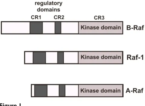
All three Raf isoenzymes are cytosolic serine/threonine protein kinases that exhibit a high degree of sequence sim-ilarity. The enzymes contain three domains termed CR1, CR2 and CR3. The N-terminal CR1 contains a Ras-binding subdomain and a cysteine-rich subdomain, both required to bind to activated Ras (Ras-GTP) at the cell membrane. CR2 is rich in serine and threonine residues and nega-tively regulates the biological activity of the catalytic domain. CR3 contains the catalytic protein kinase domain (figure 1).
Raf connects cellular stimulation with intracellular signal-ing pathways. Raf translocates to the plasma membrane as a result of receptor tyrosine kinase stimulation that leads to a subsequent activation of Ras. Following activation, Raf phosphorylates and activates mitogen-activated pro-tein kinase (MAP) kinase (MEK) which in turn phospho-rylates and activates the MAP kinases extracellular signal-regulated protein kinases ERK1 and ERK2 [1]. Raf func-tions therefore as a vital link between activated Ras and ERK. The activated protein kinases ERK1/2 are able to translocate into the nucleus and change the gene
expres-sion pattern via phosphorylation of gene regulatory pro-teins. Thus, activation of Raf is essential for activating the Raf/MEK/ERK signaling pathway and many functions attributed to Raf activation are executed by the subsequent activation of MEK and ERK. A microarray analysis con-firmed that the transcriptional response to Raf activation almost completely depends on MEK activation [2]. In line with this, MEK is the only generally acknowledged sub-strate for Raf [1,3].
Lessons from Raf-deficient mice
Gene ablation experiments involving the genes encoding the Raf isoforms Raf-1, A-Raf, and B-Raf revealed diver-gent phenotypes, indicating that Raf isoforms are not always able to compensate for each other. In particular, distinct essential functions are served by Raf-1 and B-Raf in embryonic development [4]. Nevertheless, a functional redundancy among the Raf family proteins exists and only phenotypes requiring the activity of a distinct Raf isoform are found. Inactivation of the Raf-1 and B-Raf-encoding genes revealed that Raf-1 and B-Raf play essential anti-apoptotic roles [5,6]. B-Raf is necessary for survival of embryonic motoneurons and sensory neurons [7]. Several
Published: 21 April 2009
Cell Communication and Signaling 2009, 7:8 doi:10.1186/1478-811X-7-8
Received: 20 February 2009 Accepted: 21 April 2009
This article is available from: http://www.biosignaling.com/content/7/1/8
© 2009 Thiel et al; licensee BioMed Central Ltd.
review articles have been published that discuss these mouse models in detail [4,8-10]
Gain-of-function mutants of Raf
Two strategies have been used to express constitutively active Raf. The translocation of Raf to the plasma mem-brane via binding to Ras-GTP is the key event in Raf acti-vation [1]. Thus, a method to express a constitutively active Raf-1 relies in the tethering of Raf to the plasma membrane. This Raf-1 mutant termed Raf-CAAX carries at the C-terminus an isoprenylation sequence derived from K-Ras [11,12]. The artifical targeting of Raf-1 to the plasma membrane leads to an activation of the enzyme in a Ras-independent manner and shows 30-fold higher kinase activity in growth-factor-deprived cells.
Alternatively, expression of the catalytic domain of either Raf-1, B-Raf, or A-Raf as a fusion protein with the hor-mone binding domain of the estrogen receptor [ER] gen-erates a hormone-regulated constitutively active Raf. The ΔRaf:ER fusion protein remains in an inactive state in the absence of hormone, but is rapidly activated by the addi-tion of hormone [13]. Figures 2A and 2B outline the strat-egy of using steroid-binding domains for regulating the function of Raf in cis [14]. In the absence of hormone, a heat-shock protein such as Hsp90 binds to the estrogen receptor domain and inhibit the catalytic function of the Raf-estrogen receptor fusion protein by steric hindrance, thus keeping the protein in an inactive state. Treatment
with estrogen triggers the dissociation of the heat-shock proteins, leading to a reversal of repression. As a result, Raf activity can be hormonally controlled. The use of the estrogen receptor mutant ERTamoxifen Mutant allows the use of the synthetic ligand, 4-hydroxytamoxifen (4OHT) for induction. The encoded hormone binding domain of the estrogen receptor contains a glycine residue at position 525, instead of an arginine. As a result, the receptor is largely insensitive to 17β-estradiol, but is readily activated by 4OHT [15]. The activation of the ERK signaling path-way can be visualized by Western blot analysis using anti-bodies that specifically recognize the phosphorylated forms of ERK1 and ERK2 (figure 2C). In this review, we will summarize some of the findings obtained with cells expressing Raf-estrogen receptor fusion proteins, which we consider the most important ones.
Role of Raf in the regulation of proliferation
The fact that Raf is activated following stimulation of the cells with mitogens (i.e. EGF, PDGF, IGF) indicates that these enzymes are involved in the regulation of cell growth and proliferation. Accordingly, expression of the hormone-regulated form of Raf-1, ΔRaf-1:ER, induced cell proliferation in NIH 3T3 fibroblasts that was accompa-nied by an upregulation of cyclin D1 and a repression of p27KIP, a cyclin-dependent protein kinase inhibitor [16]. Expression of conditionally active forms of A-Raf and B-Raf in NIH 3T3 cells revealed differences between the indi-vidual Raf isoforms. While the activation of both Δ A-Raf:ER and ΔB-Raf:ER induced the activation of MEK and ERK protein kinases, ΔB-Raf:ER activated MEK with the highest efficiency [17]. A microarray analysis performed with human epithelial cells underlined the importance of MEK activation by Raf [2]. Moreover, the fact that activa-tion of ΔRaf:ER strongly induced the expression of growth factors of the EGF growth factor family suggests the exist-ence of an autocrine loop through the activation of the EGF receptor: Activation of ΔRaf:ER triggers the stimulata-tion of the EGF receptor. As a result, the Raf-MEK-ERK sig-naling pathway is activated, leading to further synthesis of EGF growth factors [2,18].
In keratinocytes, activation of the EGF receptor triggers proliferation of the cells and involves the ERK signaling pathway (figure 3A) [19]. The importance of the ERK sig-naling pathway for growth of human keratinocytes was further demonstrated with HaCaT keratinocytes express-ing ΔA-Raf:ER. Proliferation of the cells was induced with 4OHT and completely inhibited by pretreatment with the MAP kinase kinase inhibitor PD98059 (figure 3B), indi-cating that the mitogenic activity of ΔA-Raf:ER is mediated by the activation of ERK. A comparison between the kinet-ics of ERK phosphorylation and activation by EGF or 4OHT revealed major differences in the duration of ERK activation (figure 3C). While EGF induced a strong phos-Modular structure of Raf
Figure 1
Modular structure of Raf. The Raf isoforms Raf-1, A-Raf and B-Raf share three conserved domains termed CR1, CR2 and CR3. CR1 contains a Ras-binding subdomain and a cysteine-rich subdomain, both required to bind to activated Ras (Ras-GTP) at the cell membrane. CR2 is rich in serine and threonine residues and negatively regulates the biological activity of the catalytic domain. This domain binds also regu-latory 14-3-3 proteins. CR3 encompasses the protein kinase domain.
Raf-1
B-Raf
A-Raf
CR3
CR1 CR2
regulatory domains
Kinase domain
Kinase domain
phorylation of ERK within 1 hour after stimulation,
phos-phorylated ERK was barely detected in ΔA-Raf:ER
expressing keratinocytes that had been incubated for 1 hour with 4OHT. Phosphorylated ERK was, however, detected in these cells 4 and 8 hours after induction of Δ A-Raf:ER. These results indicate that the kinetics of ERK acti-vation (transient versus sustained) are of minor impor-tance for the induction of the mitogenic program of keratinocytes by A-Raf. This observation is in contrast to the role of ERK in neuronal survival where a sustained activation of ERK is required for neuroprotection (see below).
In a human breast epithelial cell line, activation of ΔRaf:ER triggered the expression of genes encoding regu-lators of cell proliferation, including cyclin D1, and induced a transient increase in S phase cells. However, Raf activation did not induce growth factor-independent pro-liferation [18], in contrast to the situation encountered with ΔA-Raf:ER expressing keratinocytes. These data indi-cate that cell-type specific variations are important for the biological outcome of Raf activation.
Although the activation of a conditional form of Raf can promote DNA synthesis and cellular proliferation, other reports show that it can also provoke cell cycle arrest. Expression of ΔRaf-1:ER in small lung cancer cells induced a growth inhibitory pathway that is accompanied by the induction of the cyclin-dependent protein kinase inhibi-tor p27KIP and a decrease in cdc2 protein kinase activity [20]. In prostate cancer cells, activation of ΔRaf-1:ER induced expression of the cyclin-dependent protein kinase inhibitor p21KIP and an accumulation of the cells in G1, thus leading to growth suppression [21]. Likewise, ΔRaf-1:ER and ΔB-Raf:ER elicited a G1 arrest in NIH 3T3 cells that was accompanied by an upregulation of the cyc-lin-dependent protein kinase inhibitor p21KIP. In contrast,
Figure 2
A
ERTM
CR1 CR2
Raf ER
protein X
HSP90
inactive
+ hormone
HSP90
ER
protein X active
B
-
+
-
+
HT22
Raf-1:ER
4-OHT
P-ERK2
C
regulatory domains
Kinase domain
Kinase domain
CR3
Raf:ER
Design and biological activity of a conditionally active forms of Raf
Figure 2
activation of ΔA-Raf:ER promoted the entry of quiescent NIH 3T3 cells into the S-phase of the cell cycle. A catalyti-cally potentiated form of ΔA-Raf:ER, however, induced cell cycle arrest and enhanced p21KIP expression, similarly to ΔB-Raf:ER or ΔRaf-1:ER [22]. These data suggest that the catalytical activity and the duration of the signaling of Raf might determine the role of these enzymes in the pro-gression of the cell cycle. In addition, cell type-specific dif-ferences are essential for Raf induction and impairment of the growth capacity of the cells.
Anti-apoptotic role of Raf
Raf-1-deficient embryos are growth retarded and apop-totic cells are found in different tissues [6,7]. Raf-1-defi-cient fibroblasts are hypersensitive to apoptotic stimuli such as serum withdrawal or Fas/Fas ligand interaction. Thus, it was concluded that the major function of Raf-1 is to counteract apoptosis [7]. Also B-Raf-deficient embryos die because of vascular defects due to apoptotic death of differentiated endothelial cells [23].
The activation of the MEK/ERK signaling pathway by Raf has been correlated with inhibition of programmed cell death. The ERK signaling pathway has been described to play an important role as a main antagonist of various apoptosis-inducing challenges [[24,25]; reviewed in ref [26]]. Activation of the ERK signaling pathway suppresses the proapoptotic activity of stress-activated JNK/p38 tein kinases in PC12 pheochromocytoma cells, thus pro-tecting the cells from NGF withdrawal-induced cell death [27]. In line with this, BDNF-elicited ERK activation pro-tects cortical neurons against a challenge with the topoi-somerase I inhibitor campthothecin [28]. In addition, it has been shown that activation of the ERK signaling path-way via treatment of the cells with either EGF or 12-O -tet-radecanoylphorbol-13-acetate may lead to an inactivation of caspase-9 due to a direct phosphorylation of Thr125 of caspase-9 by ERK. This phosphorylation blocks caspase-9 processing and the subsequent activation of caspase-3 [29].
The survival of cells requires the presence of survival fac-tors, and the lack of this trophic support is one of the best-studied signals for induction of cell death. In Rat-1 fibrob-lasts, overexpression of B-Raf protected the cells from apoptosis, induced by growth factor withdrawal. Treat-ment with the MEK inhibitor PD98059 blocked the anti-apoptotic activity of B-Raf, indicating that the activation of the Raf-MEK-ERK signaling pathway is necessary for the anti-apoptotic role of B-Raf in Rat-1 fibroblasts [30].
Experiments using HT22 immortalized neurons derived from the hippocampal region of the CNS showed that stimulation with BDNF rescues the cells from serum with-drawal-induced cell death when the BDNF receptor TrkB
Figure 3
50 100 150 200 250
EGF
BrdU
incorporation
(%
of
control)
+ +
+
-PD98059
50 100 150 200 250
+
BrdU
incorporation
(%
of
control)
PD98059 +
+
4OHT
-HaCaT-
A-Raf:ER
0.25 1 4 8
-P-ERK2
A-Raf:ER
HaCaT
[h] EGF
0.25 1 4 8
-HaCaT
P-ERK2
A
B
C
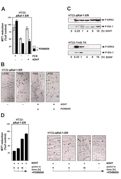
is expressed. An analysis of intracellular signaling cascades revealed that stimulation of the TrkB receptor with BDNF leads to an activation of both the ERK and the PI3 kinase pathways. A pharmacological approach showed that the major neuronal survival-promoting signaling pathway includes an activation of PI3 kinase and AKT [31]. These and other observations [28] indicate that the signaling cascade BDNF → TrkB stimulation → PI3 kinase activa-tion → activation of AKT →→ cell survival is of general importance and is not limited to a particular neuronal population or neuronal cell line. In contrast, the neuro-protective activity of BDNF is independent of the ERK sig-naling pathway since PD98059 did not impair the BDNF-mediated protection of neurons against serum with-drawal-induced programmed cell death. To clarify the role of ERK for neuroprotection, HT22 cells were analyzed that expressed a ΔRaf-1:ER fusion protein and allowed the selective activation of the ERK signaling pathway (figure 2C). Activation of the catalytic function of Raf-1 by 4OHT rescued HT22 cells from serum withdrawal-induced cell death, as depicted in figure 4A. The neuroprotective role of ΔRaf-1:ER was confirmed by phase contrast microscopy (figure 4B). Inhibition of ΔRaf-1-induced MEK activation by PD98059 blocked the cytoprotective activity of Δ Raf-1:ER [31], indicating that the activation of ERK via MEK is the underlying cause for neuroprotection mediated by the activation of the ΔRaf-1-ER fusion protein. These results were a puzzle to be solved: BDNF-mediated neuroprotec-tion against serum withdrawal-mediated cell death was independent of ERK; in contrast, the Raf-1-estrogen recep-tor fusion protein protected the cells solely via activation of the ERK signaling pathway. To find clues to the solution of the puzzle, the kinetics of ERK activation in HT22 cells
have been investigated (figure 4C). Stimulation of TrkB expressing HT22 cells with BDNF triggered a robust, but transient activation of ERK that was not sufficient to fer protection against the loss of trophic support. In con-trast, expression of a conditionally activatable Raf-1 that induced a sustained ERK phosphorylation lasting for hours, rescued neuronal HT22 cells from serum depriva-tion-induced cell death. The phosphorylation state of the ERK substrate Elk-1 mirrored the kinetic profile of ERK activation, i.e. transient or sustained activation of ERK is translated in the nucleus into a transient or sustained acti-vation of Elk-1 (figure 4C). We then asked the question how long ERK has to be activated in order to protect HT22 cells from serum deprivation-induced apoptosis. Addition of PD98059 at different time points following treatment of the cells with 4OHT revealed that a prolonged activa-tion of ERK, lasting for hours, is necessary for neuropro-tection [31] (figure 4D). These results shed light on the fact that the kinetics of ERK activation (transient versus sustained) are of major importance for the neuroprotec-tive activity. Thus, activation of PI3 kinase by BDNF in neuronal cells represents the dominant survival pathway, whereas the ERK signaling pathway plays no or only a marginal role. However, a sustained activation of ERK, lasting for several hours, protects neurons from growth factor deprivation-induced cell death, indicating that the duration of ERK activation is of major importance for its neuroprotective biological function. Thus, stimulation of the cells with neurotrophins of elevated concentrations, or stimulation with several ligands, i.e. BDNF and EGF, may trigger a sustained activation of ERK.
The activation of an estrogen-inducible activated Raf-1 mutant ΔRaf-1:ER also prevented apoptosis induced by loss of matrix contact (anoikis), cytoskeletal integrity and serum removal in lung fibroblasts [32]. In these cells it has been shown that activation of ΔRaf-1:ER prevented the upregulation of Bim, a proapoptotic BH3-only protein of the Bcl-2 family, in serum-starved cells. This rescue relies on the activation of the ERK pathway and was independ-ent of the JNK → c-Jun and PI3 kinase → PDK → AKT pathway [33]. In human breast epithelial cells, the expres-sion of genes encoding growth factors of the EGF family as a result of ΔRaf-1:ER activation protected the cells from detachment-induced apoptosis [18]. Activation of Δ Raf-1:ER also blocked programmed cell death induced by TGFβ in MLCK epithelial cells [34]. However, activation of ΔRaf-1:ER did not provide protection against oxidative glutamate toxicity in HT22 hippocampal cells [35]. These data indicate that the anti-apoptotic function of Raf is restricted to particular apoptotic signaling pathways.
In addition to the well-established target MEK, Raf may use other effectors to inhibit programmed cell death. It has been shown that Raf-1 promotes cell survival in a Activation of a conditionally active form of A-Raf(ΔA- Raf:ER)
induces proliferation of human HaCaT keratinocytes via acti-vation of the ERK signaling pathway
Figure 3
Figure 4 (see legend on next page)
B
A
+
-25 50 75 100 125
HT22-Raf-1:ER
MTT
reduction
(%of
control)
-+PD98059
FCS 4OHT
-- - +
HT22- Raf-1:ER
4OHT
+ +
-- +
+
+ FCS - FCS - FCS - FCS
PD98059
C
4OHT HT22- Raf-1:ER
0 0.25 1 4 8 16
P-Elk-1
HT22-TrkB.TK
BDNF 0 0.25 1 4 8 16
P-Elk-1 [h]
[h] P-ERK2
P-ERK2
+ + + +
0 8 16
4OHT
-25 50 75 100 125
MTT
reduction
(%of
control)
HT22-Raf-1:ER
point in time [h] +PD98059
D
+ + + + 4OHT
0 8 16 - point in
time [h] +PD98059
MEK/ERK-independent manner via antagonizing apopto-sis signal-regulating kinase-1 (ASK-1) [36]. Raf-1 is also targeted to the mitochondria by Bcl-2 that leads to cell survival without ERK activation, probably by phosphor-ylating substrates other than MEK, such as Bcl-2 family members [37,38].
Role of Raf in cellular differentiation
Raf activation has been discussed primarily as an integral part of the ERK signaling pathway controlling cellular growth. The impact of Raf in the control of cellular differ-entiation has only been put in the limelight in recent years. The lethal phenotypes observed in either Raf-1-, A-Raf- or B-A-Raf-deficient mice sheds light on the essential role of Raf during development. Nullizygous B-Raf-/- -embryos, for example, die by embryonic day 12.5 show-ing defects in vascular endothelial cell differentiation [23]. In cell culture models, B-Raf controls myelopoiesis at multiple stages. In particular, B-Raf deficient ES cells have a quantitative defect in myeloid progenitor cell for-mation [39]. B-Raf is also crucial for T-cell development, in particular for the transition to CD4+ and CD8+ single-positive cells [40]. In immortalized cells from rat hippoc-ampus neurons, activation of ΔRaf-1:ER was shown to induce neuronal differentiation [41]. In neural stem cells, supplementation of the medium with EGF and bFGF is necessary to inhibit differentiation. Removal of the mitogens stops the cell cycle and induces differentiation [42]. Figure 5 shows that expression of the astrocytic marker GFAP is upregulated in differentiated HNSC.100 neural stem cells. This upregulation of GFAP expression was prevented in ΔRaf-1:ER-expressing neural stem cells that were stimulated by 4OHT in the absence of EGF and
bFGF in the medium [43], indicating that enhanced Raf-1 activity blocked the differentiation of the cells. Together, these data show that Raf influences developmental proc-esses, although the exact molecular mechanisms have to be determined in each cell type.
Conclusion
Results obtained with constitutively active Raf mutants has been questioned, since the lack of the regulatory domains in the Raf mutants may compromise the sub-strate specificity and the dynamic regulation of activity [9]. Nevertheless, the many data obtained using these mutants have improved our knowledge of the functions of Raf in growth control, apoptosis and differentiation.
Abbreviations
ER: estrogen receptor; ERK: extracellular signal-regulated protein kinase; 4OHT: 4-hydroxytamoxifen
Competing interests
The authors declare that they have no competing interests.
Authors' contributions
GT drafted and wrote the manuscript. ME and OR have critically revised the manuscript and prepared the figures. All authors read and approved the final manuscript.
Acknowledgements
We thank Martin McMahon, UCSF, for providing ΔRaf:ER constructs and Libby Guethlein for critical reading of the manuscript. The research of the laboratory concerning intracellular signaling cascades underlying neuronal cell death and survival is supported by the Deutsche Forschungsgemein-schaft (grant SFB 530, C14).
A conditionally active form of Raf-1 (ΔRaf-1:ER) protects HT22 neuronal cells against serum deprivation-induced cell death via activation of ERK
Figure 4 (see previous page)
References
1. Wellbock C, Karasarides M, Marais R: The Raf proteins take cen-tre stage. Nat Rev Mol Cell Biol. 2004, 5(11):875-885.
2. Schulze A, Nicke B, Warne PH, Tomlinson S, Downward J: The tran-scriptional response to Raf activation is almost completely dependent on mitogen-activated protein kinase kinase activ-ity and shows a major autocrine component. Mol Biol Cell 2004,
15:3450-3463.
3. O'Neill E, Kolch W: Conferring specificity on the ubiquitous Raf/MEK signalling pathway. Br J Cancer 2004, 90:283-288. 4. Baccarini M: Second nature: Biological functions of the Raf-1
"kinase". FEBS Lett 2005, 579:3271-3277.
5. Hülser M, Luckett J, Chiloeches A, Mercer K, Iwobi M, Giblett S, Sun XM, Brown J, Marais R, Pritchard C: MEK kinase activity is not necessary for Raf-1 function. EMBO J 2001, 20:1940-1951. 6. Mikula M, Schreiber M, Husak Z, Kucerova L, Rüth J, Wieser R,
Zat-loukal K, Beug H, Wagner EF, Baccarini M: Embryonic lethality and fetal liver apoptosis in mice lacking the c-raf-1 gene.
EMBO J 2001, 20:1952-1962.
7. Wiese S, Pei G, Karch C, Troppmair J, Holtmann B, Rapp UR, Sendt-ner M: Specific function of B-Raf in mediating survival of embryonic motoneurons and sensory neurons. Nat Neurosci
2001, 4:137-142.
8. Hagemann C, Rapp UR: Isotype-specific functions of Raf kinases.
Exp Cell Res 1999, 253:34-46.
9. Hindley A, Kolch W: Extracellular signal regulated kinase (ERK)/mitogen activated protein kinase (MAPK)-independ-ent functions of Raf kinases. J Cell Sci 2002, 115:1575-1581. 10. Galabova-Kovacs G, Kolbus A, Matzen D, Meissl K, Piazzolla D,
Rubi-olo C, Steinitz K, Baccarini M: ERK and beyond: insights from B-Raf and B-Raf-1 conditional knockouts. Cell Cycle 2006,
5:1514-1518.
11. Leevers SJ, Paterson HF, Marshall CJ: Requirement for Ras in Raf activation is overcome by targeting Raf to the plasma mem-brane. Nature 1994, 369:411-414.
12. Stokoe D, Macdonald SG, Cadwallader K, Symons M, Hancock JF:
Activation of Raf as a result of recruitment to the plasma membrane. Science 1994, 264:1463-1467.
13. McMahon M: Steroid receptor fusion proteins for conditional activation of Raf-MEK-ERK signaling pathway. Meth Enzymol
2001, 332:401-417.
14. Picard D: Steroid-binding domains for regulating the function of heterologous proteins in cis. Trends Cell Biol 1993, 3:278-280. 15. Littlewood TD, Hancock DC, Danielian PS, Parker MG, Evan GI: A modified estrogen receptor ligand-binding domain as an improved switch for the regulation of heterologous proteins.
Nucl Acids Res 1995, 23:1686-1690.
16. Kerkhoff E, Rapp UR: Induction of cell proliferation in quiescent NIH 3T3 cells by oncogenic c-Raf-1. Mol Cell Biol 1997,
17:2576-2586.
17. Pritchard CA, Samuels ML, Bosch E, McMahon M: Conditionally oncogenic forms of the A-Raf and B-Raf protein kinases dis-play different biological and biochemical properties in NIH 3T3 cells. Mol Cell Biol 1995, 15:6430-6442.
18. Schulze A, Lehmann K, Jefferies HB, McMahon M, Downward J: Anal-ysis of the transcriptional program induced by Raf in epithe-lial cells. Genes Dev 2001, 15:981-994.
Figure 5
B
A
+
+
-GFAP
GAPDH
HNSC.100
Raf-1:ER
EGF/bFGF
4OHT
+
+
-EGF/bFGF
4OHT
GFAP
GAPDH
10 20 30
0
IOD
0.0 0.5 1.5
1.0
IOD
Activation of ΔRaf-1:ER blocks differentiation along the astrocytic lineage in human neural stem cells
Figure 5
Publish with BioMed Central and every scientist can read your work free of charge "BioMed Central will be the most significant development for disseminating the results of biomedical researc h in our lifetime."
Sir Paul Nurse, Cancer Research UK
Your research papers will be:
available free of charge to the entire biomedical community
peer reviewed and published immediately upon acceptance
cited in PubMed and archived on PubMed Central
yours — you keep the copyright
Submit your manuscript here:
http://www.biomedcentral.com/info/publishing_adv.asp
BioMedcentral 19. Rössler OG, Thiel G: Brain-derived neurotrophic factor-,
epi-dermal growth factor-, or A-Raf-induced growth of HaCaT keratinocytes requires extracellular signal-regulated kinase.
Am J Physiol Cell Physiol 2004, 286:C1118-C1129.
20. Ravi RK, Weber E, McMahon M, Williams JR, Baylin S, Mal A, Harter ML, Dillehay LE, Claudio PP, Giordano A, Nelkin BD, Mabry M: Acti-vated Raf-1 causes growth arrest in human small cell lung cancer cells. J Clin Invest 1998, 101:153-159.
21. Ravi BK, McMahon M, Yangang Z, Williams JR, Dillehay LE, Nelkin BD, Mabry M: Raf-1-induced cell cycle arrest in LNCaP human prostate cancer cells. J Cell Biochem 1999, 72:458-469.
22. Woods D, Parry D, Cherwinski H, Bosch E, Lees E, McMahon M: Raf-induced proliferation or cell cycle arrest is determined by the level of Raf activity with arrest mediated by p21Cip1. Mol
Biol Cell 1997, 17:5598-5611.
23. Wojnowski L, Zimmer AM, Beck TW, Hahn H, Bernal R, Rapp UR, Zimmer A: Endothelial apoptosis in Braf-deficient mice. Nat
Genet 1997, 16:293-297.
24. Holmström TH, Chow SC, Elo I, Coffey ET, Orrenius S, Sistonen L, Eriksson JE: Suppression of Fas/APO-1-mediated apoptosis by mitogen-activated kinase signaling. J Immunol 1998,
160:2626-2636.
25. Tran SEF, Holmström TH, Ahonen M, Kähäri V-M, Eriksson JE: MAP/ ERK overrides the apoptotic signaling from Fas, TNF, and TRAIL receptors. J Biol Chem 2001, 276:16484-16490.
26. Ballif BA, Blenis J: Molecular mechanisms mediating mamma-lian mitogen-activated protein kinase (MAPK) kinase (MEK)-MAPK cell survival signals. Cell Growth Differ 2001, 12:397-408. 27. Xia Z, Dickens M, Raingeaud J, Davis RJ, Greenberg ME: Opposing
effects of ERK and JNK-p38 MAP kinases on apoptosis. Sci-ence 1995, 270:1326-1331.
28. Hetman M, Kanning K, Cavanaugh JE, Xia Z: Neuroprotection by brain-derived neurotrophic factor is mediated by extracellu-lar signal-regulated kinase and phosphatidylinositol 3-kinase.
J Biol Chem 1999, 274:22569-22580.
29. Allan LA, Morrice N, Brady S, Magee G, Pathak S, Clarke PR: Inhibi-tion of caspase-9 through phosphorylaInhibi-tion at Thr 125 by ERK MAPK. Nat Cell Biol 2003, 5:647-654.
30. Erhardt P, Schremser EJ, Cooper GM: B-Raf inhibits programmed cell death downstream of cytochrome c release from mito-chondria by activating the MEK/Erk pathway. Mol Cell Biol
1999, 19:5308-5315.
31. Rössler OG, Giehl KM, Thiel G: Neuroprotection of immortal-ized hippocampal neurons by brain-derived neurotrophic factor and Raf-1 protein kinase: Role of extracellular signal-regulated protein kinase and phosphatidylinositol 3-kinase. J
Neurochem 2004, 88:1240-1252.
32. Le Gall M, Chambard JC, Breittmayer JP, Grall D, Pouysségur J, Van Obberghen-Schilling E: The p42/p44 MAP kinase pathway pre-vents apoptosis induced by anchorage and serum removal.
Mol Biol Cell 2000, 11:1103-1112.
33. Weston CR, Balmanno K, Chalmers C, Hadfield K, Molton SA, Ley R, Wagner EF, Cook SJ: Activation of ERK1/2 by ΔRaf-1:ER* represses Bim expression independently of the JNK or PI3K pathways. Oncogene 2003, 22:1281-1293.
34. Lehmann K, Janda E, Pierreux CE, Rytömaa M, Schulze A, McMahon M, Hill CS, Beug H, Downward J: Raf induces TGFβ production while blocking its apoptotic but not invasive responses: a mechanism leading to increased malignancy in epithelial cells. Genes Dev 2000, 14:2610-2622.
35. Rössler OG, Bauer I, Chung HY, Thiel G: Glutamate-induced cell death of immortalized murine hippocampal neurons: neuro-protective activity of heme oxygenase-1, heat shock protein 70, and sodium selenite. Neurosci Lett 2004, 362:253-257. 36. Chen J, Fujii K, Zhang L, Roberts T, Fu H: Raf-1 promotes cell
sur-vival by antagonizing apoptosis signal-regulating kinase 1 through a MEK-ERK independent mechanism. Proc Natl Acad
Sci USA 2001, 98:7783-7788.
37. Wang HG, Rapp UR, Reed JC: Bcl-2 targets the protein kinase Raf-1 to mitochondria. Cell 1996, 87:629-638.
38. Zhong J, Troppmair J, Rapp UR: Independent control of cell sur-vival by Raf-1 and Bcl-2 at the mitochondria. Oncogene 2001,
20:4807-4816.
39. Kamata T, Kang J, Lee TH, Wojnowski L, Pritchard CA, Leavitt AD:
A critical function for B-Raf at multiple stages of
myelopoie-sis. Blood 2005, 106:833-840.
40. Tsukamoto H, Irie A, Senju S, Hatzopoulos AK, Wojnowski L, Nishimura Y: B-Raf-mediated signaling pathway regulates T cell development. Eur J Immunol 2008, 38:518-527.
41. Kuo W-L, Abe M, Rhee J, Eves EM, McCarthy SA, Yan M, Templeton DJ, McMahon M, Rosner MR: Raf, but not MEK or ERK, is suffi-cient for differentiation of hippocampal neuronal cells. Mol
Cell Biol 1996, 16:1458-1470.
42. Villa A, Snyder EY, Vescovi A, Martínez-Serrano A: Establishment and properties of a growth factor-dependent, perpetual neu-ral stem cell line from the human CNS. Exp Neurol 2000,
161:67-84.
43. Ekici M, Hohl M, Schuit F, Martínez-Serrano A, Thiel G: Transcrip-tion of genes encoding synaptic vesicle proteins in human neural stem cells: chromatin accessibility, histone methyla-tion pattern, and the essential role of REST. J Biol Chem 2008,