Cite as
Gomez CD, Aguilera P, Ortiz-Plata A, et al. Aged garlic extract and S-allylcysteine increase the GLUT3 and GCLC expression levels in cerebral ischemia. Adv Clin Exp Med.
2019;28(12):1609–1614. doi:10.17219/acem/110328
DOI
10.17219/acem/110328
Copyright
© 2019 by Wroclaw Medical University This is an article distributed under the terms of the Creative Commons Attribution 3.0 Unported (CC BY 3.0) (https://creativecommons.org/licenses/by/3.0/)
Address for correspondence
Mónica Espinoza-Rojo
E-mail: [email protected]
Funding sources
Carlos Daniel Gómez was financed by CONACyT (scholarship No. 12296) and by Movilidad Santander Universia.
Conflict of interest
None declared
Acknowledgements
We thank Travis Ashworth for his contribution to the revi-sion of English grammar in the manuscript, and Juan Miguel Mendoza Bello for his technical assistance.
Received on February 6, 2018 Reviewed on March 6, 2018 Accepted on June 27, 2019
Published online on December 16, 2019
Abstract
Background. During cerebral ischemia, energy restoration through the regulation of glucose transporters and antioxidant defense mechanisms is essential to maintain cell viability. Antioxidant therapy has been considered effective to attenuate brain damage; moreover, the regulation of transcription factors that positively regulate the expression of glucose transporters is associated with this therapy. Recently, it has been reported that the use of antioxidants such as S-allylcysteine (SAC), a component of aged garlic extract (AGE), improves survival in experimental models of cerebral ischemia.
Objectives. The aim of this study was to determine the effect of AGE and SAC on the level of mRNA expres-sion of the main neuronal glucose transporter (GLUT3) and the glutamate cysteine ligase catalytic subunit (GCLC) in rats with transient focal cerebral ischemia.
Material and methods. Cerebral ischemia was induced in male Wistar rats by middle cerebral artery occlusion (MCAO) for 2 h. The animals were sacrificed after different reperfusion times (0–48 h). Animals injected with AGE (360 mg/kg, intraperitoneally (i.p.)) and SAC (300 mg/kg, i.p.) at the beginning of reperfu-sion were sacrificed after 2 h. The mRNA expresreperfu-sion level was analyzed in the fronto-parietal cortex using quantitative polymerase chain reaction (qPCR).
Results. Two major increases in GLUT3 expression at 1 h and 24 h of reperfusion were found. Both treat-ments increased GLUT3 and GCLC mRNA levels in control and under ischemic/reperfusion injury animals. Conclusions. This data suggests that SAC and AGE might induce neuroprotection, while controlling reac-tive oxygen species (ROS) levels, as indicated by the increase in GCLC expression, and regulating the energy content of the cell by increasing glucose transport mediated by GLUT3.
Key words: antioxidants, cerebral ischemia, glucose transporters
Aged garlic extract and S-allylcysteine increase the GLUT3
and GCLC expression levels in cerebral ischemia
Carlos Daniel Gomez
1,B,C,F, Penélope Aguilera
2,B,C,F, Alma Ortiz-Plata
3,B,F, Felipe Nares López
2,C,D,F,
María Elena Chánez-Cárdenas
2,D,E,F, Eugenia Flores-Alfaro
4,C,F, Martha Eugenia Ruiz-Tachiquín
5,C,F, Monica Espinoza-Rojo
6,A,B,D,F1 Department of Physiology and Pharmacology, University of Calgary, Canada
2 Laboratory of Cerebral Vascular Pathology, National Institute of Neurology and Neurosurgery “Manuel Velasco Suárez”, Mexico City, Mexico 3 Laboratory of Experimental Neuropathology, National Institute of Neurology and Neurosurgery, Mexico City, Mexico
4 Laboratory of Clinical and Molecular Epidemiology, Faculty of Biological and Chemical Sciences, Universidad Autónoma de Guerrero, Chilpancingo, Mexico 5 Medical Research Unit on Human Genetics, Pediatrics Hospital, Mexican Institute of Social Security (IMSS), Mexico City, Mexico
6 Laboratory of Molecular and Genomic Biology, Faculty of Biological Chemical Sciences, Universidad Autónoma de Guerrero, Chilpancingo, Mexico
A – research concept and design; B – collection and/or assembly of data; C – data analysis and interpretation; D – writing the article; E – critical revision of the article; F – final approval of the article
Damage induced by cerebral ischemia and restoration of blood flow (reperfusion) has been linked to mitochondrial dysfunction and the concomitant production of noxious lev-els of reactive oxygen species (ROS).1 Reactive oxygen species
are released during mitochondrial respiration, causing tissue injury and cellular dysfunction.2,3 Therefore, the reduction
of ROS by antioxidants has been considered as a potential therapeutic method that, through the regulation of the cel-lular redox state, might ameliorate ischemic injury.4–6
Interestingly, antioxidant agents activate the hypoxia inducible factor-1α (HIF-1α) in cultured brain endothe-lial cells, astrocytes and neurons subjected to normoxia, hypoxia or ischemia.7–9 This factor increases anaerobic
glycolysis through the upregulation of glucose transport-ers (GLUTs), which possess hypoxia response elements (HRE) in their promoters,10,11 and this upregulation
facili-tates cell survival by maintaining adenosine triphosphate (ATP) production.9,12 Glucose transporters are essential for
an adequate supply of energy in neurons, principally under conditions where the energy demand is high as occurs in hypoxia and brain ischemia.13 The glucose transporter 3
(GLUT3) is the main transporter in neurons with the high-est affinity for glucose (Km 1.4 mmol/L),10,11 its expression
increases at the onset of cerebral ischemia, and its subse-quent decline is followed by neuronal death.14 On the other
hand, treatment with antioxidant agents also increases ex-pression levels of proteins involved in cellular redox regu-lation.9,15 The glutamate cysteine ligase catalytic subunit
(GCLC) is an enzyme that participates in the production of glutathione, an important endogenous antioxidant pep-tide.16 The glutamate cysteine ligase catalytic subunit
is in-creased in brain slices subjected to oxygen-glucose depri-vation,17 and in vitro models of chemical hypoxia-induced
neurotoxicity.18 Therefore, regulation of GLUT and GCLC
are 2 potential mechanisms by which antioxidants might prevent the deleterious effects of ROS in cerebral ischemia.
In line, some antioxidant agents, such as aged garlic ex-tract (AGE), and its main active component, S-allylcysteine (SAC), have been shown to prevent brain cell death and to improve neurologic deficit induced in experimental models of cerebral ischemia/reperfusion.5,6,19,20 The effects
of AGE and SAC in brain ischemia have been associated with their high antioxidant potential; nevertheless, their complete mechanism of action is not well understood. We assume that these antioxidants act in association with the regulation of GLUT3 expression to increase the en-trance of glucose to the cell to augment ATP concentration, but are also associated with the regulation of GCLC mRNA to achieve a reduced cellular redox state, which is charac-terized by a high glutathione concentration. Therefore, we have determined the temporal expression patterns of GLUT3 induced by cerebral ischemia/reperfusion and the possible changes following reperfusion in the presence of AGE and SAC. We also have evaluated the effect of these antioxidants on GCLC expression in our model of cere-bral ischemia/reperfusion. Our purpose was to identify
a possible mechanism by which antioxidants participate in neuroprotection in the middle cerebral artery occlusion (MCAO) model of ischemia/reperfusion.
Material and methods
Reagents
Aged garlic extract Kyolic® was obtained from
Waku-naga of America Co., Ltd. (Mission Viejo, USA), and SAC was synthesized by the reaction of L-cysteine with allyl-bromide and purified by recrystallization from ethanol-water, according to a previous report.21 TRIzol Reagent,
SuperScript® III First Strand Synthesis SuperMix and
ramdom hexamer primers were obtained from Invitrogen Life Technologies (Carlsbad, USA); commercial prede-signed TaqMan Probes system was performed in a 7500 Real-time PCR System (Applied Biosystems, Foster City, USA) using specific assay GLUT3 (Rn00567331_M1) and GCLC (Rn00563101_M1) (Applied Biosystem). 18S ribo-somal RNA (18S rRNA, assay ID: Hs99999901_s1, Applied Biosystems). All other reagents were obtained from known commercial sources.
Experimental animals
Adult male Wistar rats weighing 280–320 g were in-cluded in this study. The mRNA expression encoding for GLUT3 and GCLC was studied in 3–8 animals per group. Nine animal groups were subjected to transient focal ce-rebral ischemia induced by MCAO during 2 h,22 and then
were sacrificed by decapitation after different times (0, 1, 2, 3, 4, 6, 10, 24, and 48 h) of reperfusion (n = 4). Briefly, animals were anaesthetized with isoflurane (2.5–3.0%). The left common carotid artery was exposed at the level of the external and internal carotid artery bifurcation. A 3-0 nylon monofilament was inserted into the exter-nal carotid artery and advanced into the interthe exter-nal carot-id artery at a depth of about 17 mm to block the origin of the middle cerebral artery (MCA). Body temperature was kept at 37 ±0.5°C during the procedure. Two hours after the induction of ischemia, the filament was removed to allow reperfusion. The animals were returned to their cages and monitored until they recovered from anesthe-sia. Neurological deficit was determined 30 min before reperfusion and was scored on a 2-point scale: failure to extend right paw fully = 1; circling to right (more than 5 turns over a period of 30 s) = 1. Animals that presented neurological deficits <2 were exclude from the study.5,22
and then received a single i.p. injection of one of the above antioxidant agents at the beginning of reperfusion. All ani-mal groups administered with the antioxidant agents were sacrificed by decapitation 2 h after the treatment injection. Experimental procedures were carried out in accordance with the National Institutes of Health (NIH) Guidelines for the Care and Use of Laboratory Animals, and with the ethical guidelines established by the National Institute of Neurology and Neurosurgery “Manuel Velasco Suárez” in Mexico City, Mexico (approved project No. 20/11 (624)).
Quantitative polymerase chain reaction
Total RNA extraction was performed on the whole fronto-parietal cortex, using TRIzol Reagent (Invitrogen Life Technologies) according to the manufacturer’s in-structions, and the amount and purity were determined using a spectrophotometer. cDNA was synthesized from 5 µg total RNA, using the kit SuperScript® III First Strand
Synthesis SuperMix (Invitrogen Life Technologies). Quan-titative polymerase chain reaction (qPCR) was performed in a 7500 Real-time PCR System (Applied Biosystems), using specific assay GLUT3 (Rn00567331_M1) and GCLC (Rn00563101_M1) (Applied Biosystem). 18S ribosomal RNA (18S rRNA, assay ID: Hs99999901_s1, Applied Bio-systems) was used as a control to normalize the relative mRNA amount of the amplified genes. Reactions were done in triplicate and consisted of a denaturation cycle at 95°C for 10 min, followed by 40 cycles of denaturation at 92°C for 15 s and of annealing/extension at 60°C for 1 min. Values of cycle threshold (Ct) were determined through automated threshold analysis using SDS v. 1.3.1. software (Applied Biosystems, Waltham, USA). This soft-ware uses the comparative Ct method of relative quantifi-cation to determine relative gene expression levels.
Statistical analysis
Data analysis was performed using SPSS v. 13.0 (SPSS Inc., Chicago, USA) statistical software package. The re-sults are presented as mean and standard deviation (mean ±SD). The statistical significance of differences between groups was analyzed using analysis of variance (ANOVA) and post hoc Tukey analysis was used for sta-tistical evaluations. A value of p < 0.05 was considered significant.
Results
Effects of cerebral ischemia/reperfusion
on GLUT3 mRNA expression levels
in fronto-parietal cortex
Considering that major alterations in cerebral glu-cose concentration during ischemic injury have been
reported,23–25 temporal expression of glucose transporters
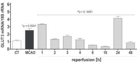
induced by cerebral ischemia/reperfusion in the fronto-parietal cortex was evaluated. Figure 1 shows that focal cerebral ischemia (MCAO group) increased the GLUT3 mRNA expression level by 224% compared with control rats (1.41 ±0.24-fold; p < 0.0001). In the case of animals subjected to 2 h of MCAO followed by different times of reperfusion, 2 major increases in the GLUT3 mRNA expression were observed. After 1 h of reperfusion, the GLUT3 mRNA expression level remained elevated and reached 31% (0.8 ±0.08-fold) more than in the MCAO group (p < 0.0001). After 2, 3 and 4 h of reperfusion, the GLUT3 expression level was reduced to 52.4%, 67.7%, and 50%, respectively (2 h reperfusion = –1.21 ±0.08-fold; 3 h of reperfusion = –0.82 ±0.17-(2 h reperfusion = –1.21 ±0.08-fold; 4 h of reper-fusion = –1.27 ±0.17-fold; reperfusion group vs MCAO, p < 0.0001) reaching the baseline levels of expression (control group). After 6 and 10 h of reperfusion, the levels of GLUT3 were even lower that baseline reaching 22.1% and 14.6% (6 h of reperfusion = –1.98 ±0.01-fold; 10 h of re-perfusion = –2.17 ±0.05-fold; reperfusion group vs MCAO, p < 0.0001). A more pronounced increase was found after 24 h of reperfusion, when the GLUT3 mRNA expression level was increased to 163% (1.62 ±0.26, 24 h of reperfu-sion vs MCAO p < 0.0001). Finally, after 48 h of reper-fusion, GLUT3 mRNA decreased again 33% compared to the MCAO group (–1.68 ±0.14, p < 0.0001).
Effects of AGE and SAC on GLUT3
mRNA expression levels in cerebral
ischemia/reperfusion
Recent evidence suggests a key role of GLUT3 as a po-tential therapeutic target in the treatment of cerebral ischemia/reperfusion.26,27 Therefore, a possible effect
of the AGE and its principal component SAC on GLUT3
Fig. 1. Effect of cerebral ischemia/reperfusion on GLUT3 mRNA
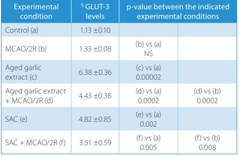
mRNA expression level in cerebral ischemia/reperfusion was tested here. Table 1 shows that both treatments in-creased the baseline expression of the mRNA encoding for GLUT3 (AGE and SAC groups). In the control rats, AGE increased GLUT3 mRNA expression level by 564.6% over basal conditions (p < 0.0001), while SAC increased the GLUT3 mRNA expression level by 426.5% (p = 0.002) (Table 1, row c and e vs a). On the other hand, AGE and SAC administration to the animals subjected to 2 h of ischemia and 2 h of reperfusion increased the GLUT3 mRNA ex-pression level by 333% (p = 0.0002) in the MCAO/2R+AGE group and 263.9% in the MCAO/2R+SAC group (Table 1, row d and f vs b; p = 0.008).
Effects of AGE and SAC on GCLC
mRNA expression levels in cerebral
ischemia/reperfusion
Considering that oxidative stress generated during the ischemia/reperfusion process aggravates the dam-age, we evaluated the effect of AGE and SAC on GCLC mRNA expression level to determine a possible mechanism by which antioxidant molecules function as regulators in neuroprotection. Table 2 shows that both AGE and SAC administration induced a significant increase in the ex-pression level of GCLC mRNA over control values in physi-ological conditions. The AGE treatment increased GCLC mRNA expression level by 306% (p = 0.006) in the control group + AGE, and by 292% (p = 0.001) in the control group + SAC (Table 2, row c and e vs a). In another observation, the increase induced by AGE and SAC on GCLC expression
level was still present in animals subjected to brain isch-emia, in the MCAO/2R+AGE group, the GLUT3 mRNA increased expression level 268% (p = 0.02), and 206% (p = 0.02) in the MCAO/2R+SAC group vs the MCAO/2R group (Table 2, row d and f vs b)
Discussion
The present study shows that in the fronto-parietal cor-tex dissected from animals exposed to transitory focal cerebral ischemia, the expression level of GLUT3 mRNA is increased after 2 h of injury and continued until 1 h of reperfusion period. We found that early transient in-crease in GLUT3 mRNA expression is presumably an acute response to brain ischemia, which could be explained by the activation of the HIF-1α, a potent inducer of GLUTs expression under hypoxia/ischemia situations. The HIF-1α mediates transcriptional regulation of glycolytic genes that possess hypoxia-response elements (HRE) in their promot-ers, including GLUT3.11 During ischemia, deficiency
of en-ergy substrates induces the activation of cAMP response element-binding protein (CREB), which is also an impor-tant transcriptional factor of the GLUT3 gene.10,27,28
Cou-pled to this, CREB is activated by the PI3K/Akt pathway,29
a mechanism of neuroprotection in cerebral ischemia/ reperfusion injury.30
In addition to neurons, it is possible that astrocytes con-tribute to the increase in GLUT3 expression. It has been demonstrated that in cultured astrocytes, under ischemic/ reperfusion conditions, the ischemic stress increased
Table 1. Single and combined effects of cerebral ischemia/reperfusion
and antioxidant agents on GLUT-3 mRNA levels in brain cortex
Experimental condition
§ GLUT-3
levels p-value between the indicated experimental conditions
Control (a) 1.13 ±0.10
MCAO/2R (b) 1.33 ±0.08 (b) vs (a)NS
Aged garlic
extract (c) 6.38 ±0.36 (c) vs (a)0.00002 Aged garlic extract
+ MCAO/2R (d) 4.43 ±0.38
(d) vs (a) 0.0002
(d) vs (b) 0.0002
SAC (e) 4.82 ±0.85 (e) vs (a)0.002
SAC + MCAO/2R (f) 3.51 ±0.59 (f) vs (a) 0.005
(f) vs (b) 0.008
§ main neuronal glucose transporter (GLUT3) levels are expressed
in arbitrary units of GLUT-3 mRNA/18S rRNA Results are the mean ±SD (n = 3–8).
Middle cerebral artery occlusion (MCAO)/2R – animals subjected to middle cerebral artery occlusion were sacrificed after 2 h of reperfusion. NS – not statistically significant difference between the indicated experimental conditions.
Aged garlic extract (AGE) and S-allylcysteine (SAC) doses were 360 mg/kg and 300 mg/kg body wt, respectively.
AGE and SAC were administered at the beginning of reperfusion and animals were sacrificed after 2 h of reperfusion.
Table 2. Single and combined effects of cerebral ischemia/reperfusion
and antioxidant agents on GCLC mRNA levels in brain cortex
Experimental condition
§ GCLC
levels p-value between the indicated experimental conditions
Control (a) 1.00 ±0.03
MCAO/2R (b) 1.09 ±0.13 (b) vs (a)NS
Aged garlic
extract (c) 3.06 ±0.38 (c) vs (a)0.006 Aged garlic extract
+ MCAO/2R (d) 2.92 ±0.45
(d) vs (a) 0.01
(d) vs (b) 0.02
SAC (e) 2.92 ±0.16 (e) vs (a)0.001
SAC + MCAO/2R (f) 2.25 ±0.22 (f) vs (a) 0.005
(f) vs (b) 0.02
§ glutamate cysteine ligase catalytic subunit (GCLC) levels are expressed
in arbitrary units of GCLC mRNA/18S rRNA. Results are the mean ±SD (n = 3–8).
Middle cerebral artery occlusion (MCAO)/2R – animals subjected to middle cerebral artery occlusion were sacrificed after 2 h of reperfusion. NS – not statistically significant difference between the indicated experimental conditions.
Aged garlic extract (AGE) and S-allylcysteine (SAC) doses were 360 mg/kg and 300 mg/kg body wt, respectively.
the expression levels of GLUT3, in order to enhance in-tracellular glucose storage during reperfusion, appar-ently as a protective mechanism against lethal ischemic stress.27–31 Possibly, this transcriptional activation
gener-ates a prolonged protective effect. Interestingly, from 2 h to 10 h of reperfusion, the level GLUT3 decreased, and of 2 h to 4 h had normalized relative to the control, possibly through HIF inactivation, since HIF-1α under normoxic conditions is rapidly hydroxylated by prolyl hydroxylase and degraded.32 Furthermore, during early reperfusion,
the sudden increase in glucose might induce a decrease in GLUT expression, as a compensatory mechanism due to the excess supply of glucose that occurs with blood flow recovery.27
The second increase in GLUT3 mRNA expression observed at 24 h of reperfusion may be associated with the need for glucose to maintain the energy demands for cell repair, since during reperfusion there is an increase of ROS that induces damage and neuronal necrosis.5 It has
been shown that the increase in GLUT3 expression is re-lated to increased glucose utilization.10,14,25,27
This data also shows that antioxidant agents such as SAC and possibly other components that have been identified in AGE are effective in enhancing GLUT3 and GCLC mRNA expression under ischemic and 2 h reperfusion. Previous studies have reported that AGE and SAC delays the appearance of neuronal damage and prevents cogni-tive impairments in some models of cerebral ischemia/ reperfusion, possibly associated with their antioxidant po-tential.4–6,19 Supporting this hypothesis in cultures of brain
cells, some antioxidant agents increased the expression of GLUTs associated with a reduced redox state.7–9
The in-crease of GLUT3 mRNA induced by AGE and SAC may be explained by the ability of some antioxidants to regu-late signaling pathways. Antioxidants such as resveratrol induce Akt phosphorylation; this protein kinase is also involved in the activation of CREB.33 Thus, it is likely that
GLUT3 plays a key neuroprotective role in brain ischemia as an endogenous mechanism and as antioxidant’s mecha-nism of action.
Comparison of the effect exerted by AGE and SAC on the mRNA baseline levels of GLUT3 shows that AGE is the most effective. The fact that AGE possesses a major effect suggests that components of the extract besides SAC are contributing to the outcome. Previously, we showed that AGE scavenges O2˙‾, ONOO‾, OH˙, and ROO˙.34
Although several studies have suggested that SAC, the ma-jor compound in AGE, is the principal compound respon-sible for the inhibition of oxidative damage in the ischemic brain,35 a wide variety of other potential antioxidant
com-pounds present on AGE, such as sulfur comcom-pounds,36 may
be responsible for the upregulation of GLUT3 observed in our study.
Brian ischemia is known to induce oxidative stress that ultimately may lead to brain cell death. One strat-egy to slow the progression of brain damage is to prevent
the formation and action of free radicals. To this end, maintenance of the reduced glutathione pool, which is the main intracellular antioxidant, is critical to cell survival during brain ischemic injury.37 Interestingly,
we found that AGE and SAC showed a positive inductive effect on the expression of the GCLC in the fronto-parietal cortex during focal cerebral ischemia and 2 h of reper-fusion. This result could be explained by the activation of Nrf2, since it has been shown that some antioxidants increase the activity of the Nrf2 pathway in animal models of stroke and induce protection against ischemia injury.2
Nrf2 activates a number of Nrf2-dependent genes encode antioxidant proteins, as GCLC and GCLM (glutamate-cysteine ligase), both enzymes involved in the synthesis of glutathione.2,16,38
Therefore, the present findings show the temporal course of GLUT3 mRNA expression in brain cortex during cere-bral ischemia/reperfusion. This study also determined that the treatment with AGE and SAC increased the baseline expression of GLUT3, which may significantly account for their protective effect during brain ischemia. Furthermore, our results suggest that, in addition to the intrinsic antioxi-dant capability of AGE and SAC, the mechanism by which these antioxidants exert their neuroprotective effect may be by enhancing cellular antioxidant systems expression and facilitating the GLUT3 activity after an ischemic/ reperfusion.
References
1. Starkov A, Chinopoulos C, Fiskum G. Mitochondrial calcium and
oxi-dative stress as mediators of ischemic brain injury. Cell Calcium. 2004;
36(3–4):257–264.
2. Zhang R, Xu M, Wang Y, Xie F, Zhang G, Qin X. Nrf2: A promising
ther-apeutic target for defensing against oxidative stress in stroke. Mol
Neurobiol. 2017;54(8):6006–6017.
3. Fraser PA. The role of free radical generation in increasing
cerebro-vascular permeability. Free Radic Biol Med. 2011;51(5):967–977.
4. Numagami Y, Sato S, Ohnishi ST. Attenuation of rat ischemic brain damage by aged garlic extracts: A possible protecting mechanism
as antioxidants. Neurochem Int. 1996;29(2):135–143.
5. Aguilera P, Chánez-Cardenas ME, Ortiz-Plata A, et al. Aged garlic extract delays the appearance of infarct area in a cerebral ischemia model, an effect likely conditioned by the cellular antioxidant
sys-tems. Phytomedicine. 2010;17(3–4):241–247.
6. Ashafaq M, Khan MM, Raza SS, et al. S-allyl cysteine mitigates oxida-tive damage and improves neurologic deficit in a rat model of focal
cerebral ischemia. Nutr Res. 2012;32(2):133–143.
7. Wilson WJ, Poellinger L. The dietary flavonoid quercetin modulates
HIF-1α activity in endothelial cells. Biochem Biophys Res Commun.
2002;293(1):446–450.
8. Zhang B, Tanaka J, Yang L, et al. Protective effect of vitamin E against focal brain ischemia and neuronal death through induction
of tar-get genes of hypoxia-inducible factor-1. Neuroscience. 2004;126(2):
433–440.
9. Doung TT, Chami B, McMahon AC, et al. Pre-treatment with the syn-thetic antioxidant T-butyl bisphenol protects cerebral tissues from
experimental ischemia reperfusion injury. J Neurochem. 2014;130(6):
733–747.
10. Simpson IA, Dwyer D, Malide D, Moley KH, Travis A, Vannucci SJ. The facilitative glucose transporter GLUT3: 20 years of distinction.
Am J Physiol Endocrinol Metab. 2008;295(2):E242–E253.
11. Barron CC, Bilan PJ, Tsakiridis T, Tsiani E. Facilitative glucose trans-porters: Implications for cancer detection, prognosis and treatment.
12. Wu F, Wu J, Nicholson AD, Echeverry R, et al. Tissue-type plasmino-gen activator regulates the neuronal uptake of glucose
in the isch-emic brain. J Neurosci. 2012;32(29):9848–9858.
13. Park SM, Lee JC, Chen BH, et al. Difference in transient ischemia induced neuronal damage and glucose transporter-1
immunore-activity in the hippocampus between adult and young gerbils. Iran
J Basic Med Sci. 2016;19(5):521–528.
14. Vannucci SJ, Seaman LB, Vannucci RC. Effects of hypoxia-ischemia on GLUTI and GLUT3 glucose transporters in immature rat brain.
J Cereb Blood Flow Metab. 1996;16(1):77–81.
15. Ide N, Lau BH. Garlic compounds minimize intracellular oxidative
stress and inhibit nuclear factor-kappa b activation. J Nutr. 2001;
131(3s):1020S–1026S.
16. Chen Y, Dong H, Thompson DC, Shertzer HG, Nebert DW, Vasiliou V. Glutathione defense mechanism in liver injury: Insights from animal
models. Food Chem Toxicol. 2013;60:38–44.
17. Rawal AK, Muddeshwar MG, Biswas SK. Rubia cordifolia, Fagonia creti
ca linn and Tinospora cordifolia exert neuroprotection by modulating the antioxidant system in rat hippocampal slices subjected
to oxy-gen glucose deprivation. BMC Complement Altern Med. 2004;4:11.
18. Guan D, Su Y, Li Y, et al. Tetramethylpyrazine inhibits CoCl2-induced neurotoxicity through enhancement of Nrf2/GCLc/GSH and
sup-pression of HIF1α/NOX2/ROS pathways. J Neurochem. 2015;134(3):
551–565.
19. Numagami Y, Ohnishi ST. S-allylcysteine inhibits free radical produc-tion, lipid peroxidation and neuronal damage in rat brain ischemia.
J Nutr. 2001;131(3s):1100S–1105S.
20. Qu Z, Mossine VV, Cui J, Sun GY, Gu Z. Protective effects of AGE and its
components on neuroinflammation and neurodegeneration. Neuro
mol Med. 2016;18(3):474–482.
21. Maldonado PD, Alvarez-Idaboy JR, Aguilar-Gonzalez A, et al. Role of allyl group in the hydroxyl and peroxyl radical scavenging
activ-ity of S-allylcysteine. J Phys Chem B. 2011;115(45):13408–13417.
22. Longa EZ, Weinstein PR, Carlson S, Cummins R. Reversible middle
cerebral artery occlusion without craniectomy in rats. Stroke. 1989;
20(1):84–91
23. Yager JY, Brucklacher RM, Vannucci RC. Cerebral energy metabo-lism during hypoxia-ischemia and early recovery in immature rats.
Am J Physiol. 1992;262(3 Pt 2):H672–H677.
24. Thoren AE, Helps SC, Nilsson M, Sims NR. The metabolism of 14
C-glu-cose by neurons and astrocytes in brain subregions following focal
cerebral ischemia in rats. J Neurochem. 2006;97(4):968–978.
25. Vannucci RC, Yager JY, Vannucci SJ. Cerebral glucose and energy utilization during the evolution of hypoxic-ischemic brain damage
in the immature rat. J Cereb Blood Flow Metab. 1994;14(2):279–288.
26. Espinoza-Rojo M, Iturralde-Rodriguez KI, Chanez-Cardenas ME, Ruiz Tachiquin ME, Aguilera P. Glucose transporters regulation
on isch-emic brain: Possible role as therapeutic target. Cent Nerv Syst Agents
Med Chem. 2010;10(4):317–325.
27. Patching SG. Glucose transporters at the blood-brain barrier:
Func-tion, regulation and gateways for drug delivery. Mol Neurobiol. 2017;
54(2):1046–1077.
28. Rajakumar A, Thamotharan S, Raychaudhuri N, Menon RK, Devaskar SU. Trans-activators regulating neuronal glucose transporter isoform-3
gene expression in mammalian neurons. J Biol Chem. 2004;279(25):
26768–26779.
29. Pugazhenthi S, Nesterova A, Sable C, et al. Akt/Protein kinase B up-regulates Bcl-2 expression through cAMP-response
element-bind-ing protein. J Biol Chem. 2000;275(15):10761–10766.
30. Ma Y, Lu C, Li C, et al. Overexpression of HSPA12B protects against cerebral ischemia/reperfusion injury via a PI3K/Akt-dependent
mechanism. Biochim Biophys Acta. 2013;1832(1):57–66.
31. Iwabuchi S, Kawahara K. Inducible astrocytic glucose transporter-3 contributes to the enhanced storage of intracellular glycogen
dur-ing reperfusion after ischemia. Neurochem Int. 2011;59(2):319–325.
32. Bruick RK, McKnight SL. A conserved family of prolyl-4-hydroxy lases
that modify HIF. Science. 2001;294(5545):1337–1340.
33. Shin JA, Lee KE, Kim HS, Park EM. Acute resveratrol treatment
modu-lates multiple signaling pathways in the ischemic brain. Neurochem
Res. 2012;37(12):2686–2696.
34. Cervantes MI, de Oca Balderas PM, Gutierrez-Baños J, et al. Compar-ison of antioxidant activity of hydroethanolic fresh and aged garlic
extracts and their effects on cerebral ischemia. Food Chem. 2013;
140(1–2):343–352.
35. Colin-Gonzalez AL, Santana RA, Silva-Islas CA, Chanez-Cardenas ME, Santamaria A, Maldonado PD. The antioxidant mechanisms underly-ing the aged garlic extract- and S-allylcysteine-induced protection.
Oxid Med Cell Longev. 2012;2012:907162.
36. Bayan L, Koulivand PH, Gorji A. Garlic: A review of potential
thera-peutic effects. Avicenna J Phytomed. 2014;4(1):1–14
37. Zimmermann C, Winnefeld K, Streck S, Roskos M, Haberl RL.
Antiox-idant status in acute stroke patients and patients at stroke risk. Eur
Neurol. 2004;51(3):157–161.
38. Sethy NK, Singh M, Kumar R, Ilavazhagan G, Bhargava K. Upregula-tion of transcripUpregula-tion factor NRF2-mediated oxidative stress response pathway in rat brain under short-term chronic hypobaric hypoxia.