I
WONAM
ARKIEWICZ−G
ÓRKA1, J
OLANTAA
NTONOWICZ−J
UCHNIEWICZ2,
K
RYSTYNAP
AWLAS1, L
IDIAJ
ANUSZEWSKA1Influence of Physical Training on Level
of Oxidative Damages in Kidneys of Rats Intoxicated
with Cadmium as Well as Simultaneously Cadmium
and Copper
Wpływ treningu fizycznego na poziom uszkodzeń oksydacyjnych
w nerkach szczurów intoksykowanych kadmem
oraz jednocześnie kadmem i miedzią
1Department of Hygiene, Silesian Piasts University of Medicine in Wrocław, Poland
2Department of Internal, Occupational Diseases and Hipertension, Silesian Piasts University of Medicine
in Wrocław, Poland
Adv Clin Exp Med 2007, 16, 4, 479–491 ISSN 1230−025X
ORIGINAL PAPERS
© Copyright by Silesian Piasts University of Medicine in Wrocław
Abstract
Background.The toxic effect of exposure to cadmium and copper is a result of their concentrations and interac− tions in the body. Free radical mechanisms and disorders of zinc metabolism have been implicated in the damage of kidneys caused by Cd and Cu. Physical exercise causes the oxidative stress and also changes the metal distrib− ution and disturbs its metabolism. On the other hand, moderate physical training influences favourably on the antioxidative protection and can reduce the toxic effect caused by cadmium and copper.
Objectives. The aim of this study was to investigate the influence of both physical training and intoxication with cadmium, copper, and cadmium and copper simultaneously on the concentrations of metals (Cd, Zn, Cu) in the blood and kidneys; the concentration of indicators of oxidative stress (malonyldialdehyd (MDA), sulfhydryl groups) in the kidneys of rats; and the changes in some antioxidant components such as: superoxide dismutase (SOD), glutathione peroxidase (GPx), catalase (CAT) and reduced glutathione (GSH).
Material and Methods. Fifty−four rats (female, Buffalo strain) were split into 9 groups of 6 rats each. One group represented the control and eight groups were intoxicated with cadmium (50 ppm Cd in drinking water) and/or cop− per (0.5 or 2.5 Cu mg/kg m.b., intraperitioneally, three times a week). Half of the intoxicated groups were subject− ed to physical training. The experiment was carried out over 15 weeks.
Results.The training decreased the concentration of Cd in the blood and increased the concentration of zinc and copper in kidneys. The administration of metals, both alone or in combination with physical training, caused a sig− nificant decline of renal content of the sulfhydryl groups (SH groups) and a rise in the content of MDA. The high− est increase of Cd concentration was observed in untrained rats intoxicated with cadmium only. Injecting copper reduced the concentration of cadmium, but increased the oxidative processes. In trained rats intoxicated only with cadmium, the concentration of Cd in the kidney was lower, and the concentration of GSH, SH groups and the activ− ity of CAT were higher, in comparison with untrained rats.
Conclusions.Physical training partially alleviates the oxidative stress induced by metals and increases the con− centration of zinc (a nutrient which protects against toxicity of Cd) in kidneys, especially in rats treated only with Cd. The combination of physical training, cadmium and a high dosage of copper, considerably intensifies the oxidative stress in the kidneys of rats (Adv Clin Exp Med 2007, 16, 4, 479–491).
Key words:cadmium, copper, zinc, physical training, oxidative stress.
Streszczenie
In industrial areas people are exposed to sev− eral heavy metals simultaneously. The interaction between metals in the human body can occur at absorption level, when binding with specific pro− teins, during transport, distribution in the body, detoxification and excretion. These interactions can lead to an increase in the toxic effects or alle− viation the toxic action of metals.
Cadmium is a widespread, environmental pol− lutant and a very toxic heavy metal. Cadmium is a poor electron acceptor and donor under physio− logical conditions, however oxidative stress has been implicated in cadmium induced organ dam− age and dysfunction. Cd is known to deplete glu− tathione and other endogenous antioxidants (vita− min C, Se) and to decrease antioxidative enzymes activity, which results in enhanced production of reactive oxygen species (ROS) [1, 2].
Cadmium also induces enzyme dysfunction by metal enzyme substitution or cadmium/apoen− zyme interaction. Disorders of copper, zinc and other biogenic metal metabolisms also play a very important role in the oxidative processes caused by cadmium [3–5].
Copper, like cadmium, is a component of the pollutants acting on people in industrialized soci− eties. Copper is capable of catalyzing Haber−Weiss and Fenton reactions, which create a very reactive hydroxyl radical. Many of the toxic effects of cop− per, such as lipid peroxidation, enzyme inactivation and nucleic acids damage, are related to its role in the generation of oxygen free radicals [6, 7].
Cytotoxicity of cadmium and cooper is also the result of the binding of thiol protein groups by these metals. It increases the proteins sensitive to
oxidative attack and in consequence causes its inhibitions [2, 7].
On the other hand, copper is an essential trace element involved in a variety of important meta− bolic processes, such as cellular respiration, neu− rotransmitter functions, melanin and connective tissue biosynthesis and iron metabolism. Copper is also an element, which takes part in the antioxida− tive defence of the human body. Cu serves as a cofactor of superoxide dismutase, which is a very important antioxidative enzyme [8]
Copper can reduce cadmium intake. The sup− plementation of copper alleviates the toxic effects caused by cadmium. The mechanism for this pro− tection has also been postulated to be due to the induction of metallothionein (MT) synthesis by copper. The cysteine−rich protein sequesters cad− mium and results in a lesser distribution of cadmi− um to the critical particulate fraction and induces a tolerance to the toxicity of cadmium. Cad− mium–copper interactions are dependent on the doses of those elements. Disorders of copper metabolism play an important role in the cadmium induction oxidative processes [9, 10].
Depending on the concentration, copper can be a kind of protection and reduce the cadmium toxicity or cooperates with cadmium to cause an increase of damages in the tissues.
Zinc is a microelement, which also protects against cadmium toxicity. Liu et al [10] show that the administration of zinc or copper 24 hours before cadmium−thionein (Cd−MT) intoxication reduces damage to the kidney caused by Cd. Treatment with zinc or copper causes an induction of metalothionein synthesis and increase its con−
burza metabolizm i rozmieszczenie metali. Jednocześnie jednak umiarkowany trening fizyczny korzystnie wpływa na obronę antyoksydacyjną i może łagodzić skutki toksyczne wywoływane przez kadm i miedź.
Cel pracy.Zbadanie wpływu treningu fizycznego i zatrucia organizmu kadmem, miedzą oraz kadmem i miedzią jednocześnie na stężenie metali (Cd, Zn, Cu) we krwi i nerkach, stężenie wskaźników stresu oksydacyjnego w ner− kach szczurów (malonylodialdehyd (MDA), grupy SH) i aktywności/stężenia niektórych antyoksydantów, takich jak: dysmutaza ponadtlenkowa (SOD), peroksydaza glutationowa (GPx), katalaza (CAT) i zredukowany glutation (GSH).
Materiał i metody. Pięćdziesiąt cztery szczury (samiczki, szczepu Buffalo) podzielono na 9 grup (po 6 sztuk w każdej grupie): jedna stanowiła grupę kontrolną, a pozostałym 8 grupom podano kadm (50 Cd ppm w wodzie do picia) i/lub miedź (0,5 lub 2,5 Cu mg/kg m.c., dootrzewnowo, 3 razy w tygodniu). Połowę grup intoksyko− wanych metalami poddano treningowi fizycznemu. Eksperyment trwał 15 tygodni.
Wyniki. Trening zmniejszał stężenie Cd we krwi i zwiększał stężenie Zn i Cu w nerkach. Podawanie samych me− tali oraz w połączeniu z treningiem znacząco zmniejszało zawartość grup SH i zwiększało stężenie MDA w ner− kach. Największe stężenie Cd w nerkach zaobserwowano u zwierząt nietrenowanych, którym podano tylko kadm. Podawanie miedzi zmniejszało stężenie kadmu w nerkach, ale nasilało procesy oksydacyjne. U szczurów trenowa− nych, którym podano tylko kadm stężenie Cd w nerkach było istotnie mniejsze, a stężenie GSH, grup SH i aktyw− ność CAT były większe w porównaniu z grupą nietrenowaną.
Wnioski.Trening częściowo łagodzi powodowany przez metale stres oksydacyjny oraz zwiększa stężenie cynku (chroniącego przed toksycznością kadmu) w nerkach, zwłaszcza u szczurów intoksykowanych tylko kadmem. Tre− ning, kadm i miedź (większa dawka) działając jednocześnie, znacznie nasilają stres oksydacyjny w nerkach (Adv Clin Exp Med 2007, 16, 4, 479–491).
centration in the kidney. Therefore, there is an increased probability that toxic cadmium ions remain bound with metalothionein.
Increased Zn supplies may reduce Cd absorp− tion and accumulation, and prevent or reduce the adverse actions of Cd, whereas Zn deficiency can intensify Cd accumulation and toxicity. The Zn status of the body is important in relation to the development of Cd toxicity [11].
Physical exercise causes the oxidative stress and also changes the metals distribution and dis− turbs their metabolism [12]. So, physical activity can be a problem in cadmium− and copper−conta− minated environment conditions. On the other hand, systematical training influences favourably on the antoxidative protection in the different tis− sues. Exercises influence the increase of reduced glutathione concentration and on the activities of antioxidative enzymes [13, 14], factors which play an important role in the protection against copper toxicity and which can mitigate the toxic effects indicated by cadmium.
Moreover, Rodriges et al. [15] showed that physically−active people have lower blood levels of cadmium and higher levels of Zn and Cu than people, that do not do physical exercise.
The aim of this study was to investigate the influence of intoxication with cadmium, copper and cadmium and copper simultaneously and phys− ical training on the concentrations of metals (Cd, Zn, Cu) in blood and kidneys of rats, concentration of indicators of oxidative stress (malonyldialdehyd (MDA), SH groups) in kidneys and the changes in some antioxidant components such as: superoxide dismutase (SOD), glutathione peroxidase (GPx), catalase (CAT) and reduced glutathione (GSH).
The most important purpose of these investi− gations was the evaluation of possible influences of moderate training on the reinforcement of antioxidative defence mechanisms in the kidney and the allieviation of oxidative damages caused by cadmium and copper.
Material and Methods
Female Buffalo rats weighing 120–150g were used in this study. The animals were housed in a ventilated room, at 22 ± 2°C, under natural light− ing conditions and had free access to standard rat feed and water. Rats were randomly divided into 9 groups of 6 rats each: One represented the con− trol and the remaining eight groups were exposed to cadmium and/or copper for 15 weeks. The ani− mals in the cadmium treatment groups received drinking water containing 50 ppm Cd (as CdCl2).
Copper (as solution CuSO4) was injected to rats
intraperitonealy (i.p.), in two doses: 0.5 or 2.5 mg Cu/kg mass body, three times a week. The control animals received clear, tap water and were inject− ed intraperitioneally with physiological saline (0.9% NaCl). Additionaly, animals which were intoxicated only with cadmium received physio− logical saline intraperitionally.
Half of the intoxicated groups were subjected to physical training. Exercise training was given by subjecting the rats to treadmill running, through 15–20 min (speed 15–20 m/min), 3 days a week. Rats were adapted to the exercise for one week before starting intoxication with metals. The pro− tocol of the rats’ treatment is given in Table 1.
Approximately 72 h after the last training ses− sion all animals were anaesthetized by a intramus− cular ketaminum injection. Blood was collected via tubes (both with and without EDTA as an anti− coagulant) after a cardiac puncture, and then the animals were sacrificed through the dislocation of cervival circles. Plasma samples were prepared by centrifugation (2500 × g, 10 min) from the blood. Kidneys were removed, cut into pieces and stored at –80°C until used.
The experimental design was approved by the Local Bioethics Committee for Animal Experi− ments in Wrocław, Poland.
Biochemical Assays
The concentrations of reduced glutathione (GSH) and malondialdehyde (MDA), and the activities of superoxide dismutase (SOD) and glu−
Table 1.The protocol of the rats treatment
Tabela 1. Procedury, którym poddano poszczególne grupy szczurów
Group of rats Treatment (Grupy (Procedury) szczurów)
(n = 6)
C (control) tap water to drink + NaCl (i.p)
Cd 50 ppm Cd in drinking water + NaCl (i.p) Cu2.5 tap water to drink + 2.5 mg Cu/ kg
m.b (i.p)
CdCu0.5 50 ppm Cd in drinking water + 0.5 mg Cu/kg m.b (i.p)
CdCu2.5 50 ppm Cd in drinking water + 2.5 mg Cu/kg m.b (i.p)
CdT 50 ppm Cd in drinking water + NaCl (i.p) + training
Cu2.5T tap water to drink + 2.5 mg Cu/kg m.b (i.p) + training
CdCu0.5T 50 ppm Cd in drinking water + 0.5 mg Cu/kg m.b (i.p) + training
tathione peroxidase (Gpx) in the kidney’s homo− genate were measured spectrophotometrically, according to instructions from OxisResearch kits (GSH−400 Assay No 21011, MDA−586Assay No 21044, SOD−525 Assay No 21010 and GPx−340 Assay No 21017, respectively). Catalase activity was measured in a homogenate of tissues in a phosphate buffer, pH 7.0. Catalase activity was assayed following the decrease of H2O2at 240 nm,
by the method of Aebi [16]. One unit of CAT is defined as 1 µmol of H2O2degraded/min/mg pro−
tein, at 25°C.
The concentration of total sulphydryl groups in kidney homogenates was assayed spectrophoto− metrically as described by Ellman [17]. 5.5−dithio− bis 2−nitrobenzoic acid (DTNB) reacts with SH groups to yield disulfide and a yellow product – 2− nitro−5−thiobenzoic acid, which has a maximal absorbance at 412 nm. A 10% homogenate of the tissues was made in 5mM sodium phosphate buffer, pH 8.0. After centrifugation, 0.3 ml aliquots of supernatant (with known concentra− tions of protein) were mixed with 0.3 ml 10% sodium dodecyl sulfate (SDS) and with 0.3 ml of DTNB (0.0198g diluted to 50 ml phosphate buffer). Samples were diluted to 3.3 ml with phos− phate buffer, and incubated for 60 min at 37°C. Absorbance was measured at 412 nm against sam− ple−free and DTNB−free blanks. GSH was used as a standard.
Protein was assayed using a commercial kit (Sigma Diagnostics, procedure No. P5656) accor− ding to the method of Lowry. Bovine serum albu− min was used as standard.
Cd, Cu and
Zn Determination
Cadmium was measured in the whole blood and the kidney. Copper and zinc was measured in the kidney and serum. Kidney samples of known weight were subjected to mineralization in con− centrated nitric acid (65% HNO3, spectral pure,
BAKER, No 9598), under pressure 45–42 Atm, using the mineralizer Uni Clever (PLASMA− TRONICA). Reference materials (Beef Liver CRM NCS ZC 85005 (T) were mineralized with each run. The concentrations of metals (Cd, Zn) were measured by the Atomic Absorption Spec− trophotometer (AAS) (Spectrophotometer SOLAAR M6, Thermo Elemental). Cd concentra− tion in the whole blood (after dilutions in 5% HNO3) and the kidney was determined by AAS
method with electro thermal atomization in a graphite cuvette, at length of wave λ= 228.8 nm. Zn and Cu concentration in the serum and kidney
was determined by AAS with flame atomization in an air−acetylene burner, at length of waves: λ = 213.9 nm (Zn) and λ= 324.8.9 nm (Cu).
Statistical Analysis
Statistical analysis for differences among rats in the experimental groups was performed by Student t−test and by Cochran−Coxa test (at het− erogeneous variances in analyzed groups of data) using STATISTICA 5.1 (for Windows) computer software. When the data did not follow the Gaussian distribution, the Mann−Whitney non parametric test was used. Data are expressed as means ± SD. Pearson’s or Spearman’s correlation analysis (for non−parametric data) was conducted for the relationship between blood and renal levels of metals (Cd, Zn, Cu) and between renal concen− trations of biochemical parameters and levels of metals in the blood and kidney. Values of p < 0.05 were considered statistically significant.
Results
The activities of antioxidant enzymes in kid− neys of control and experimental rats are shown in Table 2 (mean ±SD).
In the kidneys of rats exposed to cadmium (Cd group) and cadmium and copper simultaneously (both doses of copper: CdCu0.5 and CdCu2.5 groups) for 15 weeks a significant decrease of catalase activities compared to the control group could be observed.
Physical training caused a significant increase of catalase activity in rats intoxicated with cadmi− um alone (CdT) and co−exposed to cadmium and a lower dose of copper (CdCu0.5T) compared to analogical, untrained groups (Cd, CdCu0.5), by 24% and 55%, respectively. In contrast, in rats co− exposed to cadmium and a higher dose of copper (CdCu2.5T) CAT activity decreased with training and was lower by about 55% compared to untrained rats (CdCu2.5) CAT activity in the CdT− group was lower than in CdCu0.5T and higher compared to the CdCu2.5T group. CAT activity in trained rats co−exposed to cadmium and a lower dose of copper (CdCu0.5T) was higher (about 54%) compared to the rats injected with a bigger dose of copper (CdCu2.5T group).
physical training and intoxicated with cadmium and a higher dose of copper (CdCu2.5T ) glu− tathione peroxidase activity had fallen compared to the analogical, untrained rats(CdCu2.5) and the untrained animals injected with a bigger dose of copper alone (Cu2.5), by 22 and 19%, respective− ly, p < 0.05.
The kidney activities of SOD increased as a result of Cd and/or Cu administration and physi− cal training in all experimental groups: Cd, Cu2.5, CdCu0.5, CdCu2.5, CdT, Cu2.5T and CdCu2.5T, compared to the control (C) by 60, 104, 49, 88, 40, 89 and 70%, respectively, p < 0.05. There were no significant differences in SOD activities in kidneys among the other experimental groups of rats.
Concentration of
Malondialdehyde,
Reduced Glutathione
and Thiol Groups
Malonylodialdehyde (MDA), reduced glu− tathione (GSH) and sulphydryl groups (SH) con− centrations in the kidneys of the control and exper− imental rats are shown in Table 3 (mean ±SD).
A significant decline in the concentrations of renal reduced glutathione in comparison with the control group (C) were observed in the trained and untrained rats intoxicated with metals (except trained animals, co−exposed to cadmium and a higher dose of copper – CdCu2.5): Cd, Cu2.5, CdCu0.5, CdCu2.5, CdT, Cu2.5T, CdCu0.5T, by
Table 2.Effect of physical training on activities of antioxidant enzymes (CAT, GPx, SOD) in kidney of rats intoxicated with cadmium, copper and copper and cadmium simultaneously (different doses of Cu), mean ±SD
Tabela 2. Wpływ treningu fizycznego na aktywność enzymów antyoksydacyjnych w nerkach szczurów, którym podano kadm, miedź oraz kadm i miedź jednocześnie (różne dawki Cu), średnia ± SD
No Groups of rats CAT GPx SOD
(Lp.) (Grupy szczurów) U/mg protein mU/mg protein U/mg protein
n = 6 (U/mg białka) (mU/mg białka) (U/mg białka)
1. C 8.943 ±0.404 650.33 ±143.00 316.73 ±80.99
2. Cd 6.933a ±0.684 587.11 ±54.13 508.44a±141.48
3. Cu2.5 8.074 ±0.897 594.97 ±75.5 645.82a±138.46
4. CdCu0.5 6.852a ±0.417 516.64 ±50.61 472.64a±158.33
5. CdCu2.5 7.599a±0.736 622.65 ±83.35 597.12a ±129.69
6. CdT 8.625*±0.950 533.31±69.12 443.07a±111.70
7. Cu2.5T 8.336±0.673 517.56±66.14 597.40a ±125.94
8. CdCu0.5T 10.636a, b, d, * ±1.419 495.69a±66.41 444.65±132.61
9. CdCu2.5T 6.906a, c, d, e ±0.433 482.35a, * ±58.51 540.10a±128.61
Groups of rats: C – control, Cd – intoxicated with cadmium, Cu2.5 – intoxicated with copper (2.5 mg Cu/kg b.m.), CdCu0.5 – intoxicated with copper (0.5 mg Cu/kg b.m.), CdCu2.5 – intoxicated with cadmium and copper (2.5 mg Cu/kg b.m.), CdT – intoxicated with cadmium, subjected to physical trained, Cu2.5T – intoxicated with copper (2.5 mg Cu/kg b.m.), trained, CdCu0.5T – intoxicated with copper (0.5 mg Cu/kg b.m.), trained, CdCu2.5T – intoxicated with cadmium and copper (2.5 mg Cu/kg b.m.), trained.
a, b, c, d, e, * – significant differences between groups of rats, p < 0,05; a– control group C significantly different from intoxi−
cated with metals groups: Cd, CdCu0.5, CdCu2.5, CdT, CdCu0.5T, CdCu2.5T, b – intoxicated with cadmium group: Cd sig−
nificantly different from intoxicated with cadmium and copper groups: CdCu0.5, CdCu2.5, CdCu0.5T, CdCu2.5T, c – CdT
significantly different from CdCu0.5T, CdCu2.5T, d– CdCu0.5 significantly different from CdCu2.5, p < 0.05 and
CdCu0.5T, significantly different from CdCu2.5T, p < 0.05, e– Cu2.5 significantly different from CdCu2.5 and Cu2.5 T
significantly different from CdCu2.5T, p < 0.05, * – untrained groups: Cd, CdCu0.5, CdCu2.5 significantly different from analogical groups, subjected to physical training: CdT, CdCu0.5T, CdCu2.5T.
Grupy szczurów: C – kontrolna, Cd – intoksykowana kadmem, Cu2.5 – intoksykowana miedzią (2.5 mg Cu/kg m.c), CdCu0.5 – intoksykowana kadmem i miedzią (0.5 mg Cu/kg m.c), CdCu2.5 – intoksykowana kadmem i miedzią (2.5 mg Cu/kg m.c), CdT – intoksykowana kadmem, trenowana, Cu2.5T – intoksykowana miedzią (2,5 mg Cu/kg m.c), trenowana, CdCu0.5T – intoksykowana kadmem i miedzią (0,5 mg Cu/kg m.c), trenowana CdCu2.5T – intoksykowana kadmem i miedzią (2,5 mg Cu/kg m.c), trenowana.
a, b, c, d, e, * – różnice statystycznie istotne między grupami danych, p < 0,05; a– między grupą kontrolną C a grupami
intoksykowanymi metalami: Cd, Cu2.5, CdCu0.5, CdCu2.5, CdT,Cu2.5T,CdCu0.5T, CdCu2.5T, p < 0,05, b– między grupą
intoksykowaną kadmem Cd a grupami intoksykowanymi kadmem i miedzią: CdCu0.5, CdCu2.5, CdCu0.5T, CdCu2.5T, p < 0,05, c– między grupą CdT a grupami: CdCu0.5T, CdCu2.5T, p < 0,05, d– między grupą CdCu0.5 a grupą CdCu2.5, p <
0,05 oraz między grupą CdCu0.5T a grupą CdCu2.5T, p < 0.05, e– między grupą Cu2.5 a grupą CdCu2.5, p < 0,05 oraz
22, 11, 13, 24, 15, 13 and 15%, respectively, p < 0.05. The administration of cadmium and a lower dose of copper (CdCu0.5) simultaneously caused an increased concentration of renal GSH in com− parison with the group treated with Cd alone. However, no significant differences in GSH con− centration was observed between the group exposed to cadmium alone (Cd) and the group co− exposed to cadmium and a higher dose of copper (CdCu2.5). Treatment with both metals simultane− ously (groups: CdCu0.5 and CdCu2.5) caused a decrease in the concentrations of GSH in com− parison with the group intoxicated only with cop− per (larger dose) (Cu2.5).
In rats co−exposed to cadmium and a lower dose of Cu (CdCu0.5) the concentration of GSH was significantly higher in comparison with the group treated with cadmium and a higher dose of Cu (CdCu2.5). By contrast, in rats subjected to physical training the renal concentration of GSH in the group intoxicated with cadmium and a lower dose of Cu (CdCu0.5T) was lower than in the group intoxicated with cadmium and a higher dose of Cu (CdCu25T). In both the group of rats intox− icated with cadmium−only, and the group co− exposed to cadmium and a higher dose of copper, physical training significantly enhanced the renal content of GSH compared to untrained rats, by 9 and 31% respectively, p < 0.05.
The administration of metals alone or in com− bination with physical training (groups: Cd, Cu2.5, CdCu0.5, CdCu2.5, CdT, Cu2.5T, CdCu0.5T, CdCu2.5T) caused a significant decline in the renal content of sulphydryl groups (−SH), by 19, 17, 14, 15, 11,5, 18, 18 and 22%, respectively (p < 0.05) in comparison with the control group.
The results show that physical training slight− ly, but statistically−significantly, elevated the renal concentration of SH content in rats exposed to cadmium−only (CdT), by 7% (p < 0.05) compared to their untrained co−partners (Cd). There were no significant differences in the concentrations of the SH groups between the other, trained groups of rats and analogical untrained groups. In rats sub− jected to physical training, the concentration of sulphydryl groups was significantly higher in the group treated only with cadmium (CdT) compared to the groups intoxicated with both metals (groups: CdCu0.5T, CdCu2.5T), by 8 and 12%, respective− ly (p < 0.05).
The groups exposed to metals−only, or in com− bination with physical training (groups: Cd, Cu2.5, CdCu0.5, CdCu2.5, CdT, Cu2.5T, CdCu0.5T, CdCu2.5T) raised the concentration of malonylodialdehyde (MDA) by 40%, 42%, 31%, 40%, 40%, 31% and 49% respectively, (p < 0.05) compared to the control group (C).
The rats treated with cadmium−only (Cd) exhibited a significantly lower level of MDA in kidneys than rats intoxicated with both metals simultaneously (CdCu0.5, CdCu2.5), (p < 0.05).
The renal concentration of MDA in the CdCu2.5T group was higher than in the analogical untrained group (CdCu2.5) (p < 0.05).
Cd, Cu and Zn
Concentration
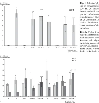
The results of measuring the concentrations of cadmium, zinc and copper in the kidneys are shown in Figure 1.
Table 3. Effect of physical training on concentration of MDA, GSH and SH groups in kidney of rats intoxicated with cad− mium, copper and copper and cadmium simultaneously (different doses of Cu), mean (± SD)
Tabela 3. Wpływ treningu fizycznego na stężenie MDA, GSH i grup SH w nerkach szczurów szczurów intoksykowanych kadmem, miedzią oraz kadmem i miedzią jednocześnie (różne dawki Cu), średnia (±SD)
No Groups of rats GSH SH−groups MDA
(Lp.) (Grupy szczurów) µmol/g tissue (Grupy SH) µmol/g tissue
n = 6 (µmol/g tkanki) µmol/g protein (µmol/g tkanki)
(µmol/g białka)
1. C 3.268 ± 0.174 186.38 ± 27.49 36.63 ± 6.63
2. Cd 2.557a± 0.087 151.40a± 10.56 43.54 ± 6.66
3. Cu2.5 2.911a± 0.201 154.67a± 8.27 51.07a± 4.97
4. CdCu0.5 2.841a, b± 0.238 160.28a± 10.06 51.92a, b± 4.15
5. CdCu2.5 2.493a, d, e± 0.117 158.41a± 14.26 47.96a, b± 5.80
6. CdT 2.792a, * ± 0.181 165.01a, * ± 8.41 51.45a± 6.41
7. Cu2.5T 2.856a± 0.215 152.87a± 9.12 44.10 ± 10.11
8. CdCu0.5T 2.787a, b± 0.218 152.32a, c± 10.48 48.15a± 10.87
9. CdCu2.5T 3.266b, c, d, e,* ± 0.230 145.28a, c± 11.87 54.51a, b, * ± 2.82
In all the groups of rats intoxicated with cad− mium (groups: Cd, CdCu0.5, CdCu2.5, CdT, CdCu0.5T, CdCu2.5T) the concentrations of Cd in the kidney were significantly higher compared to the control group.
In both trained and untrained rats co−exposed to cadmium and copper (CdCu0.5, CdCu2.5, CdCu0.5T, CdCu2.5T) the cadmium level was sig− nificantly lower in comparison to the group treat− ed only with cadmium (Cd), (p < 0.05).
In the groups treated only with copper, the concentration of cadmium in the kidney was simi− lar to the control group.
In rats intoxicated with cadmium alone, train− ing significantly increased the renal level of Cd (by 26%) in comparison with their untrained co− partners. In rats intoxicated with copper alone and co−exposed to cadmium and copper simultaneous− ly, exercise did not significantly alter the concen− tration of Cd in the kidneys.
The concentration of zinc in the kidneys was higher in the following groups: Cd, CdT and CdCu2.5T than the control group. The highest zinc
concentration was observed in the group exposed to cadmium and subjected to physical effort (CdT).
Training significantly elevated the renal level of Zn only in rats intoxicated with cadmium−only and those co−exposed to cadmium and a higher dose of copper. The concentration of zinc in the CdT group was higher by 60% than the Cd group. The zinc level in the CdCu2.5T group was higher by 45% than the CdCu2.5 group. In rats subjected to physical training and co−exposed to Cd and Cu (2.5 mg Cu/kg m.b.) the zinc concentration was higher in comparison with trained rats intoxicated with copper−only (the Cu2.5T group).
In the kidneys of rats exposed to metals (apart from the group intoxicated with Cd−only) the con− centration of copper was significantly increased compared to the control group, (p < 0.05).
The copper content in rats co−exposed to cad− mium and copper (the CdCu0.5, CdCu0.5T, CdCu2.5, and CdCu2.5T groups) was higher than in untrained rats intoxicated only with cadmium (Cd), (p < 0.05).
Fig. 1.Effect of physical train− ing on concentration of metals (Cd, Zn, Cu) in kidney of rats intoxicated with cadmium, cop− per and cadmium and copper simultaneously (different doses of Cu), mean ±SD: a) concen− tration of cadmium in kidney, b) concentration of zinc and copper in kidney
Ryc. 1.Wpływ treningu fizycz− nego na stężenie metali w ner− kach szczurów intoksykowanych kadmem, miedzią oraz kadmem i miedzią jednocześnie (różne dawki Cu), średnia ±SD: a) stę− żenie kadmu w nerkach, b) stę− żenie cynku i miedzi w nerkach 0
5 10 15 20 25 30 35 40
C Cd Cu2.5 CdCu0.5 CdCu2.5 CdT Cu2.5T CdCu0.5T CdCu2.5T
Cd
a b e a
a b
a*
a b e a b µ
µ g/g wet tissue g/g mokrej tkanki
groups of rats grupy szczurów
0 10 20 30 40 50 60 70
K Cd Cu2.5 CdCu0.5 CdCu2.5 CdT Cu2.5T CdCu0.5T CdCu2.5T
Zn Cu
a
a
a b a b d
b
a*
a* a
b
a b a b*
a b c d e* µ
µ
g/g wet tissue g/g mokrej tkanki
groups of rats grupy szczurów a)
The resuts show that exercise training signifi− cantly elevated the level of copper in the kidney.
The highest concentration of copper (five times higher than in the control group) – was observed in trained rats co−exposed to cadmium and a higher dose of copper (CdCu2.5T), p < 0.05. In groups subjected to physical effort, groups CdT and CdCu2.5T, the concentration of copper was significantly higher in comparison to analogical, untrained groups: Cd and CdCu2.5.
Moreover, in both trained and untrained rats co−exposed to cadmium and a lower dose of cop− per (CdCu0.5T and CdCu0.5) it could be observed that the concentration of copper was twice as high compared to the control group (p < 0.05).
In trained rats intoxicated only with cadmium (CdT) a higher concentration of Cu compared to the control group (p < 0.05) could be found.
In both untrained rats intoxicated with a high dose of copper−only (Cu2.5), and those co− exposed to cadmium and higher dose of copper (CdCu2.5), the content of copper in the kidney
was four− and three−times higher respectively in comparison to the control group (C), p < 0.05.
The concentration of copper in the CdCu2.5T group was higher (by 30%) compared to the Cu2.5 group, (p < 0.05).
The levels of copper in rats co−exposed to cad− mium and a higher dose of Cu (CdCu2.5, CdCu2.5T) were higher compared to the analogi− cal rats co−exposed to cadmium and a lower dose of Cu (CdCu0.5, CdCu0.5T), (p < 0.05).
However, no significant differences in Cu con− centration was observed between trained rats intoxicated only with cadmium (CdT) and trained rats co−exposed to cadmium and a lower dose of copper (CdCu0.5T).
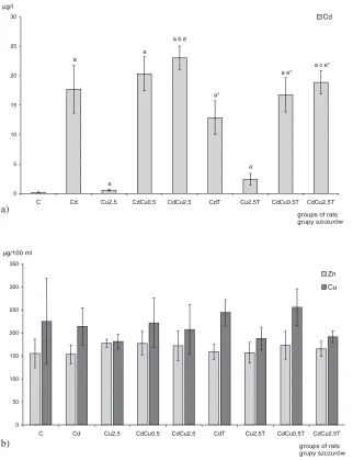
The results of measuring the concentrations of cadmium in the whole blood and zinc and copper in serum are shown in Figure 2.
Serum cadmium levels were significantly higher in trained and untrained rats, both intoxi− cated with cadmium or co−exposed to cadmium and copper, than in the control group.
Fig. 2. Effect of physical train− ing on concentration of metals
(Cd, Zn, Cu) in blood of rats intoxicated with cadmium, cop− per and copper and cadmium simultaneously (different doses of Cu), mean ±SD: a) concen− tration of cadmium in blood of rats, b) concentration of zinc and copper in serum of rats
Ryc. 2.Wpływ treningu fizy− cznego na stężenie metali we krwi szczurów
intoksykowanych kadmem, miedzią oraz kadmem i miedzią jednocześnie (różne dawki Cu). Średnia (±SD): a) stężenie kadmu we krwi szczurów, b) stężenie cynku i miedzi w surowicy szczurów
0 5 10 15 20 25 30
C Cd Cu2.5 CdCu0.5 CdCu2.5 CdT Cu2.5T CdCu0.5T CdCu2.5T
Cd
a b e
a
a
a
a*
a e*
a
a c e* µg/l
groups of rats grupy szczurów
0 50 100 150 200 250 300 350
C Cd Cu2.5 CdCu0.5 CdCu2.5 CdT Cu2.5T CdCu0.5T CdCu2.5T
Zn
Cu
µg/100 ml
groups of rats grupy szczurów a)
The highest blood cadmium concentration was found in untrained rats intoxicated with cadmium and a higher dose of copper (CdCu2.5).
The results show that physical training signif− icantly alters the blood levels of cadmium. Training significantly decreased the blood levels of Cd in rats both intoxicated with cadmium−only and co−exposed to Cd and Cu, in comparison with their untrained co−partners.
The serum concentration of Cu and Zn in experimental groups was similar to the control group.
Correlation Coefficients (
r
−
values) Between Blood and
Renal Levels of Metals (Cd,
Zn, Cu)
The r−values and levels of significant (p) were extracted in Table 4. Positive, significant correla− tions were found between the renal and blood con− tent of cadmium (r = 0.59, p = 0.000004) and also between the renal concentration of cadmium and the renal concentration of zinc (r = 0.56, p = 0.000017).
Correlation Coefficients
(
r
−values) Between Renal
Biochemical Parameters
and Levels of Metals
(Cd, Zn, Cu) in Blood
and Kidney
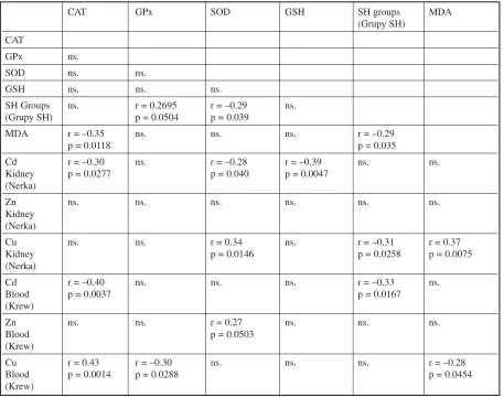
The r−values and levels of significant (p) were extracted in Table 5. Negative correlations were found between the renal activity of catalase and the concentrations of cadmium in the kidney (r = –0.30, p = 0.0277) and in the blood (r = –0.40, p = 0.0037) and also between the renal activity of cata− lase and renal concentrations of MDA (r = –0.35, p = 0.0118).
Positive correlations were observed between the blood content of Cu and the renal activity of catalase (r = 0.43, p = 0.0014), however negative correlations were found between the blood content of Cu and the renal activity of GPx (r = –0.30, p = 0,0288).
Negative correlations were found between the renal activity of SOD and the renal concentrations of cadmium (r = –0.28, p = 0.040) and also between the renal activity of SOD and the content of SH groups (r = –0.29, p = 0.039).
Table 4.Correlations between levels of metals (Cd, Zn, Cu) in blood and kidney (rats from all groups)
Tabela 4. Zależności między stężeniami metali (Cd, Zn, Cu) we krwi i nerkach (szczury ze wszystkich grup łącznie)
Cd Zn Cu Cd Zn
Kidney Kidney Kidney Kidney Kidney
(Nerka) (Nerka) (Nerka) (Nerka) (Nerka)
Cd Kidney (Nerka)
Zn r = 0.56
Kidney p = 0.000017 (Nerka)
Cu ns. ns.
Kidney (Nerka)
Cd r = 0.59 ns. ns.
Blood p = 0.000004 (Krew)
Zn ns. ns. ns. ns.
Blood (Krew)
Cu ns. ns. ns. ns. ns.
Blood (Krew)
Renal SOD activity and the copper content in kidney were found to show a significant, positive correlation (r = 0.34, p = 0.0146).
Renal GSH content and the renal concentra− tions of cadmium were found to show a signifi− cant, negative correlation (r = –0.39, p = 0.0047).
Moreover, negative correlations were observed between the renal content of SH groups and the renal concentration of MDA (r = –0.29, p = 0.035); the renal concentration of copper (r = –0,31, p=0,0258) and also the cadmium concentration in the blood (r.= –0.33, p = 0.0167)
Positive correlations were found between the renal content of copper and the renal concentration of MDA (r = 0.37, p = 0.0075), however negative correlations were found between the blood content of Cu and the renal concentration of MDA (r = –0.28, p = 0.0454).
Discussion
Cadmium is a widespread and very toxic metal. It promotes oxidative stress and afterwards con− tributes to the development of serious pathological changes in the kidney because of its long retention in this organ [1, 2]. In industrial areas, people are exposed to several heavy metals simultaneously. Interactions between metals can lead to increasing or alleviating toxic effects caused by metals.
According to expectations, the results of our investigations show that the ingestion of cadmium chloride (50 ppm Cd in drinking water) for a peri− od of 15 weeks caused an increase in Cd concen− tration in the blood, the accumulation of cadmium in the kidney and the elevation of renal lipid per− oxidation.
In rats co−exposed to cadmium and copper (both higher and lower doses) we observed
Table 5.Correlations between renal biochemical parameters and levels of metals (Cd, Zn, Cu) in blood and kidney
Tabela 5.Zależności między stężeniami metali (Cd, Zn, Cu) i badanymi parametrami biochemicznymi we krwi i nerkach szczurów (szczury ze wszystkich grup łącznie)
CAT GPx SOD GSH SH groups MDA
(Grupy SH) CAT
GPx ns.
SOD ns. ns.
GSH ns. ns. ns.
SH Groups ns. r = 0.2695 r = –0.29 ns.
(Grupy SH) p = 0.0504 p = 0.039
MDA r = –0.35 ns. ns. ns. r = –0.29
p = 0.0118 p = 0.035
Cd r = –0.30 ns. r = –0.28 r = –0.39 ns. ns.
Kidney p = 0.0277 p = 0.040 p = 0.0047
(Nerka)
Zn ns. ns. ns. ns. ns. ns.
Kidney (Nerka)
Cu ns. ns. r = 0.34 ns. r = –0.31 r = 0.37
Kidney p = 0.0146 p = 0.0258 p = 0.0075
(Nerka)
Cd r = –0.40 ns. ns. ns. r = –0.33 ns.
Blood p = 0.0037 p = 0.0167
(Krew)
Zn ns. ns. r = 0.27 ns. ns. ns.
Blood p = 0.0503
(Krew)
Cu r = 0.43 r = –0.30 ns. ns. ns. r = –0.28
Blood p = 0.0014 p = 0.0288 p = 0.0454
(Krew)
a decrease in the cadmium concentration in kid− neys and an increase of Cd concentration in the blood. It means that copper prevents the accumu− lation of cadmium in kidneys. Other researchers had already observed this phenomenon [18].
In our study, we did not find any differences in renal and blood cadmium concentration depending on the dose of copper.
The training program used in our investiga− tions caused a decrease in cadmium concentration in rats’ kidneys and blood. These results are con− sistent with other authors’ conclusions [15, 19]. It seems that systematic physical effort interferes with cadmium accumulation in kidneys and /or accelerates its elimination from the organism.
The influence of physical training on the decrease of cadmium concentration in kidneys was significant in the case of rats which were intoxi− cated only with cadmium. No changes were observed after physical training in the case of rats intoxicated with cadmium and copper (both high− er and lower doses) simultaneously. However, at this stage of the study, it is difficult to conclude the role of copper in these processes.
The administration of cadmium alone, as well as the administration of cadmium and a higher dose of copper simultaneously, caused the modification of zinc metabolism and an increase of its concentration in kidneys. There was a significant, positive correla− tion between Cd and Zn concentrations in kidneys (r = 0.56, p = 0.000017). Oishi et al [20] obtained similar results. This phenomenon can be explained by various mechanisms. The increase of zinc con− centration in the kidney mobilizes the defence sys− tem against cadmium toxicity. Metalothionein and zinc act as antioxidants and influence the stabilisa− tion of tissue membranes. Zinc is also a component of Zn, Cu SOD (Zn, Cu dismutase superoxide), which neutralises reactive superoxide radicals [11]. A higher increase in the renal concentration of zinc was found in kidneys of animals subjected to the physical training. Mishima et al [21], suggest that protective−acting mechanisms of zinc against cadmium toxicity depend on zinc concentration. In the case of higher concentrations, zinc inductes the synthesis of MT and at lower concentrations, in spite of a significant protective effect, the rise of this protein concentration was not observed.
According to these authors, the protective effect of low Zn doses results from the similarities between physical and chemical specificities of zinc and cadmium.
Zinc imitating cadmium ions, accelerate the stabilization of equality between internal and external concentration of cadmium and it is also possible that zinc disturbs the process of cadmium to passing through biological membranes.
The aforementioned research [21] suggest that protective mechanisms, independent from MT, play a more important role in natural physiological conditions.
Milnerowicz [19] has observed that lower Cd concentration in kidneys, during physical training, was accompanied by the lowering of MT content.
It is possible that the mobilization of zinc in kidneys, during physical effort, lowers the toxicity of cadmium resulting from mechanisms indepen− dent from MT.
Systematic physical effort also influences cop− per metabolism. In the case of rats intoxicated only with cadmium, physical training caused the increase of copper concentration in kidneys. It can be considered also as a positive effect because copper, similar to zinc, is an important component of the antioxidant defence system and induction MT synthesis, which bind reactive cadmium ions.
In animals intoxicated with cadmium and a lower dose of copper, the renal concentration of copper did not change significantly during train− ing.
However, in the case of rats co−exposed to cadmium and a higher dose of copper, physical effort caused a further increase in the renal content of copper.
It seems that physical effort escalates patho− logical changes induced by cadmium and a high dose of copper, and additionally it disrupts copper metabolism and enlarges the retention of Cu in the kidney.
In rats exposed to cadmium and cadmium and copper simultaneously (both higher and lower doses) we observed a significant decrease of cata− lase.
The depletion of catalase activity in the kidney correlated with the rise of blood and renal concen− tration of cadmium and also with the rise in renal concentration of products of lipid peroxidation (MDA).
These dependences confirm the influence of cadmium on the decrease of catalase activity and induce oxidative processes. The inhibitory effect of cadmium on catalase activity was observed in other investigations [22].
The data show that exercise training on tread− mill increased the renal activity of catalase in rats treated with cadmium and co−exposed to cadmium and a lower dose of copper. A higher growth in the activity of catalase was observed in the group co− exposed to cadmium and a lower dose of copper. By contrast, in rats co−exposed to cadmium and a higher dose of copper, CAT activity decreased with training.
Cu synergistically affects an increase of catalase activity. In the presence of a high renal concentra− tion of copper, training additionally intensified the inhibition of CAT activity.
The activity of GPx decreased in the kidneys of trained rats exposed to cadmium and copper. In the case of rats intoxicated with a higher dose of copper, it might also be a consequence of an inter− action between Cd, Cu and physical effort.
However, in the case of rats intoxicated with a smaller dose of copper, a fall in activity of glu− tathione peroxidase under the influence of training can be explained by the compensatory rise of cata− lase.
In trained rats exposed to cadmium alone the activity of SOD was lower than in untrained co− partners.
The activity of SOD increased in groups exposed to a higher dose of copper alone, and also in group co−exposed to cadmium and a higher dose of copper, and did not exhibit any further changes during training.
The rise of SOD activity and the significant neg− ative correlation between the activity of this enzyme and the concentrations of SH−groups (r = –0.29, p = 0.039) indicate a considerable generation of superoxide radical, which causes oxidative dam− ages of thiol group.
The activation of the antioxidative defence system connected with the rise of SOD activity was insufficient therefore in rats intoxicated with a higher dose of copper, where a significant eleva− tion in the concentration of lipid peroxidation products could be observed. The level of oxidative stress (measured as MDA) concentration was greater in rats subjected to physical training than in their untrained co−partners.
The highest activity of SOD (twice higher than in the control group) was found in rats intoxicated only with a high dose of copper. An increase of renal SOD activity positively correlated with renal copper (r = 0.34, p = 0.0146) and negatively with cadmium content in the kidney(r = –0.28, p = 0.040).
These associations indicate an important role of renal copper in both the antioxidative defence system and oxidative processes.
During the intoxication with metals, we observed a decrease of GSH content in kidney Glutathione status shown to have an impact on the ability of the body to handle heavy metals such as cadmium and copper. Glutathione is an important antioxidant. When glutathione status is elevated or increased by supplementation, the tissues were able to stop the damage caused by the lipid perox− ides induced to the metals [23–25].
The negative correlation between the content of GSH and the concentration of cadmium in the
kidney confirms the escalation of oxidative processes by Cd and the participation of reduced glutathione in the neutralization of toxic Cd ions. The data of the present study shows that exercise training significantly increased the concentration of GSH but only in rats intoxicated with Cd alone and co−exposed to cadmium and a high dose of copper
The increase of GSH content in the kidneys of trained rats co−exposed to cadmium and a high dose of copper, can be explained by the inhibition of GPx activity, which uses reduced glutathione as a substrate.
According to expectations, the level of oxida− tive stress (measured as the elevation of MDA concentration and the decline of SH−groups) increased in rats treated with metals.
The depletion of SH−groups’ concentration correlated with the increase in the renal concentra− tion of copper and MDA and with blood cadmium concentration.
A positive correlation was found between the renal concentration of MDA and copper content in kidneys. However, negative correlations were found between the renal concentration of MDA and blood content of Cu.
The highest increase of products of lipid per− oxidation and the highest decline of SH−groups were observed in the kidneys of rats subjected to physical training and co−exposed to cadmium and a high dose of copper.
The results of this study show that physical training significantly alters blood and renal levels of cadmium. Training significantly decreased plas− ma Cd levels in rats intoxicated with cadmium or co−exposed to cadmium and copper in comparison with their untrained equivalens. Physical training reduces the concentration of cadmium in kidneys and also increases the renal concentration of zinc (a nutrient which protects against cadmium toxici− ty), especially in animals exposed to cadmium alone. Systematical physical exercise intensifies the antioxidant defence system. We observed a higher concentration of reduced glutathione, SH−groups and activity of CAT in trained rats compared to their untrained equivalents.
In rats which were intoxicated by cadmium and a higher dose of copper, training only enlarged the oxidative stress induced by these metals and lowered the activity of antioxidative enzymes such as GPX and CAT.
However, physical training intensifies the antioxidant defence system, but does not reduce lipid peroxidation.
The results of this study show that the influ− ence of physical training on oxidative processes and antioxidant defences depend on doses of met− als and their concentrations in tissues.
References
[1]WHO, Cadmium, Environmental Health Criteria, 134, 1992.
[2] Jarup L: Health effect of cadmium exposure – a review of literature and risk estimate. Scan J Work Environ Health 1998. 24 (Suppl.), 1–52.
[3] Stohs SJ, Bagchi D:Oxidative mechanisms in the toxicity of metal ions. Free Rad Biol Med 1995, 18, 321–336. [4] Peraza MA, Ayala−Fierro F, Barber DS, Casarez E, Rael LT: Effects of micronutrients on metal toxicity.
Environ Health Perspect 1998, 106 (Suppl 1), 203–216.
[5] Satarug S, Baker JR, Reilly PEB, Moore MR, Williams DJ:Changes in zinc and copper homeostasis in human liver and kidneys associated with exposure to environmental cadmium. Hum Exp Toxicol 2001, 20, 205–213. [6]International Programme on Chemical Safety, Environmental Health Criteria. 200−COPPER (raport – praca
zbiorowa). WHO, Geneva 1998.
[7] Bremner I: Manifestation of copper excess. Am J Clin Nutr 1998, 67 (Suppl.), 1069–1073.
[8] Uauy R, Olivares M, Gonzalez M:Essentiality of copper in humans. Am J Clin Nutr 1998, 67 (Suppl.), 952–959. [9] Peraza MA, Ayala−Fierro F, Barber DS, Casarez E, Rael LT: Effects of micronutrients on metal toxicity.
Environ Health Perspect 1998, 106 (Suppl. 1), 203–216.
[10] Liu XY, Jin TY, Nordberg GF, Rännar S, Sjöström M, Zhou Y:A multivariate study of protective effects of Zn and Cu against nephrotoxicity induced by cadmium metallotionein in rats. Toxicol Appl Pharmacol 1992, 114, 239–245.
[11] Brzóska MM, Moniuszko−Jakoniuk J:Interaction between cadmium and zinc in the organizm. Review Food Chem Toxicol 2001, 967–980.
[12] Speich M, Pineau A, Ballereau F:Minerals, trace elements and related variables in athletes and during physical activity. Clin Chim Acta 2001, 312, 1–11.
[13] Johnson P:Antioxidant enzyme expression in health and disease: effects of exercise and hypertension. Review. Comp Biochem, Physiol Part C 2002, 133, 493–505.
[14] Elsayed NM:Antioxidant mobilization in response to oxidative stress: a dynamic environmental−nutritional inter− action. Nutrition 2001, 17, 828–834.
[15] Rodriguez TJ, Pinilla GE, Maynar MM, Garcia Monco C, Sanches MA:Evaluation of the influence of phys− ical activity on the plasma concentrations of several trace metals. Eur J Appl Physiol 1996, 73, 3–4, 299–303. [16] Aebi H:Catalase in vitro.Meth Enzymol 1984, 105, 121–126.
[17] Ellman GL:Tissue sulfhydryl groups. Arch Biochem Biophys 1959, 82, 70–77.
[18] Shaikh ZA, Blazka ME, Endo T:Metal Transport in Cells: Cadmium uptake by rat hepatocytes and renal corti− cal epithelial cells. Environ Health Perspect 1995, 103 (Suppl. 1), 73–75.
[19] Milnerowicz H, Nowak P, Wochyński Z, Sobiech KA: Wpływ kadmu i wysiłku fizycznego na wybrane mar− kery w tkankach szczura. I Metalotioneina. Nowa Med. 2000, 12, 108, 1–3.
[20] Oishi S, Nakagawa JI, Ando M:Effects of cadmium administration on the endogenous metal balance in rats. Biol Trace Elem Res 2000, 76, 257–278.
[21] Mishima A, Yamamoto C, Fujiwara Y, Kaji T: Tolerance to cadmium cytotoxicity is induced by zinc through non−metallothionein mechanisms as well as metallothionein induction in cultured cells. Toxicology 1997, 118, 85–92.
[22] Jurczuk M, Brzóska MM, Moniuszko−Jakoniuk J, Gałażyn−Sidorczuk M, Kulikowska−Karpińska E: Antioxidant enzymes activity and lipid peroxidation in liver and kidney of rats exposed to cadmium and ethanol. Food Chem Toxicol 2004, 42, 967–980.
[23] Freedman JH, Cricolo MR, Peisach J: The role of glutation in copper metabolizm and toxicity. J Biol Chem 1989, 264, 10, 5598–5605.
[24] Bartosz G:Druga twarz tlenu. Wydawnictwo Naukowe PWN, Warszawa 1999.
[25] Congiu L, Chicca M, Pilastro A, Turchetto Tallandini L:Effects of chronic dietary cadmium on hepatic glu− tathione levels and glutathione peroxidase activity in starlings. Arch Environ Contam Toxicol 2000, 38, 357–361.
Address for correspondence:
Iwona Markiewicz−Górka Department of Hygiene
Silesian Piasts University of Medicine J. Mikulicza−Radeckiego 7
50−345 Wrocław Poland
Tel.: +48 71 784 15 08
Conflict of interest: None declared