Turkish Journal of Fisheries and Aquatic Sciences 16: 463-469 (2016)
www.trjfas.org ISSN 1303-2712 DOI: 10.4194/1303-2712-v16_2_26
RESEARCH PAPER
© Published by Central Fisheries Research Institute (CFRI) Trabzon, Turkey in cooperation with Japan International Cooperation Agency (JICA), Japan
MRF Gene Family in Schizothorax Prenanti: Molecular Cloning, Tissue
Expression, and mRNA Expression in Muscle Development
Introduction
Growth rate is one the economically important trait in animals and involves the recruitment and hypertrophy of muscle fibers (Johnston, 1999). Myogenesis is regulated by a group of muscle-specific transcription factors that is known as the Myogenic Regulatory Factors (MRFs), including myoblast determination protein (MyoD), myogenin (MyoG), Myogenic factor 5 (Myf5) and Myogenic factor 6 (Myf6; also known as MRF4) (Braun and Gautel, 2011).
MRFs are involved in muscle development through muscle fiber formation during embryonic development to their postnatal maturation and function (Te Pas et al., 1999). The MRFs are specifically expressed in skeletal muscle linage and can convert several cell lines to differentiated skeletal muscle (Weintraub et al., 1991). However, every gene has gained a unique expression pattern and specialist function in initiating or maintaining myogenesis. Myf5 and MyoD are myogenic determination factors and activate myoblast determination in proliferating myoblasts before overt differentiation. On the contrary, MyoG and Myf6 are myogenic
differentiation factors that contribute to the differentiation of myoblasts and play an important role in the downstream molecular pathway of Myf5 and MyoD, although Myf6 partly acts at both the determination and differentiation levels (Buckingham and Vincent, 2009; Bryson-Richardson and Currie, 2008; Bentzinger et al., 2012). Up to date, several reports have described that the functions of MyoD gene family in fish, such as Zebrafish (Danio rerio) (Lin et al., 2006; Yaniv et al. 2011), Tilapia (Oreochromis niloticus) (Zou et al., 2015) and Atlantic salmon (Salmo salar L.) (Macqueen et al., 2007), seem to be similar to those reported in mammals. The Schizothorax prenanti (S.prenanti), an endemic cold-water fish, is popular cultured fish in Southwest China and has become an important food fish because of its good meat quality and wide acceptance in the markets. However, this fish displays remarkably low growth rates in nature (Song et al., 2006). Given the critical roles that MRFs play in myogenesis and the expression of MRFs is associated with skeletal muscle growth, it is important to study the roles of MRFs in regulating the muscle development to accelerate the growth rate in the S. prenanti and the research about this in this fish is
Yaqiu Lin
1,*, Jishu Zhou
2, Ruiwen Li
3, Xianrong Xiong
2, Pengfei Wu
11 Southwest University for Nationalities, College of Life Science and Technology, Chengdu, 610041, P.R. China. 2Northwest A & F University, College of Animal Science and Technology, Yangling 712100, P.R. China. 3 Chengdu Woman-Child Central Hospital, Reproductive and endocrine laboratory, Chengdu 610091,P.R. China.
* Corresponding Author: Tel.: +86.028 85528275; E-mail: [email protected]
Received 28 July 2015 Accepted 01 May 2016
Abstract
The myogenic regulatory factors (MRFs) play key roles in growth and muscle development in mammals and some fish, however previous studies about the cloning, tissue expression and mRNA expression in muscle development of MRFs genes in Schizothorax prenanti (S.prenanti) was scarce. In order to know how MRFs genes were expressed in different tissues and development period, we cloned the complete CDS of the MRF gene family from S.prenanti for the first time, including MyoD,
MyoG, Myf5 and Myf6. Amino acid sequence alignment indicated that the S.prenanti MRF gene family has a basic helix-loop-helix (HLH) domain. Quantitative real-time RT-PCR analysis indicated that MyoD, MyoG, Myf5 and Myf6 mRNA was highly expressed in red muscle and white muscle. The mRNA expression of MRF family in muscle during different developmental stages (larvae, juvenile and adult) in vivo were also investigated. The results showed that MyoG and Myf5
expression in muscle were significantly associated with muscle development. Our findings provided molecular characterization and expression profile of the MRF gene family in S.prenanti and enhanced understanding about the MRFs in muscle growth and development in fish.
scarce. In order to study the characterization of MRFs of S. prenanti and provide a foundation for understanding the molecular control of skeletal muscle growth in fish species, MRFs, including MyoD, MyoG, Myf5 and Myf6, were cloned and characterized and tissue-specific expression patterns as well as the expression profile of MRFs in muscle development was also investigated.
Materials and Methods
Fish
The S. prenanti used in this study were commercially obtained and reared temporally at LU SHAN Farm (Yaan, Sichuan Province, China). The three adult fish were used for the gene cloning. The red muscle and white muscle from these fish were removed on ice, and rapidly frozen in liquid nitrogen, and stored at 80 oC for RNA isolation. The S. prenanti was obtained from the same broodstock with the first group and was used to investigate mRNA expression patterns of various tissues (n=6) and during different developmental stages(n=6, for each). Three different developmental stages were chosen: larvae (8.4 ± 0.9 g/tail, 3 month old), juvenile (50 ± 3.5 g/tail, 10 months old) and adult (500 ± 12 g/tail, 2 year old). S. prenanti were maintained in indoor cylindrical fiberglass tanks at ambient temperature of around 15 ± 3 oC with a natural dark/light cycle.
Total RNA Extraction and cDNA Synthesis of the First Chain
Total RNA was isolated from fish red muscle and white using TRIzol Reagent (Invitrogen, Carlsbad, CA, USA) according to the instructions of the manufacturer. The purity and the concentration of total RNA were measured by a spectrophotometer at 260 and 280 nm. The ratios of absorption (260/280 nm) for all samples were approximately 2.0. The integrity was tested by electrophoresis in formaldehyde agarose gels. Corresponding cDNAs were synthesized by reverse transcription using the M-MLV reverse transcriptase (Thermo, Shanghai, China)
Molecular Cloning and Sequence Analysis of MRFs Genes in S. prenanti
In order to obtain the full-length S.prenanti MyoD, MyoG, Myf5, and Myf6 sequences, degenerate primers (Table 1), designed based on the most conserved regions of the fish MyoD, MyoG, Myf5, and Myf6 sequences available in the GenBank. The PCR parameters were 39 cycles at 94 ℃ for 30 s, 56-59.5 ℃ for 30s, and 72 ℃ for 2 min, with an additional initial 3-min denaturation at 94 ℃ and a 10-min final extension at 72 ℃.
Products of the degenerate PCR reactions were electrophoresed on 1.2 % agarose gel. Bands of
expected size was purified with the PCR Purification Kit (TIAN GEN, Beijin, China). The purified fragments were then cloned into a PMD19-T vector following the manufacture’s instructions (TaKaRa, Dalian, China) and propagated in E. coli DH5α. For each fragment, five clones were sequenced in both directional by Shanghai Sangon Biological Engineering Technology (Shanghai, China).
Sequence and Phylogenetic Analysis
Sequence alignment and analysis were conducted using the BLAST sequence analysis service of the National Center for Biotechnology Information (http://www.ncbi.nlm.nih.gov). Multiple protein sequence alignments were aligned using the ClustalW program. The predicted protein sequence
was analyzed by ORF Finder
(http://www.ncbi.nlm.nih.gov/projects/gorf/).
Phylogenetic tree was constructed by neighbor-joining method from a distance matrix calculated with the Mega 5.0 software.
Tissue Expression of MyoD, MyoG, Myf5, and Myf6 Genes in S. prenanti
Six S. prenanti were randomly selected and killed by anesthetized with MS222 (100 mg L-1). Tissue samples (brain, heart, liver, kidney, spleen, red muscle and white muscle) for RNA collection were quickly frozen in liquid nitrogen and stored at −80 °C until RNA analysis. Total RNA was extracted as described above. The extracted and purified total RNA was treated with RNase-free DNase to prevent the genomic DNA amplification. One microgram of total RNA were used to synthesize the first-strand cDNA by RT-PCR as described above. Quantitative real-time PCR (qPCR) assays were carried out in a quantitative thermal cycler (MyiQ™ 2 Two Color Real-Time PCR Detection System, BIO-RAD, USA) with a 20 μL reaction volume containing 10 μL SYBR®Premix Ex Taq™ II (TaKaRa, Japan), 1 μL of diluted cDNA (10-fold), 10 mM each of forward and reverse primers 0.64 μL, and 7.2 μL H2O. Primers are given in Table 1. The procedure was: 1 cycle of 94℃
for 2 min, 28 cycles of 94℃ for 30 s, 60℃ for 50 s, and 72℃ for 1 min, and a final extension of 10 min at 72℃. Actin mRNA was used as the internal control.
Expression Patterns of MRFs Genes in 3 Growth Stages of S. prenanti
Total RNA of muscle in three different growth stages of S. prenanti (n=6, for each) were extracted respectively and the expression patterns of MRFs were determined by qPCR, as described above.
Statistical Analysis
expressed as mean ± standard error (SE) and were analyzed using one-way analysis of variance (ANOVA). Differences were considered to be significant if P< 0.05.
Results
Cloning and Sequence Analysis of MRFs Genes in
S. prenanti
Complete CDS sequences of MyoD (JQ793894.1), MyoG (KM196536.1), Myf5 (JQ793895.1) and Myf6 (KM196535.1) genes in S. prenanti were obtained by RT-PCR. These genes contained an open reading frame of 825 bp, 762 bp,723 bp 720 bp respectively and encoded a predicted protein of 274, 253, 240, 239 amino acids respectively. High conservation, Helix-loop-helix domain, was founded among these genes (Figure 1).
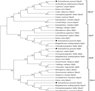
The phylogenetic analysis based on the deduced amino acid of the cloned S. prenanti MyoD, MyoG, Myf5, Myf6 and other fish's showed that the sequence s of fish MyoD, MyoG, Myf5 and Myf6 were conserv ative, suggesting that the cloned S. prenanti MyoD, M yoG, Myf5, and Myf6 were indeed these genes (Figure 2).
Tisscue Expression of MRFs in S. prenanti
The mRNA expression levels of MyoD, MyoG, Myf5 and Myf6 gene in 7 tissues were analyzed by qPCR. The MRFs genes were significantly highly expressed in both red muscle and white muscle and they were little expressed in heart, liver, spleen, kidney and brain (Figure 3).
Expression Patterns of MRFs in 3 Growth Stages of S. prenanti
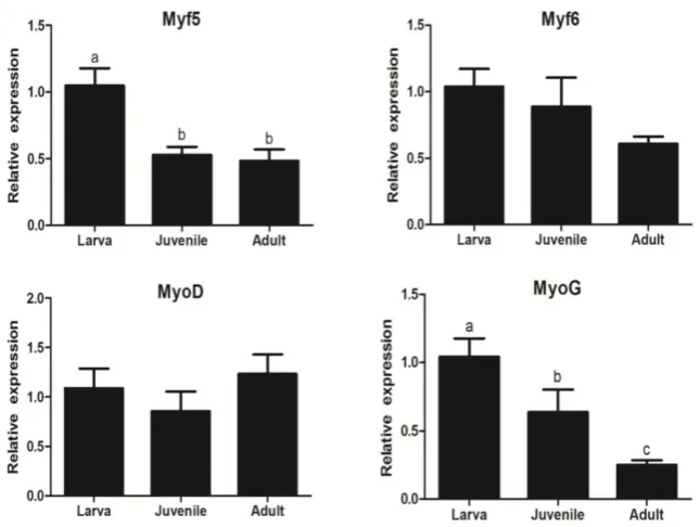
The MyoD, MyoG, Myf5 and Myf6 expression in
muscle of S. prenanti were detected in 3 growth stages of fish. The mRNA expression of MyoD and Myf6 was not significantly different in this 3 growth stages, while the Myf5 gene expression levels in larvae were significantly higher than that in juvenile and adult growth stages and MyoG gene expression also decreased from larvae to adult, indicating that Myf5 and MyoG is more sensitive to the growth of fish than the other two genes (Figure 4).
Discussion
The important roles of MRF gene family in mammalian skeletal myogenesis have been well studied, including MyoD, MyoG, Myf5 and Myf6. The MRF genes, a family of HLH transcription factors, has the HLH
domain that can bind to an E-box sequence (CANNTG) which exists in the promoters or enhancers to activate the transcription of these muscle-related genes to regulate the growth and muscle development (Yafe et al., 2008). In the present study, the complete CDS sequences of S. prenanti MyoD, MyoG, Myf5 and Myf6 gene were cloned. Amino acid sequence alignment indicated that the S.prenanti MRF gene family also has a basic helix-loop-helix (HLH) domain. MyoD, MyoG, Myf5 and Myf6 were mainly expressed in muscle and play a major role in muscle development (Zhu et al., 2016). Our result, their high expression in muscle of S. prenanti, was in keeping with this. The high conservation of the HLH region in vertebrates and their highly expressed in muscle demonstrate that these four transcription factors may keep maintaining the fundamental role in muscle development in S. prenanti.
The four MRF gene family members are divided into two functional groups: the primary includes MyoD and Myf5, and the secondary includes MyoG and Myf6. The MyoD and Myf5 are required for the
Table 1. Specific Primers used for cDNA cloning and qPCR of MRF gene family from Schizothorax prenanti
Names Sequences Annealing temperature(℃) Utilizations
MyoD-F1 5'- ACACATAAAGATGGAGTTGTCG -3' 56
56
For cloning
MyoD-R1 5'- CAGCAGTGGATCGGAATAGT -3'
Myf5-F1 5'- ATGGACGTATTCTCTACATCCC -3'
Myf5- R1 5'- TCACAGGACGTGGTAGACTG -3'
MyoG-F1 5'- TCTCGCAAGAACCGCAAAG -3' 57.5
MyoG -R1 5'- TATGGTCGGTGAAAGGTGGTC -3'
Myf6-F1 5'-ATGATGGACCTGTTTGAGACC -3' 59.5
Myf6-R1 5'-TCACTTCTCTGAGATCTGGCTG -3'
MyoD-F2 5'-TCTGAGAAACGCCATTAGTTACATC -3' 61
For qPCR
MyoD-R2 5'-GCTGTCATAACTGTTCCGTCTTCT-3'
Myf5-F2 F:5'- TCTGAAGAGGACGAGCACAT - 3' 57.8
Myf5-R2 R:5' - AAGACGCTGACTGGGGTT - 3'
MyoG -F2 5' - TTTACGAAGGCGGCGATAAC - 3' 62
MyoG-R2 5'- AGTGCTGCTGCTCCTGGTGA - 3'
Myf6-F2 5' -TGCGATGGGCAGTGTCTTATG -3' 62
Myf6-R2 R:5' -CAGCCTCTGGTTCGGATTGG -3'
β-actin-F 5’-GATTCGCTGGAGATGATGCT-3’ 58
Endogenous control
determination of skeletal myoblasts, whereas the MyoG and Myf6 act as differentiation factors (Megeney and Rudnicki 1995; Wyszynska-Koko et
al., 2006). In this study, we found different MRFs expression patterns in muscle at different developmental stages. In mice, the MyoD and Myf5
Figure 1. The protein sequence alignment of the MRF gene family in S. prenanti using ClustalW. The helix-loop-helix domain are boxed.
Figure 2. Phylogenetic analysis of MRF gene family. Phylogenetic tree based on protein sequences was constructed by neighbour-joining methodwith Mega 5.0 software. The strength of branch relationships was assessed by bootstrap
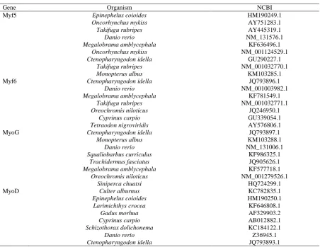
replication (N = 1000 replicates). S. prenanti MyoD, Myf5, Myf6 and MyoG were indicated by ‘◆’. Accession numbers
were highly expressed before embryonic day 21, indicating that these genes regulated satellite cells and induce muscle development (Holterman et al. 2007; Kanisicak et al. 2009; Schnappet al. 2009). Our results showed that Myf5 gene expression levels in larvae were very significantly higher than that in
juvenile and adult, whereas the mRNA expression of MyoD was not distinctly different at different developmental stages. This indicates that Myf5 plays a more critical role during the early determination of muscle in S. prenanti, although Myf5 and MyoD have redundant functions in myoblast determination and
Figure 3. Analysis of MyoD, Myf5, Myf6 and MyoG gene expression among different tissues in Schizothorax prenanti by quantitative realtime PCR (mean±SEM, n =6), respectively. Data were normalized to housekeeping gene (β-actin) expressed as a ratio of the control.
can compensate for the functional loss of each other (Kablar et al., 1998; Parker et al., 2003). MyoG is related to muscle differentiation, while Myf6 has the function of maintaining the status of differentiated myofibers (Liu et al., 2012). In our study, MyoG showed decreasing expression from larvae to adult and the Myf6 was not distinctly different at different developmental stages. These data indicates that muscle differentiation is predominance from larvae to adult. Different MRF gene family expression pattern in muscle at different developmental stages showed their unique function in growth and muscle development of S. prenanti. And their unique expression pattern may be associated with the characteristics of slow growth of S. prenanti.
In conclusion, MyoD, MyoG, Myf5 and Myf6 of S. prenanti have been identified and the structural features have been characterized. Quantitative expression of MyoD, MyoG, Myf5 and Myf6 was highly expressed in red muscle and white muscle, suggesting that these genes could play a major role in muscle. The expression profiles of MRF gene family in muscle development were observed. These data emphasize the need to further explore the complex physiological processes involved in the growth and muscle development in fish.
Acknowledgments
This work was supported by Applied Basic Research Program of Sichuan Province, China (No.2014JY0088), National Natural Sciences Foundation of China (No.31201990) and the Fundamental Research Funds for the Central Universities (2014NZYTD01).
References
Bentzinger, C.F., Wang, Y.X. and Rudnicki, M.A. 2012. Building muscle: molecular regulation of myogenesis. CSH Perspect Biol, 4(2): a008342. doi: 10.1101/cshperspect.a008342;
Braun,T. and Gautel, M.2011.Transcriptional mechanisms regulating skeletal muscle differentiation, growth and homeostasis. Nat Rev Mol Cell Biol, 12: 349-361. doi:10.1038/nrm3118;
Bryson-Richardson, R.J. and Currie, P.D. 2008. The genetics of vertebrate myogenesis. Nat Rev Genet, 9: 632-646. doi: 10.1038/nrg2369;
Buckingham, M. and Vincent, S.D.2009. Distinct and dynamic myogenic populations in the vertebrate embryo. Curr Opin Genet Dev, 19: 444-453. doi: 10.1016/j.gde.2009.08.001;
Table 2. Accession numbers of the MyoD, Myf5, Myf6 and MyoG genes used in the analysis
Gene Organism NCBI
Myf5 Epinephelus coioides HM190249.1
Oncorhynchus mykiss AY751283.1
Takifugu rubripes AY445319.1
Danio rerio NM_131576.1
Megalobrama amblycephala KF636496.1
Oncorhynchus mykiss NM_001124529.1
Ctenopharyngodon idella GU290227.1
Takifugu rubripes NM_001032770.1
Monopterus albus KM103285.1
Myf6 Ctenopharyngodon idella JQ793896.1
Danio rerio NM_001003982.1
Megalobrama amblycephala KF781549.1
Takifugu rubripes NM_001032771.1
Oreochromis niloticus JQ246950.1
Cyprinus carpio GU339054.1
Tetraodon nigroviridis AY576806.1
MyoG Ctenopharyngodon idella JQ793897.1
Monopterus albus KM103288.1
Danio rerio NM_131006.1
Squaliobarbus curriculus KF986325.1
Trachidermus fasciatus JQ905626.1
Megalobrama amblycephala KF577718.1
Oreochromis niloticus NM_001279526.1
Siniperca chuatsi HQ724299.1
MyoD Culter alburnus KC782835.1
Epinephelus coioides HM190250.1
Larimichthys crocea KF646808.1
Gadus morhua AF329903.2
Cyprinus carpio AB012882.1
Schizothorax dolichonema KC184122.1
Danio rerio Z36945.1
Hinits, Y., Williams, V.C., Sweetman, D., Donn, T.M., Ma, T.P., Moens, C.B. and Hughes, S.M. 2011. Defective cranial skeletal development, larval lethalit y and haplo insufficiency in MyoD mutant zebrafish.
Dev Biol, 358:102-112.
doi:10.1016/j.ydbio.2011.07.015;
Holterman, C.E., Le Grand, F., Kuang, S., Seale, P. and Ruanicki, M.A. 2007. Megf10 regulates the progression of thesatellite cell myogenic program. J Cell Biol,179:911-922. doi: 10.1083/jcb.200709083; Johnston, I.A. 1999. Muscle development and growth:
potential implications for flesh quality in fish. Aquaculture, 177: 99-115.doi: 10.1016/S0044-8486(99)00072-1;
Kablar, B., Asakura, A., Krastel, K., Ying, C. and May, L.L. 1998. MyoD and Myf-5 define the specification of musculature of distinct embryonic origin. Biochem Cell Biol,76: 1079-1091. doi: 10.1139/bcb-76-6-1079; Kanisicak, O., Mendez, J.J., Yamamoto, S., Yamamoto, M. and Goldhamer, D.J. 2009. Progenitors of skeletal musclesatellite cells express the muscle determination gene,MyoD. Dev Biol, 332:131-141. doi: 10.1016/j.ydbio.2009.05.554;
Lin, C.Y., Yung, R.F., Lee, H.C., Chen, W.T., Chen, Y.H. and Tsai, H.J.2006.Myogenic regulatory factors Myf5 and MyoD function distinctly during craniofacial myogenesis of zebrafish. Dev Biol Nov, 299:594-608. doi:10.1016/j.ydbio.2006.08.042;
Liu, H., Wang, J., Si, J., Jia, J., Li, L., Han, C., Huang, K., He, H. and Xu, F. 2012. Molecular cloning and in silico analysis of the duck (Anas platyrhynchos) MEF2A gene cDNA and its expression profile in muscle tissues during fetal development. Genet Mol Biol, 35:182-190. doi: 10.1590/S1415-47572012005000023;
Macqueen, D.J., Robb, D. and Ian, A. 2007. Johnston. Temperature influences the coordinated expression of myogenic regulatory factors during embryonic myogenesis in Atlantic salmon (Salmo salar L.). J Exp Biol, 210: 2781-2794. doi:10.1242/jeb.006981; Megeney, L.A. and Rudnicki, M.A..1995.
Determinationversus differentiation and the MyoD-family of transcriptionfactors. Biochem. Cell Biol, 73: 723-732. doi:10.1101/sqb.2008.73.064;
Parker, M.H, Seale, P. and Rudnicki, M.A. 2003. Looking back to the embryo: defining transcriptional networks in adult myogenesis. Nat Rev Genet, 4: 497-507. doi:10.1038/nrg1109;
Schnapp, E., Pistocchi, A.S., Karampertsou, E., Foglia, E.,
Lamia, C.L., Cotelli, F. and Cossu, G.. 2009. Induced earlyexpression of mrf4 but not myog rescues myogenesis inthe MyoD/myf5 double-morphant zebrafish embryo.J Cell Sci, 122:481-488. doi: 10.1242/jcs.038356;
Song, J., Song, Z., Yue, B. and Zheng, W.2006.Assessing genetic diversity of wild populations of Prenantä9s schizothoracin, Schizothorax prenanti, using AFLP markers. Environ Biol Fish, 77: 79-86. doi: 10.1007/s10641-006-9056-x;
Te Pas, M.F., Soumillion, A., Harders, F.L., Verburg F.J. and van den Bosch, T.J., Galesloot, P.and Meuwissen, T.H. 1999. Influences of myogenin genotype on birth weight,growth rate, carcass weight, backfat thickness and lean weight of pigs. J Anim Sci, 77: 2352-2356. doi:/1999.7792352x;
Weintraub, H., Davis,R., Tapscott, S., Thayer, M., Krause, M., Benezra, R., Blackwell, T.K., Turner, D., Rupp, R. and Hollenberg, S. 1991. The myoD gene family: Nodal point during specification of the muscle cell lineage. Science, 251:761-766. doi: 10.1126/science.1846704;
Wyszyn´ ska-Koko, J., Pierzchala, M., Flisikowski, K., Kamyczek, M., Ro´ zycki M. and Kuryl,J.P.2006. Polymorphisms incoding and regulatory regions of the porcine MYF6 and MYOG genes and expression of the MYF6 gene in m.longissimus dorsi versus productive traits in pigs. J Appl Genet, 47: 131-138. . doi: 10.1007/BF03194612;
Yafe, A., Shklover, J., Weisman-Shomer, P., Bengal, E. and Fry, M. 2008. Differential binding of quadruplex structures ofmuscle-specific genes regulatory sequences by MyoD, MRF4 and myogenin. Nucleic Acids Res, 36: 3916-3925. doi: 10.1093/nar/gkn340; Yokoyama,S. and Asahara, H.2011. The myogenic
transcriptional network. Cell Mol Life Sci, 68:1843-1849. doi: 10.1007/s00018-011-0629-2;
Zou, G.W., Zhu, Y.Y., Liang, H.W. and Zhong, L.2015.Association of pituitary adenylate cyclase-activating polypeptide and myogenic factor 6 genes with growth traits in Nile tilapia (Oreochromis niloticus). Aquacult Int. doi: 10.1007/s10499-015-9878-7;