This is an open access journal, and articles are distributed under the terms of the Creative Commons Attribution-Non Commercial-ShareAlike 4.0 License, which allows others to remix, tweak, and build upon the work non-commercially, as long as appropriate credit is given and the new creations are licensed under the identical terms.
© 2019 Journal of Advanced Pharmacy Education & Research | Published by SPER Publication
96
Aloe Vera protect the Rat's Lung after Cigarettes Smoke
inducement: A histological study
Nur Atik
1*, Yogi Umbarawan
2, Windi Nurdiawan
3, Erda Avriyanti
4, Achadiyani
1, Rovina Ruslami
11Department of Biomedical Sciences, Faculty of Medicine, Universitas Padjadjaran, Jl. Raya Bandung Sumedang KM 21, Sumedang, West Java, Indonesia, 2 Faculty of Medicine, Universitas Padjadjaran, Jl. Raya Bandung Sumedang KM 21, Sumedang, West Java, Indonesia,
3 Department of Obstetrics and Gynecology, Faculty of Medicine, Universitas Padjadjaran, Jl. Raya Bandung Sumedang KM 21, Sumedang, West Java, Indonesia 4Department of Dermatology and Venereology, Faculty of Medicine, Universitas Padjadjaran, Jl. Raya Bandung Sumedang KM 21, Sumedang, West Java, Indonesia.
Correspondence:Nur Atik, Department of Biomedical Sciences, Faculty of Medicine, Universitas Padjadjaran, Jl. Raya Sumedang No. 21, Sumedang, West Java, Indonesia. E-mail: n.atik @ unpad.ac.id.
ABSTRACT
Chronic obstructive pulmonary disease (COPD) becomes a global health problem nowadays. COPD occurs due to an imbalance condition between oxidants and antioxidants in the lung. Further, preventing damage of the lung that caused by free radical is needed to be done. One of the examples is by consuming natural materials that increase levels and activity of antioxidants. Laboratory experiments have been conducted using rat male (Rattus novergicus). Rats were randomly selected and grouped into three groups and each contains 8 rats. The comparison group was not given any treatment, the control group and the treatment were given cigarette smoke exposure of 8 cigarettes for 30 minutes every day. The treatment group was given A. vera gel consumption, one hour prior to the exposure of cigarette smoke. Six weeks after treatment, all of the rats were terminated and the lungs were collected and histologically prepared for stained by Haematoxylin-Eosin. The results showed that the number of alveoli epithelial cells of the cigarette smoke with A. vera group are higher compared to the cigarette smoke group only. Additionally, the diameter of the alveoli was smaller than the group given only cigarettes smoke (p <0.05). We conclude that administration of 1 ml of aloe gel orally/day given prior to exposure with cigarette smoke could protect the lungs from damage.
Keywords:COPD, Aloe vera, Cigarette smoke, Lung tissue.
Introduction
Chronic obstructive pulmonary disease (COPD) has become a common but yet very serious disease affecting global health in general. COPD in Indonesia has high morbidity and mortality. By the end of 2020, it is estimated that COPD will be one of the three most common causes of death in the world.[1-3] COPD is generally caused by exposure to cigarette smoke.[4, 5]
Smoking is a very difficult habit to eliminate, according to data from the WHO, smoking is the second leading cause of death in the world. Smoking can cause various diseases in various organs in the human body, one of which is the lungs.[2, 4, 5]
COPD occurs due to imbalance oxidant (oxygen) and antioxidant (superoxide dismutase, glutathione peroxidase) activity in the lungs due to cigarette smoke or air pollutants. The nicotine in cigarettes increases the number of free radicals in the lungs indirectly through the chemoattractant nature of nicotine in attracting neutrophil cells that produce oxygen free radicals. These free radicals will cause the inactivation of the antiprotease (alpha-antitrypsin) so that the protease-antiprotease activity becomes unbalanced, resulting in cell death (apoptosis) and connective tissue damage that caused an abnormal widening of the alveolar diameter by excessive endogenous protease activity.[6] Lung damage from exposure to cigarette smoke in COPD is irreversible, so prevention efforts need to be done before more severe damage occurs.[7] Based on the free radical oxidation mechanisms originating from cigarette smoke to the lungs, it is suspected that sufficient antioxidant can protect the lungs from damage.
A. vera has been shown to increase the levels and performance of antioxidants and has empirically been used as a medicinal plant, especially in Asia, including Indonesia.[8-10] Various studies have been done to determine the effects of A.vera on health. Many Access this article online
Website: www.japer.in E-ISSN: 2249-3379
How to cite this article: Nur Atik, Yogi Umbarawan, Windi Nurdiawan, Erda Avriyanti, Achadiyani, Rovina Ruslami. Aloe Vera protect the Rat's Lung
after Cigarettes Smoke inducement: A histological study. J Adv Pharm Edu Res 2019;9(3):96-100.
Journal of Advanced Pharmacy Education & Research | Jul-Sep 2019 | Vol 9 | Issue3 97
studies show that A. vera has many contents that can be used for health. A. vera has antitumor, antidiabetic, hepatoprotective, epithelial protective effect, wound healing effect, promotion of gastric ulcer healing, antimicrobial, radioprotective, antioxidant, and other effects that still in research.[10-12]
As an antioxidant, A. vera has several ingredients, among which are potent antioxidants such as superoxide dismutase, glutathione peroxidase, flavonoids, selenium, vitamin C, and vitamin E.[13] The purpose of this study is to help solve the problem of COPD caused by the use of cigarettes, especially by using natural materials that are widely available in Indonesia. Research has been conducted to determine the antioxidant effect of A. vera gel in protecting animal lungs from damage caused by exposure of cigarette smoke. In a previous study, we concluded that aloe vera gel increases the amount and activity of macrophage cells and Bcl2 expression in the lung that exposed with cigarette smoke so it is good for protecting the lung after exposure to cigarette smoke. Similarly, research conducted by Koul et al, the results showed good results despite the different test parameters with our study.[10, 14]
To strengthen the information about A. vera benefits on lungs protection following exposure to cigarette smoke, we performed this study to know the effect of A. vera in lung protection by observing the number of epithelial alveoli cells in the lung and the diameter of alveoli.
Materials and Methods
Research material
A. vera that used had wide and healthy leaves. Part of the A. vera used was the inside of the leaf or called as a gel. A. vera leaves were cut in the lower end of the leaf, then washed with clean water. The leaves then placed in a container in a standing position for 10 minutes in order to remove the sap. The sap must be removed first so that the absorption of gel could be maximal. After 10 minutes, the leaves are slashed by a knife until the semisolid slimy part (gel) was visible. After slashing, the gel is taken by using a spoon then placed in a clean container. The gel was mashed, then given to rats as much as 1 cc /rat /day using a nasogastric tube. Other necessary ingredients are 0.9% NaCl physiologic solution, formalin solution, alcohol, HE staining, and paraffin.
Generation of rats’ model
This experimental study was using rats as a subject. All animal procedures conformed to the institutional guidelines and were approved by the Animal Care and Ethical Committee, Faculty of Medicine, Universitas Padjadjaran. Animals were bred at the animal house of Department of Pharmacology. They were housed under 12:12-h light–dark cycle and free access to food pellets and water.
The rats were treated as previously described.[10] Briefly, rats were divided into two groups each comprising of eight rats and exposed to cigarette smoke for six weeks. The first group was
exposed to non- filtered cigarette smoke while the second group was exposed to both non-filtered cigarette smoke and A. vera gel
Histological analysis
The rats were fixed by perfusion with 3% (w/v) paraformaldehyde in a 0.1 M phosphate buffer (pH 7.4) and processed as previously described.[11, 15, 16] We performed haematoxylin and eosin (HE) from lung organ.
The sample unit of the study was the alveolus from rat lung. Each group was made 1 object glass preparation that consists 8 incisions for each glass. Next, every single rat was taken 4 incisions using systematic sampling to be observed.
Results and Discussion
Qualitative histological observation
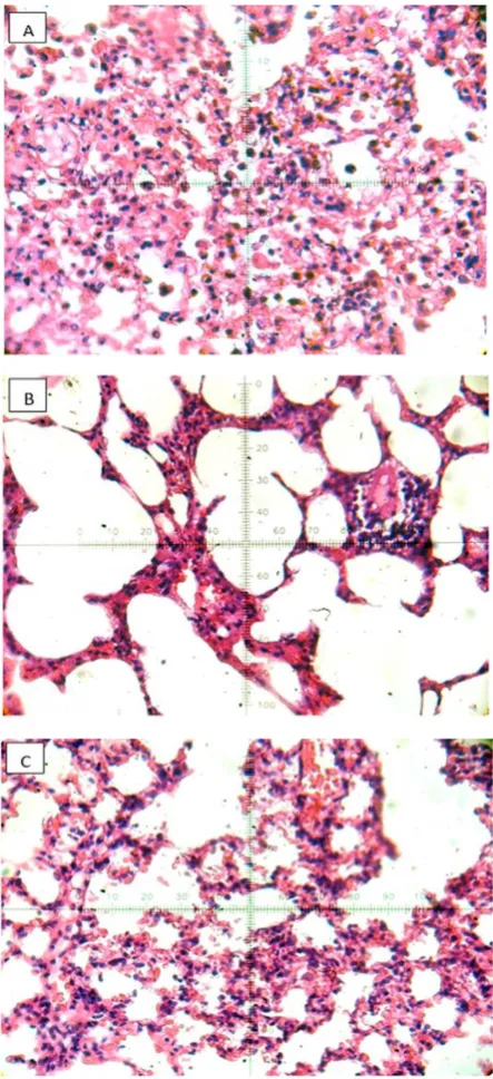
The microscopic images of rats lung tissue in the cigarette smoke group (figure 1a) show a large and dense number of epithelial alveoli cells, the boundary between one alveolus and the other alveoli can still be seen, and two layers of cells separating an alveolus from another alveolus may still be distinguished. Macrophages can be easily found between the epithelial cells and in the middle of the alveoli chamber.
In microscopic images of cigarette smoke group (control group) lung tissue (Figure 1b) it can be clearly seen that the spread of cells is uneven and sparse, the epithelial cells separating one alveolus from another alveolus consisting of only one cell layers, and alveoli cavities show widening. Macrophages are rarely seen, especially in the alveoli cavity.
98 Journal of Advanced Pharmacy Education & Research | Jul-Sep 2019 | Vol 9 | Issue3
Figure 1: Histology of lung from the rats using haematoxylin
and eosin (H&E) staining.
The rats were exposed with cigarette smoke for 6 weeks. Cigarette smoke exposed group (B) showed decreasing the number of alveoli epithelial cells and widening in diameter of alveoli compare to non-cigarette smoke exposed group (A). Furthermore, the number of alveoli epithelial cells and diameter of alveoli in cigarette smoke and A. vera group were recovers (C). Magnification: 400x.
Quantitative histological analysis
The result of epithelial cells number, measurement of alveoli diameter, and its statistical analysis between comparison group and control group are shown in table 1.
The data showed that the comparison group had more alveolar epithelial cells and smaller diameter size than the control group. To determine whether the differences in the number of alveoli
epithelial cells between groups were significant, statistical tests were performed using the Mann-Whitney test. The purpose of this analysis is to prove whether there is a significant difference in the number of epithelial cells and the mean alveoli diameter between the two groups that were compared.
Mann-Whitney test results show p-value of 0.005 for the number of epithelial cells and 0.000 for the diameter of the alveoli (p <0.05). This shows that there is a significant difference between the number of epithelial cells and the diameter of the alveoli between the comparison group and the control group. This concluded that exposure to cigarette smoke can decrease the number of epithelial cells and increase the diameter of rats lungs alveoli.
The result of epithelial cells number, measurement of alveoli diameter, and Mann-Whitney test between control group and treatment group are shown in table 2. Furthermore, the treatment group had more alveoli epithelial cells and smaller diameter size compared with the control group.
To determine whether the differences in the number of alveoli epithelial cells between groups were significant, statistical tests were performed using the Mann-Whitney test (Table 2). The purpose of this analysis is to prove whether there is a significant difference in the number of epithelial cells and the mean alveoli diameter between the two groups that were compared.
Table 1. The outcome of cigarette smoke exposure to rats lung tissue
Parameter Median (range) P value*
P K
Number of alveoli epithelial cells 402 (262-508) 338 (274-543) 0,005
Size of alveoli diameter 22 (12-51) 28 (15-70) 0,000 * Mann-Whitney test
The comparison of alveoli epithelial cells and diameter of alveoli between non-cigarette smoke exposed group (P) and cigarette smoke exposed group (K).
Table 2. The success rate of A. vera gel protect the lungs tissue against damage that caused by cigarette smoke
Parameter Median (range) P value* K Pr
Number of alveoli epithelial cells 338 (274-543) 382,5 (196-591) 0,013
Size of alveoli diameter 28 (15-70) 23 (11-60) 0,000 * Mann-Whitney test
The comparison of alveoli epithelial cells and diameter of alveoli between cigarette smoke exposed group (K) and cigarette smoke exposure & A. vera (Pr).
Journal of Advanced Pharmacy Education & Research | Jul-Sep 2019 | Vol 9 | Issue3 99
abnormal widening of the alveoli diameter due to exposure of cigarette smoke.
The result is similar to the previous research that we have done. With the same treatment in experimental animals, we concluded that A.vera gel increases the amount and activity of macrophage cells and Bcl2 expression in lungs that have been induced by cigarette smoke.[10] This condition is good to protect the lungs from cigarette smoke.
Our study showed that giving 1 ml of aloe gel orally / day before cigarette smoke exposure
could protect the lungs of rats from damage. Cigarette smoke is a very important problem for human health because it causes various deadly diseases. According to WHO statistics, smoking is the second leading cause of death in the world. One of the most common diseases is COPD. Previous study showed that passive smoking has a 1.48 times higher risk of developing COPD than non-exposed to cigarette smoke. COPD is caused by free radicals found in cigarette smoke.[2, 3, 5]
Within this observation, we found that A.vera protect the rat’s lung after exposer to cigarette smoke.[10] In the rat lung tissues of the group exposed to cigarette smoke showed the number of alveoli epithelial cells were decreased and alveoli diameter is widened. Mann-Whitney test results showed that there was a significant difference in the number of epithelial cells and the alveoli diameter between the A. vera group compare to the control group. This suggests that giving cigarette smoke for 6 weeks in rats could cause lung damage that characterized by decreasing in number of epithelial cells and abnormal widening of the alveoli diameter. It could be explained due to free radical and nicotine in cigarette smoke.[5-7] Free radicals in cigarette smoke will decrease the activity of antiproteases, α-antitrypsin 1.
α-antitrypsin 1 is an antiprotease that plays a role in degrading proteases in the form of elastase, proteinase 3, and katepsin G that produced by neutrophil cells. When the α-antitrypsin 1 activity decreases, secretion of proteinase by neutrophils in the alveoli cannot be inhibited, consequently, the proteinase will damage the lung tissue protein.[8]
Furthermore, we found that A.vera gel with antioxidant content such as superoxide dismutase, glutathione peroxidase, polyphenol, and flavonoid may increase the antioxidant level in rat lung. Forth, antioxidant will eliminated free radical from cigarette smoke and neutrophil in the lung. The resulting free radical decline is significant, as evidenced by the difference in the number of epithelial cells and the diameter of the alveoli between the control group and the treatment group.
The previous study showed that A. vera maintain the integrity of antioxidant through improved performance of superdioxide dismutase and catalase. Our previous study also proved that Aloe vera gel increases the number and cell activity of macrophages in addition to the Bcl2 expression in rat lungs which have been induced by cigarette smoke. [8,10]
Thus, our result showed that giving A. vera gel orally before exposure to cigarette smoke has an effect in protecting the lung of rats from damage that could by increasing levels of
antioxidants in the lungs and maintain the integrity of antioxidants which has existed.
Conclusion
A.vera gel consumption might be used for protecting lungs from cigarettes smoke negative effect.
Conflict of Interest
The authors declare there is no conflict of interest
Acknowledgement:
We thanks to Irfan Ahmad for technical assistance. This work was supported by grant from Ministry of Research, Technology and Higher Education of the Republic of Indonesia to Nur Atik.
References
1. Decramer M, Janssens W. Chronic obstructive pulmonary disease and comorbidities. The Lancet Respiratory Medicine. 2013 Mar 1;1(1):73-83.
2. Bloom D, Cafi ero E, Abrahams-Gessel S, et al. The global economic burden of non-communicable diseases: a report by the World Economic Forum and the Harvard School of Public Health, September 2011. Geneva, Switzerland: World Economic Forum, 2011.
3. Marques P, Collado A, Escudero P, Rius C, González C, Servera E, Piqueras L, Sanz MJ. Cigarette smoke increases endothelial CXCL16-leukocyte CXCR6 adhesion in vitro and in vivo. Potential consequences in chronic obstructive pulmonary disease. Frontiers in immunology. 2017 Dec 13;8:1766.
4. Li D, Wang J, Sun D, Gong X, Jiang H, Shu J, Wang Z, Long Z, Chen Y, Zhang Z, Yuan L. Tanshinone IIA sulfonate protects against cigarette smoke-induced COPD and down-regulation of CFTR in mice. Scientific reports. 2018 Jan 10;8(1):376.
5. Salvi S. Tobacco smoking and environmental risk factors for chronic obstructive pulmonary disease. Clinics in chest medicine. 2014 Mar 1;35(1):17-27.
6. Serban KA, Petrache I. Alpha-1 antitrypsin and lung cell apoptosis. Annals of the American Thoracic Society. 2016 Apr;13(Supplement 2):S146-9.
7. Martínez-García MÁ, Soler-Cataluña JJ, Sanz YD, Serra PC, Lerma MA, Vicente JB, Perpiñá-Tordera M. Factors associated with bronchiectasis in patients with COPD. Chest. 2011 Nov 1;140(5):1130-7.
100 Journal of Advanced Pharmacy Education & Research | Jul-Sep 2019 | Vol 9 | Issue 3
9. Ranjbar R, Arjomandzadegan M, Hosseiny H. Evaluation of antioxidant activity and growth control properties of nonoscale structure produced from Aloe vera var. Littoralis extract on clinical isolates of salmonella. Scientia pharmaceutica. 2017 Sep;85(3):28.
10. Atik N, Avriyanti E, Indrati AR. Pengaruh lidah buaya (Aloe vera L.) pada paru-paru tikus yang diinduksi asap rokok. Majalah Kedokteran Bandung. 2012 Sep 28;44(3):159-64.
11. Atik N, Nanua NK, Avriyanti E. The Aloe vera Effect on Cardiomyocytes and VEGF-A Expression in Rats After Cigarette Smoke Exposure. Advanced Science Letters. 2017 Jul 1;23(7):6658-61.
12. Atik N, Rahman JI. Perbedaan efek pemberian topikal gel lidah buaya (aloe vera l.) Dengan solusio povidone iodine terhadap penyembuhan luka sayat pada kulit mencit (Mus
musculus). Majalah Kedokteran Bandung. 2009 Jun 25;41(2).
13. Surjushe A, Vasani R, Saple DG. Aloe vera: a short review. Indian journal of dermatology. 2008;53(4):163.
14. Koul A, Bala S, Arora N. Aloe vera affects changes induced in pulmonary tissue of mice caused by cigarette smoke inhalation. Environmental toxicology. 2015 Sep;30(9):999-1013.
15. Abdurahman JI, Atik N. Perbandingan Pemberian Topikal Aqueous Leaf Extract of Carica Papaya (ALEC) dan Madu Khaula Terhadap Percepatan Penyembuhan Luka Sayat pada Kulit Mencit (Mus musculus). Majalah Kedokteran Bandung. 2010; 41(2): 76-81.