Downloaded from
http://journals.tums.ac.ir/
on Monday, August 13, 2012
Advanced Motion Correction Methods in PET
(Review Article)
Arman Rahmim PhD
Department of Radiology, School of Medicine, Johns Hopkins University, Baltimore MD, USA (Received 15 September 2005, Revised 2 October 2005, Accepted 10 October 2005)
ABSTRACT
With the arrival of increasingly higher resolution PET systems, small amounts of motion can
cause significant blurring in the images, compared to the intrinsic resolutions of the scanners. In this
work, we have reviewed advanced correction methods for the three cases of (i) unwanted patient
motion, as well as motions due to (ii) cardiac and (iii) respiratory cycles. For the first type of motion
(most often studies in PET brain imaging), conventional motion-correction algorithms have relied on
extraction of the motion information from the emission data itself. However, the accuracy of motion
compensation in this approach is degraded by the noisy nature of the emission data. Subsequently,
advanced methods, as reviewed in this work, make use of external real-time measurements of motion.
Various image-based and projection-based correction methods have been discussed and compared.
The paper also reviews recent and novel applications that perform corrections for cardiac and
respiratory motions. Unlike conventional gating schemes, in which the cardiac and respiratory gated
frames are independently reconstructed (resulting in noisy images), the reviewed methods are seen to
follow a common trend of seeking to produce images of higher quality by making collective use of all
the gated frames (and the estimated motion). As an observation, a general theme in motion-correction
methods is seen to be the use of increasingly sophisticated software to make use of existing advanced
hardware. In this sense, this field is very open to future novel ideas (hardware, and especially
software) aimed at improving motion detection, characterization and compensation.
Key Words: Motion tracking, Motion correction, Cardiac gating, Respiratory gating, 4D Image reconstruction, High resolution PET
Corresponding author: Arman Rahmim PhD, Division of Nuclear Medicine, Department of Radiology, School of Medicine, Johns Hopkins University, Baltimore MD 21287, USA,
Downloaded from
http://journals.tums.ac.ir/
on Monday, August 13, 2012
I. INTRODUCTION
Recent developments in 3D positron emission
tomography (PET) systems have enabled the
spatial resolution to reach the 2-5 mm FWHM
(Full-Width-at-Half-Maximum) range. With such
improvements in spatial resolution, even small
amounts of motion during PET imaging become
a significant source of resolution degradation. In
other words, increased spending on new
generation scanners can only be fully justified
when appropriate motion correction methods are
considered, in order to achieve the true resolution
of the scanner. To see this, one may note (1) that
the effective resolution of an image ∆eff can be
written as:
[
2 2]
12motion tomograph
eff
=
∆
+
∆
∆
(1)
where ∆tomograph denotes the intrinsic
resolution of the scanner (FWHM), and ∆motion is
the FWHM of the distribution of the patient’s
motion. With ∆tomographhaving become
comparable to (and no longer much larger than)
motion
∆ , it is therefore essential to develop and
implement accurate patient motion correction
techniques.
One must note that a number of motion
correction methods developed for SPECT are not
applicable to PET. This is because these methods
rely on the time-dependence of projections in
SPECT, due to rotating head(s), which is not the
case in PET imaging. Nevertheless, a number of
other methods implemented in SPECT imaging
are equally applicable to PET (and vice versa)
which we have reviewed in this work.
This paper has been broadly categorized into
the review and discussion of advanced correction
methods for the three cases of (i) unwanted
patient motion, as well as motions due to (ii)
cardiac and (iii) respiratory cycles. Most of the
existing literature on the first type of motion has
been investigated and implemented in brain PET
imaging, since the last two types of motion
(which are dominant in whole-body and cardiac
PET imaging) are absent in this case. Section II
therefore discusses motion-correction methods in
brain PET imaging, followed by discussions of
advanced correction methods, in sections III and
IV, for cardiac and respiratory cycle motions.
These sections first discuss existing hardware
instrumentation, followed by a review of
advanced motion-correction algorithms which
make use of such hardware to achieve
motion-compensated PET images. Finally, some important
areas of future research are discussed in section
V, followed by concluding remarks in section VI.
II. BRAIN PET IMAGING
Unlike cardiac and respiratory-related
motions, patient movements in brain imaging are
unanimously assumed to be of rigid nature (i.e.
modeled as translational and/or rotational
transformations only). As a typical PET brain
imaging session can last hours, it is not
reasonable to expect a patient to remain
motionless during this time. A number of head
restraints are nowadays common, such as
thermoplastic masks or neoprene caps, which
lower the amount of motion but do not eliminate
it. Even with head restraints, typical translations
in the range of 5-20 mm and rotations of 1-4o are
observed1, depending on the type of mask and
the duration of scan (e.g. see (2,3), and also (1)
in which a study of various types of head
movements, such as those caused by coughing
and leg crossing, has been presented).
Methods to correct for such patient
movements were in the past largely based on
correction of interscan movements. These
1. Largest translation typically occurs along the transaxial-(x)
Downloaded from
http://journals.tums.ac.ir/
on Monday, August 13, 2012
(software-based) methods involved the division
of a scan into a number of frames, followed by
spatial registration of the reconstructed images
using mathematical algorithms (e.g. see (4,5)).
Nevertheless, motion correction strategies in
emission computed tomography (ECT) that rely
exclusively on emission data itself are inadequate
for robust clinical usage, since (i) they depend on
the quality of the scan data – including noise
characteristics – and (ii) they assume the activity
distribution does not significantly change within
the frames, whereas the frames are chosen a
priori. Because of these disadvantages, this
review focuses on methods using external
real-time measurements of motion.
Instrumentation: Aside from electromagnetic
systems (which suffer from interference with
eddy currents in the metal in the PET gantry) and
acoustical devices (whose audible signal can be
unacceptable especially for neurological studies),
the following motion-tracking instruments can be
mentioned:
1) A video-camera-based surveillance system
used by Picard and Thompson (6) which used 3
LEDs attached to the head of the patient. The
system had two CCD cameras placed on the
gantry of the PET scanner.
2) The system used by Goldstein et al. (7)
based on opto-electronic position sensitive
detectors, which worked by optical triangulation
of three miniature (lamp) lights fixed to the
patient's head. However, the large space between
the cameras (1.25 m) prevents the use of this
system in PET scanners with long and narrow
gantry holes.
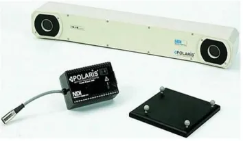
3) The nowadays-popular high-resolution
(<0.3mm) POLARIS system (1) which is an
infrared (IR) opto-electronic motion tracking
device using four IR-reflective spheres (depicted
in Fig. 1). The system has the advantages that it
is commercially available (<$15,000, Northern
Digital Inc., Waterloo, Canada), and that using
IR-light2, it is insensitive to room lighting
conditions and takes much less disk space to
store the IR-tracker output compared to optical
image sequences. However, POLARIS has the
disadvantage that the reflective spheres need to
be affixed in a precisely known geometry3, and
furthermore, similar to all aforementioned
methods, the issue of possible relative motion
between the skin and the skull, during the scan,
remains potentially problematic, making the
accuracy of these techniques questionable. It is a
topic of growing and great interest to the ECT
community to minimize or eliminate the latter
problem through innovative methods and/or
technology.
Figure 1: The POLARIS system uses four infrared-reflective spheres placed in a precisely known geometry.
Motion-correction algorithms: Assuming
accurate measurement of patient movement
during the scan, a number of approaches to
motion compensation have been proposed:
2. A system with CCD video cameras also sensitive to IR
light was used in (8); however the reflectors were affixed to a
landmark device that was rigidly attached to the teeth of the
subject’s upper jaw, which proved to be inconvenient for the
patients.
3. A proposed solution to this, as shown in
www.tru-scan.com (Tru-Scan Imaging Inc.), is the use of head-sets on
top of which a plastic board is attached which can be used to
Downloaded from
http://journals.tums.ac.ir/
on Monday, August 13, 2012
1) One method (9,3) involves dividing of
detected events into multiple acquisition frames
(MAFs). That is, with the use of an external
monitoring system, every time the displacement
of the patient is measured to be larger than a
specified threshold, the PET data are saved in a
new frame. This is then followed by correction
of the individually-reconstructed images of the
MAFs, via rotation and translation, to
compensate for the measured amount of motion
(i.e. this is an image-driven approach).
The major limitation of the MAF approach is
that by using a high motion threshold, motion
within the frames are neglected; and lowering the
motion threshold can instead result in the
acquisition of an increasing number of
low-statistic frames to be reconstructed, especially in
the presence of considerable movement. Lack of an
adequate number of acquired events in the
individual frames can in turn adversely affect the
quality of the final reconstructed images, and an
increased number of frames will also lead to
increased reconstruction times.
2) Another image-driven correction method
proposed by Menke et al. (8) involves
post-processing of the motion-blurred reconstructed
images using de-convolution operators (whose
shape is determined by the measured motion).
Nevertheless, this method has not attracted much
attention because even though it is theoretically
accurate for noise-less data, (i) the
de-convolution process amplifies the noise in the
PET data, and (ii) when the movements include
significant rotation, spatially-variant
de-convolution filters need to be employed, which
increases the computational costs and can
introduce other artifacts (8).
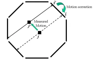
3) A more accurate approach consists of
correcting individual lines-of-response (LORs)
for motion (10) (this is an event-driven approach);
i.e. motion correction is performed by transforming
the LORs along which the events are measured
to where they would have been measured if the
object had not moved (this is shown in Fig. 2 for
the example of an octagonal scanner).
Figure 2: An event that would have been detected along LOR i is detected along LOR i’ due to motion. From the measured motion information, one can then transform LOR i’ back into LOR i.
The method was elaborated and implemented
by Menke et al. (8), and required some hardware
modification to achieve on-the-fly
motion-corrected LORs. However in that work, due to
hardware limitations, the corrected LORs where
not corrected by normalization factors that
corresponded to the original detector-pairs (along
which the events were detected), and instead the
normalization factors for the transformed LORs
were used. This normalization mismatch has
recently been shown to result in artifacts (11).
Alternatively, to solve this problem, one
requires a PET scanner either (i) equipped with
more specialized hardware to achieve accurate
on-the-fly normalization correction followed by
LOR-transformation; e.g. see (12), or (ii) capable
of acquiring data in list-mode format, so that
LOR corrections can be accurately performed
post-acquisition; e.g. see (2).
Beyond the purely event-driven approach:
The above approach neglects two issues, as
Downloaded from
http://journals.tums.ac.ir/
on Monday, August 13, 2012
(14), Thielemans et al. (15) and Buhler et al. (11),
which we shall refer to as (issue 1) and (issue 2):
(Issue 1) An event that is normally detected
can exit the PET scanner undetected because of
motion. This therefore results in a loss of events
that would normally have been detected, an
effect that is not modeled by regular
reconstruction methods.
(Issue 2) On the other hand, an event that is
normally not detected (i.e. not passing through
PET detectors) may be detected because of
motion. Therefore, after correction for motion,
some detected events may correspond to no
actual detector pairs.
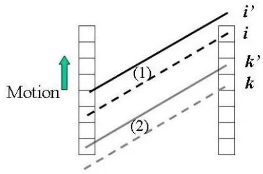
These two effects can occur in two ways:
(a) Along the axial direction of the scanner,
via translation (as shown in Fig. 3) or rotation
(not shown); or
(b) Similarly via translation or rotation, along
the transaxial direction, but only for scanners
with gaps in between the detectors (an example
of this is the High Resolution Research
Tomograph - HRRT (16) which has an octagonal
design with gaps in-between the heads). This
effect is shown in Fig. 4 (for the case of
translation).
Figure 3: Axial motion can result in (issue 1) LOR i not to be detected (i’), and (issue 2) LOR k, which is normally not detected, to be actually detected (as k). The effect is shown due to translation, but is equally valid for rotation.
Figure 4: Transaxial motion, for scanners with gaps in between the detector heads, can result in the exact same issues as shown in Fig. 3.
Presence of these two issues can imply the
need for a more accurate modeling of the
image-data relation into the reconstruction task;
otherwise, neglecting the first issue can produce
image artifacts, as demonstrated by simulation
(15,11) or experimentally (14), and neglecting
the LORs obtained in the second case can result
in a loss of signal-to-noise ratio (SNR) in the
images, as we describe later. Below we review a
number of proposed solutions to one or both of
these issues:
4) A method suggested by Thielemans et al.
(15) addressing (issue 1) involves scaling of
counts recorded in the motion-corrected
sinogram bins in order to correct for the events
that were lost due to motion, where the scale
factors are computed by averaging of LOR
weighting factors using the measured motion
information. This can be thought of as a ``motion
pre-correction'' technique applied to the sinogram
bins before the image reconstruction task.
5) The method investigated by Buhler et al.
(11), similarly addressing (issue 1), involves
Downloaded from
http://journals.tums.ac.ir/
on Monday, August 13, 2012
counts in each motion-corrected sinogram bin
by the factor tdetectable/ttotal i.e. the fraction of time
each sinogram bin could have been detected by
the scanner. Aside from the issue of
normalization correction, this method can be
shown, though not obvious, to be equivalent to
the previous method. However, this method
pre-corrects the individual measured events by
related normalization factors, whereas the
previous method, which is expected (15) to
exhibit less noise, first sums the un-normalized
motion-corrected LORs and then performs
normalization correction (by an
accurately-calculated overall factor).
The above two methods have two potential
difficulties:
(a) They may require consideration of noise
enhancement issues, as is done in (15) when
dividing by small scale factors.
(b) They address (issue 1) but not (issue 2)
because they simply discard motion-corrected
events which do not correspond to actual
detector elements. It must be noted that
neglecting such events should not result in image
artifacts (unlike neglecting (issue 1)) since the
patient will be still sampled enough by the
existing detector pairs; however it can result in a
loss of SNR in the images since some of the
measured signal with useful information is
simply discarded.
(iii) A method capable of addressing both
(issue 1) and (issue 2) has been elaborated by
Rahmim et al. (14). The approach is based on
modification of the probability system matrix of
the iterative expectation-maximization (EM)
algorithm. Momentarily neglecting various
correction terms (e.g. normalization,
attenuation), the regular histogram-mode EM
algorithm can be written as
∑
∑
∑
= = = = I i J b old ij ij I i ij old new j f p i n p p j f j f 1 1 1 ) ( ) ( ) ( ) ( (2)where fold(j)and fnew(j) are previous and
current activity-distribution image estimates in
the iterative EM algorithm, n(i) is the number of
events detected along an LOR i, and pij, often
referred to as the system matrix element, is the
probability that an emission from voxel j
(j= 1…J) is detected along an LOR i (i= 1…I).
For a motion-corrected sinogram (wherein all the
events were first corrected for motion before
histogramming), the proposed algorithm is able
to accurately address issues 1 and 2, and can be
written as
∑
∑
∫∑
= = = = = I i J b old ij ij T t I i t i ij old new j f p i n p dt p T j f j f 1 1 0 1 ) ( ) ( 1 ) ( ) ( δ (3)where T is the duration of the scan, and t
i
δ is
1 if an LOR i was detectable at time t, and 0
otherwise. Normalization correction can
subsequently be included either (i) as a
pre-correction factor (similar to (11)), or (ii) as an
intrinsic component of the system matrix element
(somewhat similar to (15)). The reader is referred
to Ref. (14) for more details.
The above approach has also been proposed
(17,14) for list-mode image reconstruction. The
list-mode reconstruction approach has a number
of general advantages compared to the
histogram-mode approach, as elaborated in
(18,19); in the context of motion correction, the
following two potential advantages can be
mentioned:
(a) Events are corrected for motion during
(and not before) the image reconstruction task,
Downloaded from
http://journals.tums.ac.ir/
on Monday, August 13, 2012
coordinates can be processed as continuous
variables, therefore improving the accuracy
(whereas time-consuming interpolations may
instead be required in histogram-mode methods,
as nicely demonstrated by (11), mainly because
sinogram bins are not continuous).
(b) Addressing (issue 2) is more convenient
in list-mode reconstruction, because in
histogram-mode methods, one would have to
extend the sinogram-space in order to record all
motion-corrected events (even those that would
not have been detected in the absence of motion),
whereas in list-mode reconstruction, such events
are very easily handled.
Furthermore, Rahmim et al. (14) have also
shown that, with appropriate modifications,
calculation of the motion-averaging term
∫ ∑
= = Tt I
i
t i ij dt p
T 0 1
1
δ can be conveniently
performed in image-space (instead of
projection-space) which for current high-resolution scanners
can improve the calculation speed significantly4
(e.g. the HRRT scanner, with no axial
compression i.e. span 1, has ~800M sinogram
bins compared to only ~14M voxels in
image-space).
III. MOTION DUE TO THE
CARDIAC CYCLE
While a spatial resolution of <5 mm is
possible with today’s PET scanners, the base of
the heart moves 9-14 mm towards the apex, and
the myocardial walls thicken from approximately
10 mm to over 15 mm between end-diastole and
4. In projection-space, Carson et al. (20) have proposed to
perform the above calculation over only a randomized subset
of the projection-space, in order to produce a fast, practical
algorithm; however, the image-space approach proposed in
(14) can yield a practical and accurate algorithm with
complete (non-randomized) processing of the LORs.
end-systole, as measured from tagged MR
images (21). Compared to the intrinsic resolution
of today’s scanners, cardiac motion can therefore
result in significantly blurred images (as seen by
Eq. 1). Most common approach in ECT to
cardiac cycle motions is gating of the data into
frames, each representing a particular cardiac
phase, as we explain next.
Instrumentation: Cardiac gating is most
commonly performed with the aid of
electrocardiograph (ECG) devices. By
convention, the R-wave (which precedes
ventricular contraction) is chosen as the gating
signal because it has the greatest amplitude, and
is therefore more easily identified in the ECG. In
scanners with the list-mode acquisition
capability, sorting of the list-mode data into
gated frames can be performed after the
acquisition (e.g. see (22)), whereas in
conventional scanners (i.e. with histogram-mode
acquisition only), on-the-fly ECG-triggered data
acquisition is employed (e.g. see (23)).
Typically, the cardiac cycle is divided into
50-100 ms time frames, and an acquisition
ranging from 5-60 minutes is acquired. Most
commonly, the obtained cardiac-gated datasets
(i.e. cardiac frames) are then independently
reconstructed (as shown in Fig. 5). This approach
is successful in nearly removing the
cardiac-motion blurring of the images; however, it can
produce images which are (much) noisier than a
reconstruction of the ungated data, since each
gated dataset contains (much) less statistics
compared to the entire dataset, and therefore, the
clinical utility of this approach is very
Downloaded from
http://journals.tums.ac.ir/
on Monday, August 13, 2012
Figure 5: In conventional gated schemes, the gated frames are independently reconstructed (in this example, N=4 gated frames are shown).
Motion-correction algorithms: The
motivation behind advanced correction methods
in cardiac imaging is two-fold:
(i) To further improve the quality of cardiac
PET images (noise, resolution) so as to enhance
identifiably of radiotracer uptake defects in the
left ventricle by the clinicians, since regions of
decreased radiotracer uptake can be indicative of
hibernating or infracted myocardial tissue (24).
This is also important when applying quantitative
measures of perfusion and metabolic parameters
in dynamic compartmental modeling studies
(25).
(ii) Measurement of motion itself can be
useful for characterizing cardiac function (26).
Measures such as ejection fraction and regional
wall thickening may be derived from a measure
of contractile motion in this way.
In this section, we neglect the problem of
cardiac motion due to the respiratory cycle,
which is discussed in the next section. Below, we
outline five important motion correction
approaches which have been proposed in the
literature. A common theme amongst these
advanced methods is that they seek to move
beyond the conventional gated scheme (as was
shown in Fig. 5) and instead seek to obtain
images which make collective use of all the
gated frames (as depicted in Fig 6). In this way,
motion information is extracted from the
measured dataset (with the exception of approach
3 which uses modeling), and is used in addition
to all the gated frames to obtain images of higher
quality.
Figure 6: In motion-correction gated schemes, individual images are reconstructed using information from the complete dataset.
The first three outlined works incorporate the
measured or modeled motion-information into
4D image reconstruction tasks, while the fourth
approach performs the motion estimation and
image reconstruction tasks simultaneously.
Approach 5, on the other hand, performs
image-based motion correction and summing of 3D
reconstructed images. These are elaborated
below:
1) In (27,28), Brankov et al. have replaced
the uniform-voxel framework with the use of
mesh modeling: an efficient image description
based on non-uniform sampling (mesh nodes are
placed most densely in image regions having fine
detail). This approach is a natural framework for
reconstruction of motion image sequences,
Downloaded from
http://journals.tums.ac.ir/
on Monday, August 13, 2012
over time5. Using a gradient-descent search
algorithm applied to initial cardiac gated images,
the authors are able to determine the motion field
vector
d
k→l(x
)
mapping a mesh element xfrom the current frame k to another frame l. The
authors have subsequently used the following
motion-compensated temporal summation when
reconstructing each frame k:
∑
= →−
=
K l l k l kf
f
1))
(
(
)
(
ˆ
x
x
d
x
(4)
where
f
l(x
)
is the image estimate for thelth frame (k=1…K), and the above expression
can be applied as (i) an inter-iteration temporal
filter in iterative reconstruction, or as (ii) a
post-reconstruction filter6. The above
summation/filtering step is therefore able to
improve the SNR obtained in the cardiac images,
since it makes use of information from other
frames also, when reconstructing a given frame
k.
Before explaining the remaining methods, we
must first explain the concept of MAP image
reconstruction: A main drawback with the
commonly-used expectation maximization (EM)
algorithms is that with further iterations the
images become increasingly noisy. To tackle
this, often a post-reconstruction smoothing filter
is used. However, post-filtering, even though
lowering the noise, also degrades image
resolution. Alternatively, maximum a posteriori
probability (MAP) methods7 have been proposed
5. See http://www.ipl.iit.edu/brankov/MIC02_4D.htm for a
very nice dynamic demonstration of this method.
6. Meanwhile, though not shown here, the authors have
added another term to the above expression in order to
account for brightening of the myocardium as it thickens due
to the partial volume effect.
7. This is also referred to as the Baysian method (originally
derived from a simple application of Bayes' rule to image
which, in the 3D-framework, seek to minimize
variations between voxels and their neighboring
voxels. A particular class of the MAP method
(first utilized by Geman and McClure (29) in
nuclear medicine), instead of maximizing the
Poisson log-likelihood function L(F), as is the
case with the regular EM algorithm, instead seek
to maximize the MAP function L(F)-βV(F),
where V(F) is a potential function that decreases
in value with less variations for neighboring voxel (β is a smoothing parameter set by the user: the higher its value, the greater the amount
of smoothing encouraged in the images).
For instance, the so-called 3D-MAP-EM
one-step-late (OSL) algorithm, introduced by Green
(30) and aimed to maximize the above MAP
function, can be written as
∑
∑
∑
= = = ∂ = ∂ + = I i J b old ij ij Ii j F F
ij old new j f p i n p f F V p j f j f old 1 1 1 ) ( ) ( ) ( ) ( ) ( β (5)
and is able to suppress noise more
successfully than the regular EM algorithm
which can be thought of as a special case of the
MAP method with β=0. An interesting
observation is that the above approach can be
extended to a 4D-MAP algorithm - e.g. see
(31,32) - in which one uses a summation of
spatial
β
sV
s(
F
)
and temporal)
(
F
V
tt
β
potential functions, in order toencourage smoothing between neighboring
voxels in both the spatial and temporal
directions. We now proceed to explain how
motion compensation has been incorporated
within the 4D-MAP framework in some of the
following works.
2) In (31), Gravier et al. initially reconstruct
reconstruction). It is also, sometimes, referred to as penalized
Downloaded from
http://journals.tums.ac.ir/
on Monday, August 13, 2012
the gated frames using the fast filtered
backprojection (FBP) algorithm, followed by
low-pass filtering to reduce the noise. They then
use the optical flow approach developed by Horn
and Schunck (33) to estimate the motion
in-between the reconstructed images. Finally, they
use the 4D-MAP-EM-OSL algorithm (4) while
defining:
∑∑
∑
= =
≠
= →
−
−
=
Kk J
j
K
k l l
k l k
t
f
j
K
j
f
F
V
1 1 1
)
(
1
1
)
(
)
(
(6)
where
f
l→k(
j
)
denotes the estimated imageintensity (in frame l) at the location
corresponding to voxel j of frame k (considering
the motion). In this way, smoothing is
encouraged between voxels in all the frame
sequences while taking the motion of the voxels
into consideration, and therefore one is able to
suppress the noise level that is normally obtained
in gated frame images.
3) In the work of Lalush et al. (32,34) in
SPECT imaging (which can be similarly applied
to PET), a similar 4D-MAP-EM-OSL approach
as above has been considered, except that motion
is modeled and assumed to be known a priori
(and not measured from initial gated images).
The motion vectors are computed by modeling
the left ventricular inner and outer walls as
ellipsoids that undergo affine transformations
(rotation, scaling, and translation) with each
frame. The exact form of the potential function is
also different in this work. It must be noted,
however, that the authors have not observed a
noticeable degradation when the motion
information is simply not included in the
4D-MAP algorithm method (which may have been
due to the limited resolution of their scanner).
Furthermore, Comparison of the above two
approaches is an interesting area of future
research, as we discuss in section V.
4) Very commonly in the literature, cardiac
motion is estimated after reconstructions of gated
frames; and in the previously mentioned
techniques, this extracted motion information is
then used in subsequent reconstructions to yield
enhanced images (i.e. improved SNRs). In (35)
however, Cao et al. have hypothesized that,
given the close link between the image
reconstruction and motion estimation steps, a
simultaneous method of estimating the two will
be better able to (i) reduce motion blur and
compensate for poor SNRs, and to (ii) improve
the accuracy of the estimated motion. Their
proposed algorithm works by two-step
minimization of a joint energy functional term
(that includes both image likelihood and
motion-matching terms). This work has also been
extended from a two-frame approach to the
complete cardiac cycle in (36). This is a very
novel approach, yet its accuracy remains to be
demonstrated (e.g. it remains to be verified that
the estimated motion matches the actual motion
of the heart as measured by, for instance, tagged
MR).
5) Klein and Huesman (37) have developed a
sophisticated motion-estimation approach which
exhibits an impressive knowledge of cardiac
anatomy, and makes use of a non-uniform elastic
material model to provide accurate estimates of
heart motion (from individually reconstructed
gated frames). The authors then continue to
perform non-rigid/deformed summing of the
gated images making use of the motion
information (i.e. image-based
motion-compensation). Alternatively, one must note that
the estimated motion can instead be directly
incorporated into 4D image reconstruction tasks,
Downloaded from
http://journals.tums.ac.ir/
on Monday, August 13, 2012
IV. MOTION DUE TO THE
RESPIRATORY CYCLE
The common approach to the problem of
respiratory blurring of PET images has been that
of respiratory gating. For instance,
respiratory-gated PET has been investirespiratory-gated in imaging of
lung cancer to reduce breathing motion artifacts
(38,39). In cardiac imaging, combined
cardiac-respiratory gating has been implemented in
human (40) and animal (22) studies.
Instrumentation: A number of instruments
are used for the purpose of measuring respiratory
motion:
1) Commonly, a pneumatic bellows is placed
around the mid-abdomen of the patient, which
monitors variation in pressure in the
belt-assembly with stretching of the belt during
respiration; e.g. see (41).
2) Another approach (42) involves the
Real-Time Position Management (RPM) Respiratory
Gating system (Varian Medical Systems), which
monitors the motion of the chest wall of the
patient by infrared tracking of the vertical
position of two reflective markers mounted on a
plastic block (stabilized on the patient’s
abdomen).
3) Livieratos et al. (43) have used an
inductive respiration monitor (RespiTrace R250,
Studley Data Systems) with a belt around the
patient’s chest.
4) In animal (mouse) imaging, a respiration
sensor (Graseby Medical Limited) has been used
by Yang et al. (22) to provide the respiratory
signal, being taped to the animal’s chest, and
connected to a high sensitivity differential
pressure transducer.
5) Finally, Beach et al. (41) have used the
POLARIS system (described in section II) during
cardiac imaging (which has the advantage of
monitoring both respiratory and unwanted
motions). Four infrared reflective markers were
placed on an elastic material band, placed around
the patient’s mid to lower abdomen.
Respiratory-Correlated Dynamic Imaging: In
the work by Nehmeh et al. (42), an alternate
method which performs respiratory
phase-isolation while not making use of gating has been
implemented (in lung imaging). A radioactive
point-source was set on the patient’s abdomen,
and the data were acquired in very short (e.g.
1-sec) consecutive time frames and were
individually reconstructed. In order to capture a
specific phase within the breathing cycle, all the
images were next analyzed, and those with the
point-source at a specific (user-selected) position
were then identified, with the corresponding
sinograms summed and reconstructed using
iterative reconstruction.
This method compared to respiratory gating,
while involving significantly more computation,
has the advantages that: (i) it does not require
tracking hardware to monitor and trace
respiratory motion (a benefit for small
institutions that do not have a gating system), (ii)
it allows reconstruction of PET images at any
breathing phase (e.g. phase-matching with the
CT image data acquired on PET/CT scanners),
and (iii) it is less susceptible to irregular
breathing and allows the exclusion of data from
irregular breathing cycles. Nevertheless, it has
the disadvantage, similar to the conventional
gating approach, that less data is used in each
reconstruction, and thus the obtained images are
more noisy.
Motion-correction algorithms: below we
review two proposed advanced methods (one
based in projection-space and one in
image-space) that seek to obtain images of higher
quality compared to images that are otherwise
obtained (i.e. by regular respiratory-gating or
respiratory-correlated dynamic imaging):
Downloaded from
http://journals.tums.ac.ir/
on Monday, August 13, 2012
performs cardiac imaging by modeling of the
respiratory motion of the heart as a rigid-body
motion, and the parameters for motion correction
are obtained from an initial series of
respiration-only gated images via edge-tracking of the left
ventricle (23). The obtained model is then
applied in the form of rigid-body transformations
(i.e. translations and rotations) on the list-mode
data event-by-event (i.e. motion correction in
projection-space). The list-mode approach allows
one to make maximal use of the time resolution
of list-mode data for interpolation of motion
parameters, thus potentially achieving higher
accuracy in respiratory motion compensation.
After correction of the data for respiratory
motion, the authors have proposed to use simple
cardiac gating for the rest of the imaging task;
however, we note that the more advanced
methods presented in the last section can instead
be used.
2) Klein et al. (44) have investigated a
twelve-parameter affine motion model for
4D-registration of different respiratory gates, which
in addition to the six parameters of rotation and
translation, allows for three scale and three skew
parameters for non-rigid motion. However, this
approach, which was based in the image-space,
was applied to doubly-gated cardiac PET
sequence as it required images with high SNR
for appropriate registration.
Rigid vs. non-rigid modeling of the
respiratory motion of the heart: While (43) has
claimed the validity of modeling (i.e.
approximating) respiratory motion of the heart as
rigid-body motion, a number of other works may
suggest that non-rigid modeling of respiratory
motion of the heart may be beneficial. To start,
we note that the non-rigidity of respiratory
motion of the heart, which is related to it being
pushed and pulled by the diaphragm and other
connected tissue, has been investigated using a
number of modalities. For instance, the gated CT
study in (45) measured on dogs, has recorded an
average change of 12% in the total end-diastolic
heart volume during forced positive pressure
inspiration at 15 cm H2O. Using
echocardiography, similar shape changes have
been found in human subjects (46).
Related work by Klein et al. (44) in PET
imaging is particularly worth noting: in that
work, quantitative measures of respiratory
motion of the heart were extracted from ten
respiratory-gated patient studies. Translations
between end-inspiration and end-expiration were
often greater than 10 mm and ranged from 1 to
over 20 mm (rigid motion). Moreover, the left
ventricle exhibited fairly large compression
factors8 (non-rigid motion) - close to 10% in a
number of cases – computed as the product of
the three extension factors along the x, y and z
directions.
The extension factors were largest along the
superior/inferior axis (~5%), which, given the
typical 80-100 mm dimension of the left
ventricle along this direction, would result in a
heart image that would be 4-5 mm too small if
motion was assumed simply rigid. Compared to
the average 10-mm thickness of the left
ventricular wall, this scaling error may therefore
be considerable. However, with the ECAT
EXACT HR scanner, only small improvements
were actually observed (44) after performing
non-rigid motion modeling, though it is expected
that in next-generation (higher-resolution)
scanners further improvements may be observed.
V. AREAS OF FUTURE RESEARCH
In this section, I shall attempt to outline few
areas of research in motion correction that still
8. The left ventricle was generally largest at inspiration and
Downloaded from
http://journals.tums.ac.ir/
on Monday, August 13, 2012
involve open questions, and important areas
which demand further inquiries and research:
1) Current motion tracking and correction
methods in brain imaging do not address the
occurrence of relative motions between the skin
and the skull during the scans. This can imply an
inaccuracy since motion-tracking lights or
reflectors only follow the motion of the surface
area to which they are attached (and not
necessarily the regions of interest inside the
brain). It is currently a topic of growing interest
to introduce novel methods of characterizing and
correcting for this issue.
2) Incorporation of accurate
coincidental-accidence (random) and scattered events
correction terms, considering patient motion, has
received little attention in the past, since
normalization/attenuation correction and LOR
transformations have been the major issues. In
this regard, we note that current random and
scatter estimation techniques simply assume a
static patient, and therefore, further attention
needs to be paid to this topic.
3) In cardiac imaging, it remains an open
question as to whether (and in which imaging
conditions) it is best to estimate cardiac motion
simultaneously with the image reconstruction
task (as is done in (35)) or before application of
an advanced image reconstruction algorithm
(that makes use of the estimated motion). It also
remains an open task to compare the qualities of
motion-information obtained from (i)
individually-reconstructed cardiac images (e.g.
as is done in (31)), or (ii) by means of modeling
(e.g. as is done in (32,34)); the comparison is not
trivial because the first approach relies on
initially noisy images while accuracy of the
second general approach in the context of
distinct individual orientations and conditions is
in question.
4) In respiratory motion correction, it remains
an area of future research to determine whether
non-rigid modeling of respiratory motion of the
heart has observable advantages compared to
rigid modeling.
5) While estimated rigid movements can be
easily corrected for in projection-space (by
simple translations and rotations of the LORs), it
is not straightforward to implement such LOR
motion compensations for non-rigid motion. This
is, for instance, the reason Klein et al. (44)
performed correction of the estimated non-rigid
respiratory motion of the heart in image-space.
However, projection-space correction methods
have the advantage that they make maximal use
of the time resolution of data (unlike
image-space method which do not assume any motion
within the gated images). Therefore, it remains
an important topic of interest as to whether it is
possible/suitable to implement non-rigid motion
compensation in projection-space.
6) The principle component analysis (PCA)
method elaborated in (47)is a very efficient and
natural framework for fast 4D image
reconstructions. The method is developed for the
motion-free object assumption however, though
it has been shown (48) to work very well in
reconstructing cardiac image sequences as well
(which can indicate that the method is somehow
able to intrinsically capture and incorporate
motion information). More work is needed in this
area to shed light on the potentials of this
technique to include accurate motion
compensation.
VI. CONCLUSION
In this work, we have reviewed advanced
correction methods in PET for the three cases of
(i) unwanted patient motion, as well as motions
due to (ii) cardiac and (iii) respiratory cycles.
Nearly all the work related to the first type of
Downloaded from
http://journals.tums.ac.ir/
on Monday, August 13, 2012
noted that use of an external motion tracking
device (and not solely relying on the emission
data) is and becoming popular for high resolution
PET imaging.
In brain PET imaging, given the rigid nature
of motion, it is seen to be more accurate to
perform motion corrections in projection-space,
instead of image-space, to make maximal use of
the time resolution of data. A number of
reviewed works have also observed and proposed
solutions to complications caused by the
motion-based interactions of LORs that are normally
detectable and those which are not (e.g. axially
out of the field-of-view or passing through
detector gaps).
In advanced cardiac and respiratory
correction schemes, this paper has observed a
general attempt to move beyond the noisy
images obtained by cardiac- and
respiratory-gated data which are individually reconstructed,
and instead, advanced techniques are seen to
make use of novel motion estimation and image
reconstruction applications to obtain images of
enhanced quality (improved SNR and
resolution). It is therefore observed from the
works reviewed in this paper that a general
theme has been the use of increasingly
sophisticated software to make use of existing
advanced hardware, and that the field of motion
correction in high resolution PET is very open to
future novel ideas (hardware, and especially
software) aimed at improving motion detection,
characterization and compensation.
VII. REFERENCES
1. Lopresti BJ, Russo A, Jones WF, Fisher T,
Crouch DG, Altenburger DE, Townsend DW.
Implementation and Performance of an
Optical Motion Tracking System for High
Resolution Brain PET Imaging. IEEE Trans
Nucl Sci. 1999; 46:2059-2067.
2. Bloomfield PM, Spinks TJ, Reed J, Schnorr
L, Westrip AM, Livieratos L, Fulton R, and
Jones T. The design and implementation of a
motion correction scheme for neurological
PET. Phys Med Biol. 2003; 48:959-978.
3. Fulton RR, Meikle SR, Eberl S, Pfeiffer J,
Constable CJ. Correction for head movement
in positron emission tomography using an
optical motion tracking system. IEEE Trans
Nucl Sci. 2002; 49:116-123.
4. Woods RP, Cherry SR, Maziotta. A rapid
automated algorithm for aligning and
reslicing PET images. J Computer Assis
Tomog. 1992; 16:620-633.
5. Friston KJ, Ashburner J, Frith CD, Poline JB,
Heather JD, Frackowiak RSJ. Spatial
registration and normalization of images.
Human Brain Mapping 1995; 2:165-189.
6. Picard Y, Thompson CJ. Digitized video
subject positioning and surveillance system
for PET. IEEE Trans Nucl Sci. 1995;
42:1024-1029.
7. Goldstein SR, Daube-Witherspoon ME,
Green MV, Eidsath A. A Head Motion
Measurement System Suitable for Emission
Computed Tomography. IEEE Trans Med
Imag. 1997; 16: 17-27.
8. Menke M, Atkins MS, Buckley KR.
Compensation Methods for Head Motion
Detected During PET Imaging. IEEE Trans
Nucl Sci. 1996; 43(1):310-317.
9. Picard Y and Thompson CJ. Motion
correction of PET images using multiple
Downloaded from
http://journals.tums.ac.ir/
on Monday, August 13, 2012
1997; 16:137-144.
10. Daube-Witherspoon ME, Yan YC, Green
MV, Carson RE, Kempner KM, Herscovitch
P. Correction for motion distortion in PET by
dynamic monitoring of patient position
(Abstract), J Nucl Med. 1990; 31:816.
11. Buhler P, Just U, Will E, Kotzerke J, van den
Hoff J. An Accurate Method for Correction
of Head Movement in PET. IEEE Trans Med
Imag. 2004; 23(8): 1176-1185.
12. Jones WF, Real-time event stream correction
for patient motion in clinical 3-D PET. IEEE
Nucl Sci Symp Conf Record. 2001;
4:2062-2064.
13. Qi J, Huesman RH. Correction of Motion in
PET using Event-Based Rebinning Method:
Pitfall and Solution (Abstract only). J Nucl
Med. 2002; 43:146P.
14. Rahmim R, Bloomfield P, Houle S, Lenox M,
Michel C, Buckley KR, Ruth TJ, Sossi V.
Motion Compensation in Histogram-Mode
and List-Mode EM Reconstructions: Beyond
the Event-Driven Approach. IEEE Trans
Nucl Sci. 2004; 51:2588-2596.
15. Thielemans K, Mustafovic S, Schnorr L.
Image Reconstruction of Motion Corrected
Sinograms, IEEE Nucl Sci Symp Conf
Record. 2003; 4:2401-2406.
16. Wienhard K, Shmand M, et al. The ECAT
HRRT: Performance and First Clinical
Application of the New High Resolution
Research Tomograph. IEEE Trans Nucl Sci.
2002; 49:104-110.
17. Qi J, Huesman RH. List mode reconstruction
for PET with motion compensation: a
simulation study. Proc. IEEE Inter Symp Biol
Imag. 2002; 413-416.
18. Rahmim A, Lenox M, Reader AJ, Michel C,
Burbar Z, Ruth TJ, Sossi V. Statistical
list-mode image reconstruction for the high
resolution research tomograph, Phys Med
Biol. 2004; 49:4239-4258.
19. Rahmim A, Cheng JC, Blinder S, Camborde
ML, Sossi V. Statistical dynamic image
reconstruction in state-of-the-art high
resolution PET. Phys Med Biol. 2005;
50:4887-4912.
20. Carson RE, Barker WC, Liow JS, Johnson
CA. Design of a motion-compensation
OSEM list-mode algorithm for
resolution-recovery reconstruction for the HRRT. IEEE
Nucl Sci Symp Conf Record. 2003;
5:3281-3285.
21. O’Dell WG, Moore CC, Hunter WC,
Zerhouni EA, McVeigh ER.
Three-dimensional myocardial deformations:
calculation with displacement field fitting to
tagged MR images. Radiology. 1995;
195:829-835.
22. Yang YF, Rending S, Siegel S, Newport DF,
Cherry SR. Cardiac PET imaging in mice
with simultaneous cardiac and respiratory
gating. Phys Med Biol. 2005;
50(13):2979-2989.
23. Stegger L. Biedenstein S, Schafers KP,
Schober O, Schafers MA. Elastic surface
contour detection for the measurement of
ejection fraction in myocardial perfusion
SPET. Eur J Nucl Med. 2001; 28:48-55.
24. Jadvar H, Strauss HW, Segall GM. SPECT
and PET in the evaluation of coronary artery
disease. Radiographics. 1999; 19(4):915-926.
25. Hutchins GD, Caraher JM, Raylman RR. A
region of interest strategy for minimizing
resolution distortions in quantitative
myocardial PET studies. J Nucl Med. 1992;
33(6):1243-1250.
26. Nichols K, Lefkowitz D, Faber R, Cooke D,
Garcia EV, Yao SS, DePeuy EG, Rozanski
A. Echocardiographic validation of gated
SPECT ventricular function measurements. J
Downloaded from
http://journals.tums.ac.ir/
on Monday, August 13, 2012
27. Brankov JG, Yang Y, Narayanan MV,
Wernick MN. Motion-Compensated 4D
Processing of Gated SPECT Perfusion
Studies. IEEE Nucl Sci Symp Conf Record.
2002; 3: 1380-1384.
28. Brankov, JG, Yang Y, Feng B, King MA,
Wernick MN. 4D smoothing of gated SPECT
images using a left-ventricle shape model and
a deformable mesh. IEEE Nucl Sci Symp
Conf Record. 2004; 5:2845-2848.
29. Geman S, McClure D. Baysian image
analysis: An application to single photon
emission tomography. Proc. Statist. Comput.
Sect (Amer. Statist. Assoc.), Washington,
DC. 1985; 12-18.
30. Green PJ. Baysian Reconstructions from
Emission Tomography Data Using a
Modified EM Algorithm. IEEE Trans Med
Imag. 1990; 9:84-93.
31. Gravier EJ, Yang Y. Motion-Compensated
Reconstruction of Tomographic Image
Sequences. IEEE Trans Nucl Sci. 2005;
52:51-56.
32. Lalush DS, Cui L, Tsui BMW. A Priori
Motion Models for Four-Dimensional
Reconstruction in Gated Cardiac SPECT.
IEEE Nucl Sci Symp Conf Record. 1996;
3:1923-1927.
33. Horn BKP, Schunck BG. Determining optical
flow. Artif. Intell. 1981; 17:185-203.
34. Lalush DS, Tsui BMW. Block-iterative
techniques for fast 4D reconstruction using a
priori motion models in gated cardiac
SPECT. Phys Med Biol. 1998, 43:875-886.
35. Cao Z, Gilland DR, Mair BA, Jaszczak RJ.
Three-Dimensional Motion Estimation With
Image Reconstruction for Gated Cardiac
ECT. IEEE Trans Nucl Sci. 2003;
50(3):384-388.
36. Gilland DR, Mair BA, Sun J. Joint 4D
Reconstruction and Motion Estimation in
Gated Cardiac ECT. Intern Conf on Fully 3D
Image Recon Rad Nucl Med 2005; 303.
37. Klein GJ, Huesman RH. Four-dimensional
processing of deformable cardiac PET data.
Med Image Anal. 2002; 6:29-46.
38. Boucher L, Rodrigue S, Lecomte R, Benard
F. Respiratory gating for 3-dimensional PET
of the thorax: feasibility and initial results. J
Nucl Med 2004; 45:214-219.
39. Nehmeh SA, Erdi YE, Ling CC, Rosensweig
KE, Schoder H, Larson SM, Macapinlac A,
Squire OD, Humm JL. Effect of respiratory
gating on quantifying PET images of lung
cancer. J Nucl Med. 2002; 43:876-881.
40. Klein GJ, Reutter BW, Ho MH, Reed JH,
Huesman RH. Real-time system for
respiratory-cardiac gating in positron
tomography. IEEE Trans Nucl Sci. 1998;
45:2139-2143.
41. Beach DB, Hendrik Pretorius P, Boening G,
Bruyant PP, Feng B, Fulton RR, Gennert
MA, Nadella S, King MA. Trans Nucl Med.
2004; 51:2693-2698.
42. Nehmeh SA, Erdi YE, Rosenzweig KE,
Schoder H, Larson SM, Squire OD, Humm
JL. Reduction of Respiratory Motion
Artifacts in PET Imaging of Lung Cancer by
Respiratory Correlated Dynamic PET:
Methodology and Comparison with
Respiratory Gated PET. J Nucl Med. 2003;
44:1644-1648.
43. Livieratos L, Stegger L, Bloomfield PM,
Schafers K, Bailey DL, Camici PG.
Rigid-body transformation of list-mode projection
data for respiratory motion correction in
cardiac PET. Phys Med Biol. 2005;
50:3313-3322.
44. Klein GJ, Reutter BW, Huesman RH.
Four-Dimensional Affine Registration Models for
Respiratory-Gated PET. IEEE Trans Nucl
Downloaded from
http://journals.tums.ac.ir/
on Monday, August 13, 2012
45. Hoffman EA, Ritman EL. Heart-Lung
Interaction: Effect on regional lung air
content and total heart volume. Ann Biomed
Eng. 1987; 15:241-257.
46. Anderson K, Vik-Mo H. Effects of
spontaneous respiration on left ventricular
function assessed by echocardiography.
Circulatin. 1984; 69:874-879.
47. Wernick MN, Infusino EJ, Milosevic M. Fast
Spatio-Temporal Image Reconstruction for
Dynamic PET. IEEE Trans Med Imag. 1999;
18:185-195.
48. Narayanan VM, King MA, Soares E, Byrne
C, Pretorius H, Wernick MN. Application of
the Karhunen-Loeve transform to 4D
reconstruction of gated cardiac SPECT
images. IEEE Nucl Sci Symp Conf Record.