Downloaded from
http://journals.tums.ac.ir/
on Monday, August 13, 2012
Dipyridamole Stress and Rest Gated
99mTc-Sestamibi Myocardial
Perfusion SPECT: Left Ventricular Function Indices and
Myocardial Perfusion Findings
Vahid Reza Dabbagh Kakhki MD
1and Hadi Jabari BS
21
Department of Nuclear Medicine,
2Department of Statistics, Imam Reza Hospital,
Mashhad University of Medical Sciences, Mashhad, Iran.
(Received 20 June 2006, Revised 23 July 2006, Accepted 19 November 2006)
ABSTRACT
Introduction: We investigated the difference in left ventricular ejection fraction (LVEF) and end-systolic
volume(ESV) measured by gated myocardial perfusion SPECT (GSPECT) in the post-dipyridamole stress and rest periods, and compared the results with the perfusion patterns found in the conventional non-gated tomograms.
Methods: 297 consecutive patients were studied with post-stress and rest 99mTc-sestamibi GSPECT using
a two-day protocol. Stress images were obtained 90 min after dipyridamole infusion and radiotracer injection. All acquisitions were analyzed visually, semi-quantitatively and quantitatively using QGS software.
Results: Patients were divided into 4 groups according to the perfusion patterns: Group-1 = no perfusion
defects (n= 129, 43.4%); Group-2 = reversible perfusion defects (n = 85, 28.6%); Group-3=Fixed defects (n =52, 17.5%); Group 4 = partially reversible perfusion defects (n =31, 10.4%). Differences between post-stress LVEF (SEF) and rest LVEF (REF) (DEF=SEF-REF) were +3.39, -6.45, -1.61, and -0.70 for groups 1, 2, 3 and 4 respectively. Post-stress stunning (>5% decrease in LVEF) was present in 49 patients (16.5%). SEF was significantly more than REF in patients with summed difference score (SDS) <5 while patients with SDS≥5 had lower SEF (54.84) than REF (60.44). No statistical significant difference was seen between post-stress end-systolic volume (SESV) and rest end-systolic volume (RESV) in patients with SDS<5. In patients with SDS≥5, SESV was significantly more than RESV.
Conclusion: LVEF as measured by GSPECT decreased slightly in post-stress period when an ischemic
insult was present, while it has a mild tendency to increase when the myocardial perfusion is normal. Not only exercise stress but also dipyridamole can cause a transient decrease in LVEF in stunned patients. It was concluded that gated study be performed in both stress and rest phases of the procedure.
Key words: Gated SPECT, Stunning, Dipyridamole, Left ventricular volume, Ejection fraction.
Iran J Nucl Med 2007; 15(27): 1-7
Downloaded from
http://journals.tums.ac.ir/
on Monday, August 13, 2012
هرود،ناﺮﻳاياﻪﺘﺴﻫﻲﻜﺷﺰﭘﻪﻠﺠﻣ
15
هرﺎﻤﺷ،
27
لﺎﺳ،
1386
Iran J Nucl Med 2007; Vol 15, No 27
INTRODUCTIONState-of-the-art SPECT myocardial perfusion imaging involves the acquisition of SPECT images in electrocardiography (ECG)-gated mode for simultaneous assessment of myocardial perfusion(1) and left ventricular function (2) by reference to left ventricular volumes and LVEF (3). This integrated approach has already proved useful clinically in tissue characterization (4) and prediction of prognosis (5).LVEF at stress or rest was shown to be a major determinant of long-term survival in patients with coronary artery disease (6). Exercise induced myocardial ischemia may be associated with post-stress reduced LVEF, probably due to stress induced myocardial stunning (7,8). Myocardial stunning or post-ischemic dysfunction is defined as a contractile dysfunction that follows a severe but relatively brief ischemic insult, persisting for some time after restoration of adequate blood flow. Although in some instances full recovery may occur within few minutes after recovery of myocardial perfusion, in some cases it may take hours, days or even weeks according to the severity of the ischemic episode (9).
Although dynamic exercise and dobutamine tests are considered to be the procedures with most capability of provoking myocardial ischemia, dipyridamole- as well as adenosine-induced myocardial stunning were recently demonstrated (10-12), confirming that vasodilators are capable of producing more than simple flow heterogeneity.
The purpose of the present study was to evaluate LVEF and ESV using gated myocardial perfusion SPECT at rest and after dipyridamole stress which is different from exercise stress tests because images are acquired late after tracer injection. We also compared the results with the perfusion patterns found in the conventional non-gated tomograms in order to evaluate post-stress myocardial stunning.
METHODS
Study population: We studied 297 patients (158
men and 139 women), ranging in age between 27 and 78 years (mean age: 56.12±10.9 years)
with known or suspected coronary artery disease referred to us for GSPECT. Forty (13.5%) of the patients had a history of coronary artery bypass graft, and 11 (3.7%) had undergone percutaneous transluminal coronary angioplasty. Study protocol: All patients underwent stress/rest GSPECT using a 2-day protocol started with a GSPECT examination after stress and continued next day with rest GSPECT images.
On the first day, 740-925 MBq 99m Tc-sestamibi was injected intravenously 4 min after the infusion of 0.142 mg/kg/min of dipyridamole for 4 minutes. Post-stress GSPECT was performed 90 min after radiotracer injection. The next day, rest GSPECT was performed 90 min after intravenous injection of 740-925 MBq 99m Tc-sestamibi. GSPECT was performed in the supine position by use of a dual-head gamma-camera in the 90°-setting (Dual-Head Variable-Angle E.CAM; Siemens) and equipped with high-resolution, low-energy collimators. Thirty two views over a 180˚ orbit were obtained from RAO 45˚ to LPO 45˚ with a zoom factor 1.46, at 25 sec per view and 8 frames per cardiac cycle. The images were stored in a 64×64 matrix in the computer and reconstructed by filtered backprojection using a Butterworth filter (cut-off value was 0.35 cycle/cm for gated data but 0.55 cycle/cm for ungated data, order =5).
Data analysis: Myocardial perfusion was
assessed visually and semi-quantitatively. The 17-segment five point scoring system was used for semi-quantitative assessment of myocardial perfusion (including six basal, six mid-ventricular and four apical segments in short axis slices and one additional mid-ventricular apical slice in the vertical long axis). Defects were scored as 0, no defect; 1, mildly reduced uptake; 2, moderately reduced uptake; 3, severely reduced uptake; and 4, absent uptake. The summed stress score (SSS), summed rest score (SRS) and the summed difference score (SDS=SSS-SRS) were calculated.
Downloaded from
http://journals.tums.ac.ir/
on Monday, August 13, 2012
هرود،ناﺮﻳاياﻪﺘﺴﻫﻲﻜﺷﺰﭘﻪﻠﺠﻣ
15
هرﺎﻤﺷ،
27
لﺎﺳ،
1386
Iran J Nucl Med 2007; Vol 15, No 27
67
.8
5
61.
54
48
.2
6
40
.1
2
61.
39
73.
74
62
.2
7
48.
97
70.
08
41.
73
0 20 40 60 80
G1 G2 G3 G4 Total
SEF REF
61.23
53.4
50.44
57.05
64.67 66.2
63.19
56.36
0 10 20 30 40 50 60 70 80
SDS<5 SDS:5-8 SDS:9-13 SDS>13
SEF REF
Statistical analysis: All analyses were done
using SPSS 10 software. Data are expressed as mean± SD. The paired t-test was used to test for significant difference between mean values as well as compare different variables in the same patient group. A P value of less than 0.05 was considered statistically significant.
RESULTS
According to the perfusion patterns found when interpreting the conventional tomograms, 129 (43.4%) had normal myocardial perfusion SPECT (group 1), 85(28.6%) patients had reversible defects (group 2: ischemia), 52(17.5%) had fixed defects (group 3: myocardial infarction alone) and 31(10.4%) patients had partially reversible perfusion defects (group 4: infarction plus ischemia). Mean SSS, SRS and SDS were 7.86±9 (0-44), 4.49±7.5 (0-41) and 3.32±4.6 (0-25) respectively. Mean SEF and REF were 61.54±17.9 (17-100) and 62.27±17.1 (20-95), respectively (P= 0.115). Mean SESV and RESV were 34.27±36.6 and 34.28±37.7 (P= 0.995). Results are summarized in Fig 1 and Fig 2.
Fig 1- Post-stress left ventricular ejection fraction (SEF) and rest ejection fraction (REF) for all groups of patients (G1: normal perfusion, G2: reversible defects, G3: fixed defects, G4: partially reversible defects).
Difference between mean SEF and mean REF (DEF) for group 1, 2, 3 and 4 were +3.39 (P<0.001), -6.45 (P<0.001),-1.61(P=0.005), and -0.70 (P=0.415), respectively. The SEF in the group 1(with normal perfusion) was
significantly more than REF, but in groups 2 (with ischemia)and 3(with infarction) was significantly lower than REF. Difference between mean SESV and mean RESV(SESVRESV) for group 1, 2, 3 and 4 were 2.84(P<0.001),+5.77(P<0.001),-2.84(P=0.609), and +0.70 (P= 0.666), respectively. The SESV in the group 1 was significantly lower than RESV, but in group 2 was significantly more than RESV.
Fig 2- Post-stress end-systolic volume (SESV) and rest end-systolic volume (RESV) for all groups of patients (G1: normal perfusion, G2: reversible defects, G3: fixed defects, G4: partially reversible defects).
Fig 3- Post-stress left ventricular ejection fraction (SEF) and rest ejection fraction (REF) in divided patients groups based on summed difference score (SDS).
22
.5
2 34
.2
7
46
.1
3
77
.6
5
28
.2
9
17
.8
8 34
.2
8
45
.4
2
20
.7
2
80
.5
0 10 20 30 40 50 60 70 80 90
G1 G2 G3 G4 Total
Downloaded from
http://journals.tums.ac.ir/
on Monday, August 13, 2012
هرود ،ناﺮﻳا يا ﻪﺘﺴﻫ ﻲﻜﺷﺰﭘ ﻪﻠﺠﻣ 15
هرﺎﻤﺷ ، 27 لﺎﺳ ، 1386
Iran J Nucl Med 2007; Vol 15, No 27
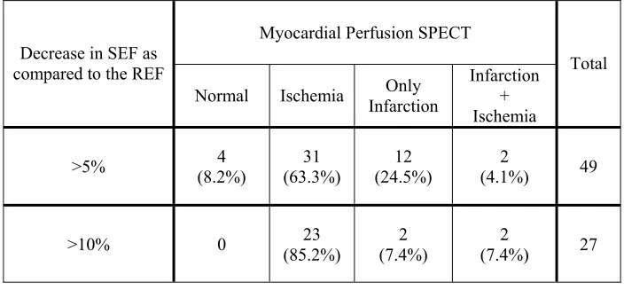
For better evaluation how severity and extent of ischemia affect the SEF, a SDS of greater than 4 (SDS ≥5) was arbitrarily considered as clinically significant ischemia (n=91, 30.6%). SEF was significantly more than REF (64.67±18.4 vs. 63.19±17.1, P<0.001) in patients with SDS<5(n=205) while patients with SDS≥5 had lower SEF than REF (54.84±14.3 vs. 60.44±17.1, P<0.001). No statistical significant difference was seen between SESV and RESV in patients with SDS<5 (32.23±38.3 vs. 34.53±39.7, P= 0.109). In patients with SDS≥5, SESV was significantly more than RESV (38.84±32.2 vs. 33.78±33.1, P<0.001). Fig 3 summarizes the result of SEF and REF indifferent patients groups that they divided based on SDS. As seen, the difference between SEF and REF is greater in patients with more SDS. Forty-nine patients (16.5%) had a decrease>5% in SEF compared with REF (DEF< -5) (Table 1). These patients had a SESV significantly more than RESV (31.76±24.6 vs. 21.57±22.2, P<0.001) while there was no statistical significant difference between SESV and RESV in all other patients (34.77±38.5 vs. 36.79±39.6, P= 0.092).
On the other hand, a decrease>10% was seen in 27 patients (Table1). All they have abnormal myocardial perfusion SPECT.
Table 1- Patients with a decrease>5% in post-stress ejection fraction (SEF) compared with rest ejection fraction (REF) (DEF<-5, DEF=SEF-REF) and patients with a decrease>10% in SEF compared with REF in different patients groups.
Myocardial Perfusion SPECT Decrease in SEF as
compared to the REF
Normal Ischemia Infarction Only Infarction + Ischemia
Total
>5% (8.2%) 4 (63.3%) 31 (24.5%) 12 (4.1%) 2 49
>10% 0 (85.2%) 23 (7.4%) 2 (7.4%) 2 27
DISCUSSION
GSPECT is largely employed in myocardial perfusion imaging because it offers the unique advantage of providing information on both perfusion and function by means of a single, simple, and inexpensive test (13). In the present study, using rest and post-dipyridamole stress GSPECT, we demonstrated increased SEF but decreased SESV as compared to rest GSPECT in patients with normal myocardial perfusion. On the other hand, reduced SEF and increased SESV were seen in patients with stress induced ischemia. Myocardial stunning is a lingering contractile dysfunction that occurs after brief
Downloaded from
http://journals.tums.ac.ir/
on Monday, August 13, 2012
هرود،ناﺮﻳاياﻪﺘﺴﻫﻲﻜﺷﺰﭘﻪﻠﺠﻣ 15
هرﺎﻤﺷ، 27 لﺎﺳ، 1386
Iran J Nucl Med 2007; Vol 15, No 27
myocardial infarction with early reperfusion, cardiac surgery and cardiac transplantation (15,16). However, myocardial stunning can also develop following silent or symptomatic ischemic episodes during common daily activities and after diagnostic stress tests with ischemic response (10,12,17,18). Although dynamic exercise and dobutamine tests are considered to be the procedures with most capability of provoking myocardial ischemia, dipyridamole-induced myocardial stunning was recently demonstrated by our group and a few others (9-11, 20, 21). Adenosine-induced stunning has also been reported (12), confirming that vasodilators are capable of producing more than simple flow heterogeneity. Although many studies showed stress-induced myocardial stunning, it is important to note that many of previous published studies included a mix of stress types (exercise and pharmocolgical stress tests) or only exercise stress test. Johnson et al. (7) reported that if ischemic patients in whom post-stress LVEF was decreased more than 5% as compared to the rest LVEF value, were considered to be stunned. Accordingly, in the present study, stunning was observed in 49(16.5%) of all patients, that 33(67.4%) of 49 patients had evidence of myocardial ischemia (ischemia or ischemia+infarction) (Table 1). Ben-Haim et al. (22)studied 236 patients using dual-isotope myocardial perfusion SPECT with gated 201Tl- SPECT at rest and post-stress gated 99m
Tc-MIBI SPECT to assess the occurrence of post-stress stunning. Their findings were similar to our results. Post-stress gated SPECT was performed 30–60 min after the injection of 99m
Tc-sestamibi at peak treadmill exercise (107 patients, 45%) or after dipyridamole infusion (n=129, 55%) (22). DEF was – 2.25 ± 5.36 and 3.42 ± 5.25 in patients with and without ischemia, respectively (P < 0.001). LVEF increases post-stress in patients with no ischemia. Post-stress stunning (>5% decrease in LVEF) was present in 68 of all 236 patients (29%) and in 58/103 (56%) patients with ischemia, after treadmill exercise or dipyridamole infusion and was more common in patients with more extensive ischemia. Post-stress stunning was observed in 37 patients who performed treadmill exercise (35%) and in 31
patients after dipyridamole infusion (24%) (22). They showed relationship between the extent and severity of ischemia (SDS score) and REF, SEF and DEF. These were all significantly reduced as the extent and severity of ischemia increased (22). Weinman and Moretti (23) have demonstrated an increase in LVEF from 63.2%±8% to 73.8%±8.2% during dipyridamole infusion in 18 normal subjects. In 62 patients with known CAD, Lee et al. (20) reported that 29% of myocardial segments had wall motion abnormalities after dipyridamole infusion, which improved at rest. The occurrence of post-dipyridamole myocardial stunning was again documented in 60% of ischemic patients after dipyridamole infusion (24). Usually coronary vasodilators do not provoke true myocardial ischemia, but may uncover a reduced flow reserve in the vascular beds perfused by stenotic lesions. In patients with severe stenosis, dipyridamole administration can cause a ‘steal’ of flow away from the myocardial bed distal to the stenosis through collateral blood vessels, leading to reduced flow, which may result in true ischemia in the presence of increased oxygen demand and therefore may also cause stunning(22). Therefore, post-stress LVEF is not synonymous with true resting LVEF in patients with ischemia, nor in those patients who had normal myocardial perfusion. In present study as seen on Figure 3, increase in SDS had been associated with more decreased SEF as compared to the REF. Santiago et al. (25) have shown early transient myocardial stunning using 201
Downloaded from
http://journals.tums.ac.ir/
on Monday, August 13, 2012
هرود،ناﺮﻳاياﻪﺘﺴﻫﻲﻜﺷﺰﭘﻪﻠﺠﻣ
15
هرﺎﻤﺷ،
27
لﺎﺳ،
1386
Iran J Nucl Med 2007; Vol 15, No 27
they had RESV less than 30 ml. In 12 other patients with fixed perfusion defects due to previous myocardial infarction, hybernation may be a possible explanation. Ben-Haim et al studied 236 patients and reported more than 5% decrease in SEF in 6 patients with normal myocardial perfusion that 4 had small heart (22). Bestetti et al studied 283 patients with gated99mTc-tetrofosmine myocardial perfusion
SPECT. They reported that increase in SESV only was seen on stunned patients (patients with SEF >5% lower than REF)(27). Also we showed that SESV in these patients significantly was more than RESV. In our study, patients with SDS≥5 had SESV significantly more than RESV while no significant difference was noticed between SESV and RESV in patients with SDS<5. Thus LVEF as measured by gated SPECT slightly but significantly decreases in the post-stress period when an ischemic insult is present, while it has a mild tendency to increase in presence of normal perfusion (11,22) .Post-stress reduction in LVEF seems to be related to an increase in end-systolic volume in stunned patients(27). The inadequate contraction may cause an increase in end-systolic volume (28). Other explanation for this finding is the presence of post-ischemic stunning as a consequence of the stress induced ischemic episode which occurs primarily in the endocardial layer (27). Ischemic stunning after dipyridamole-stress on gated SPECT may be an indicator of severe and extensive coronary artery disease, and can help the interpretation of borderline perfusion images and the elimination of false-negatives secondary to relatively balanced lesions in three-vessel disease(28,29).
CONCLUSION
We conclude that having gated SPECT in both phases of the perfusion studies may add useful information concerning cardiac function, since the post-stress study alone probably reflects stunned myocardium in patients undergoing ischemic stress tests. The SEF reduction in this population seems to be due to an increase of SESV. In this setting, the value of difference between post-stress and rest LVEF represents a new quantitative parameter derived from gated
SPECT studies (11), and it may further demonstrate to have powerful impact in prognosis since it seems to depend on the extent and severity of induced ischemia(10,22,18).
REFERENCES
1. Smanio PE, Watson DD, Segalla DL, Vinson EL, Smith WH, Beller GA. Value of gating of technetium-99m sestamibi single-photon emission computer tomographic imaging. J Am Coll Cardiol. 1997; 30:1687-92.
2. Wackers FJT. Myocardial Perfusion Imaging. In: Sandler MP, Coleman RE, Patton JA, Wackers FJT, Gottschalk A. Diagnostic Nuclear Medicine. 4th ed. Philadelphia, Lippincott Williams & Wilkins, 2003: 273-317.
3. Lipke CSA, Kühl HP, Nowak B, Kaiser HJ, Reinartz P, Koch KC, et al. Validation of 4D-MSPECT and QGS for quantification of left ventricular volumes and ejection fraction from gated 99mTc-MIBI SPECT: comparison with cardiac magnetic resonance imaging. Eur J Nucl Med Mol Imaging. 2004; 31:482-490.
4. DePuey EG, Rozanski A. Using gated technetium-99m-sestamibi SPECT to characterize fixed myocardial defects as infarct or artifact. J Nucl Med. 1995; 36:952-5.
5. Sharir T, Germano G, Kanavagh PB, Lai S, Cohen I, Lewin HC, et al. Incremental prognostic value of post-stress left ventricular ejection fraction and volume by gated myocardial perfusion single photon emission computed tomography. Circulation. 1999; 100:1035-42.
6. Lee KL, Pryor DB, Pieper KS, Harrell Jr. FE, Califf RM, Mark DB, et al. The prognostic value of radionuclide angiography in medically treated patients with coronary artery disease. Circulation. 1990; 82:1705–171.
7. Johnson LL, Verdesca SA, Aude WY, Xavier RC, Nott LT, Campanella MW, et al. Germano G. Postischemic stunning can affect left ventricular ejection fraction and regional wall motion on post-stress gated sestamibi tomograms. J Am Coll Cardiol. 1997; 30:1641–1648.
Downloaded from
http://journals.tums.ac.ir/
on Monday, August 13, 2012
هرود،ناﺮﻳاياﻪﺘﺴﻫﻲﻜﺷﺰﭘﻪﻠﺠﻣ
15
هرﺎﻤﺷ،
27
لﺎﺳ،
1386
Iran J Nucl Med 2007; Vol 15, No 27
Poststress measurements of left ventricular function with gated perfusion SPECT: comparison with resting measurements by using a same day perfusion-function protocol. Radiology. 2000; 215:529–533. 9. Braunwald E, Kloner RA. The stunned
myocardium: Prolonged, postischemic ventricular dysfunction. Circulation. 1982;66: 1146-1149.
10. Hale SL, Kloner RA. Acetaminophen and myocardial stunning after transient ischemia in rabbit hearts. J Cardiovasc Pharmacol Ther. 2005; 10: 121-129.
11. Mut F, Beretta M, Vidal I, Rener A, Alonso O, Nunez M, et al. Identification of myocardial stunning by means of gated perfusion SPECT in patients undergoing ischaemic stress myocardial tests. World J Nucl Med. 2003; 2; 122-125.
12. Dakik HA, Alam S. Myocardial stunning induced and detected by adenosine stress perfusion imaging. J Nucl Cardiol. 2001; 8:711-712.
13. Giubbini R, Rossini P, Bertagna F, Bosio G, Paghera B, Pizzocaro C, et al. Value of gated SPECT in the analysis of regional wall motion of the interventricular septum after coronary bypass grafting. Eur J Nucl Med Mol Imaging. 2004; 31:1371-7.
14. Kloner RA. Myocardial Stunning. Lancet. 1993; 341:1323-1325.
15. Rinaldi CA, Hall RJ. Myocardial stunning and hibernation in clinical practice. In J Clin Pract. 2000; 54:659-664.
16. Ambrosio G, Tritto I. Clinical manifestation of myocardial stunning. Coron Artery Dis. 2001; 12: 357-361.
17. Barnes E, Dutka DP, Khan M, Camici PG, Hall RJ. Effect of repeated episodes of reversible myocardial ischemia on myocardial blood flow and function in humans. Am J Physiol Heart Circ Physiol.2002; 282: H1603-1608.
18. Otto AC, van Staden J, van Aardt A, van Aswegen E, Joubert G, Englebrecht H. Evaluation of exercise-induced stunning using myocardial perfusion imaging. Cardiovasc J S Afr. 2001; 12:259-262. 19. Beretta M, Rener A, Vidal I, Nunez M,
Alvarez B, Mut F. Demonstration of myocardial stunning after dipyridamole stress test with gated perfusion SPECT. J Nucl Cardiol. 2001; 8: S70.
20. Lee DS, Yeo JS, Chung JK, Lee MM, Lee MC. Transient prolonged stunning induced
by dipyridamole and shown on 1- and 24-hour poststress 99mTc-MIBI gated SPECT. J Nucl Med. 2000;41: 27-35.
21. Paeng JC, Lee DS, Yeo JS, Noh CI, Kim YK, Chung JK, et al. Septal stunning by dipyridamole stress shown on quantitative gated perfusion SPECT in a child with hypertrophic cardiomyopathy. Clin Nucl Med. 2002; 27: 96-100.
22. Ben-Haim S,Gips S, Merdler A, Front A, Tamir A. Myocardial stunning demonstrated with rest and post-stress measurements of left ventricular function using dual-isotope gated myocardial perfusion SPECT. Nucl Med Commun. 2004; 25: 657-663.
23. Weinmann P, Moretti J-L. Effects of dipyridamole on left ventricular function. J Nucl Cardiol. 2000; 7:103–106.
24. Songy B, Finker F, Gray M. Post-dipyridamole myocardial stunning assessed by gated sestamibi myocardial perfusion SPECT [Abstract]. J Nucl Med. 2002; 43:190P.
25. Santiago JF, Heiba SI, Jana S, Mirzaitehrane M, Dede F, Abdel-Dayem HM. Transient ischemic stunning of the myocardium in stress thallium-201 gated SPET myocardial perfusion imaging: segmental analysis of myocardial perfusion, wall motion and wall thickening changes. Eur J Nucl Med Mol Imaging. 2002; 29:979–983.
26. Hashimoto J, Kubo A, Iwasaki R, Iwanaga S, Mitamura H, Ogawa S, et al. Gated single-photon emission tomography imaging protocol to evaluate myocardial stunning after exercise. Eur J Nucl Med. 1999; 12: 1541–1546.
27. Bestteti A, Dileo C, Alessi A, Triulzi A, Taglibue L, Tarolo GL. Post-stress end-systolic dilation: a marker of endocardial post-ischemic stunning. Nucl Med Commun. 2001;22: 685-693.
28. Kakhki VD, Zakavi SR, Sadeghi R, Yousefi A. Importance of gated imaging in both phases of myocardial perfusion SPECT: Myocardial stunning after dipyridamole infusion. J Nucl Med Technol. 2006;34:88-91
29. Hung GU, Chen CP, Yang KT. Incremental value of ischemic stunning on the detection of severe and extensive coronary artery disease in dipyridamole Tl-201 gated myocardial perfusion imaging. Int J Cardiol.2005; 105:108-110.