INTRODUCTION
The pancreas develops from the endoderm germ layer, which during early vertebrate gastrulation consists of a flat sheet of multipotent precursor cells. A series of evolutionarily conserved extrinsic signals emanating from surrounding mesodermal tissues coordinate pancreatic specification and differentiation by activating or suppressing distinct networks of transcription factors within the endodermal cells. Many of these signals and transcription factors have been identified and are well characterized in various vertebrate model systems, including zebrafish. However, much less is known about the temporal sequence of these signaling events and how they interact to control pancreas development.
The earliest and most specific marker for newly specified pancreas tissue is the expression of pancreatic and duodenal homeobox 1 (Pdx1), which later becomes restricted to the -cell lineage and duodenum (Biemar et al., 2001; Guz et al., 1995). Pdx1+cells represent a multipotent progenitor population that gives rise to all pancreatic cell types, including islet, acinar and ductal cells (Gu et al., 2002). Loss of Pdx1 function results in pancreatic agenesis (Huang et al., 2001a; Jonsson et al., 1994; Offield et al., 1996; Yee et al., 2001). During amniotic embryogenesis, two members of the vertebrate hedgehog (Hh) family of secreted proteins, sonic hedgehog (Shh) and Indian hedgehog (Ihh), are expressed at high levels throughout the early endoderm epithelium but are noticeably excluded from the foregut region that is destined
to become pancreas, which instead expresses high levels of Pdx1 (Apelqvist et al., 1997; Hebrok et al., 1998; Ramalho-Santos et al., 2000). Studies in mice and chick have established that notochord-associated signals locally repress expression of Shh in the underlying prepancreatic endoderm – a step that is necessary for the induction of Pdx1expression (Hebrok et al., 1998; Kim et al., 2000; Kim et al., 1997). Shh and Ihh bind with similar affinities to the patched 1 (Ptc1, or Ptch1) receptor (Carpenter et al., 1998), the expression of which is also absent from the pancreatic primordium (Apelqvist et al., 1997). Binding of the Hh ligand to Ptc1 mitigates Ptc1-mediated inhibition of the Hh signal transducer smoothened (Smo), thereby allowing Smo to initiate the Hh signaling cascade. Ectopic expression of Shhin the developing pancreatic endoderm of mouse and chick and increased Hh signaling in Ptc1–/–mice abolish Pdx1 expression (Apelqvist et al., 1997; Hebrok et al., 1998; Hebrok et al., 2000; Kawahira et al., 2003; Kim et al., 2000). Conversely, global inhibition of Hh signaling in the early chick embryo using the Smo antagonist cyclopamine produces extra pancreas buds with differentiated endocrine cells and promotes ectopic pancreas transformation in the stomach and intestine (Kim and Melton, 1998). Furthermore, Shh–/–;Ihh+/– mouse embryos display an increase in pancreas size and endocrine cell number (Hebrok et al., 2000). Altogether, these observations have led to the model that, in amniotes, Hh signaling has a disruptive effect on pancreas specification and that active suppression of Hh activity in the prepancreatic endoderm is a critical step for the initiation of pancreatic organogenesis. However, similar observations have not yet been extended to other vertebrates.
Although the basic structure and function of the pancreas are conserved from fish to mammals, there are small but significant differences in zebrafish with respect to pancreatic morphogenesis. Development 138, 631-640 (2011) doi:10.1242/dev.050450
© 2011. Published by The Company of Biologists Ltd
Department of Molecular, Cell and Developmental Biology, University of California, Los Angeles, CA 90095-1606, USA.
*Author for correspondence (shuolin@ucla.edu)
Accepted 26 November 2010
SUMMARY
Pancreatic organogenesis is promoted or restricted by different signaling pathways. In amniotes, inhibition of hedgehog (Hh) activity in the early embryonic endoderm is a prerequisite for pancreatic specification. However, in zebrafish, loss of Hh signaling leads to a severe reduction of -cells, leading to some ambiguity as to the role of Hh during pancreas development and whether its function has completely diverged between species. Here, we have employed genetic and pharmacological manipulations to temporally delineate the role of Hh in zebrafish endocrine pancreas development and investigate its relationship with the Bmp and retinoic acid (RA) signaling pathways. We found that Hh is required at the start of gastrulation for the medial migration and differentiation of pdx1-expressing pancreatic progenitors at later stages. This early positive role of Hh promotes -cell lineage differentiation by restricting the repressive effects of Bmp. Inhibition of Bmp signaling in the early gastrula leads to increased -cell numbers and partially rescued -cell formation in Hh-deficient embryos. By the end of gastrulation, Hh switches to a negative role by antagonizing RA-mediated specification of the endocrine pancreas, but continues to promote differentiation of exocrine progenitors. We show that RA downregulates the Hh signaling components ptc1 and smoin endodermal explants, indicating a possible molecular mechanism for blocking axial mesoderm-derived Hh ligands from the prepancreatic endoderm during the specification stage. These results identify multiple sequential roles for Hh in pancreas development and highlight an unexpected antagonistic relationship between Hh and other signaling pathways to control pancreatic specification and differentiation.
KEY WORDS: Zebrafish, Endocrine, Pancreas, Bmp, Hedgehog, Retinoic acid, Pdx1, Insulin, -cell, Endoderm
Antagonistic interactions of hedgehog, Bmp and retinoic
acid signals control zebrafish endocrine pancreas
development
Zahra Tehrani and Shuo Lin*
D
E
V
E
LO
P
M
E
N
In particular, the mammalian pancreas is specified from two distinct domains of the primitive gut tube, which subsequently evaginate to form the dorsal and ventral pancreatic buds (Murtaugh, 2007). By contrast, in zebrafish pancreatic progenitors emerge prior to gut tube formation within two bilateral rows of pdx1-expressing cells beginning at the 10-somite stage [14 hours post-fertilization (hpf)]. The most medial subset of these cells, expressing high levels of pdx1, differentiates into future dorsal bud endocrine cells, including insulin-expressing -cells (15 hpf). As the two endodermal sheets begin to converge, Pdx1+and insulin+ cells migrate medially (16 hpf). By 24 hpf, endocrine cells have coalesced at the midline into the prospective dorsal pancreatic bud. By 40 hpf, cells expressing low levels of pdx1have formed the anterior intestinal primordium and the ventral pancreatic bud, which gives rise to exocrine cells as well as to additional endocrine cells at later stages of development (Field et al., 2003).
As in amniotes, shhexpression is absent from the pancreatic endoderm of zebrafish throughout development (Roy et al., 2001); yet, zebrafish shh and smo mutants almost completely lack endocrine pancreatic expression of insulin, glucagon, nkx2.2and neurod (diIorio et al., 2002). Furthermore, the addition of cyclopamine to embryos at early gastrulation leads to severely reduced insulin expression, whereas treatment after gastrulation results in multiple clusters of insulin-expressing cells at ectopic sites anterior to the normal islet (diIorio et al., 2002). These findings imply opposite functions for Hh signaling during pancreas development in fish versus mammals. To reconcile these differences, it has been proposed that responses to Hh change over time to regulate different aspects of pancreas development (Hebrok et al., 2000); however, this idea has not been thoroughly investigated in vivo.
In addition to the Hh pathway, the retinoic acid (RA) signaling pathway is also a crucial regulator of pancreas formation; however, nothing is known about how the two pathways interact. The vitamin A derivative RA acts as a global mediator of anteroposterior (A/P) patterning in the developing embryo (Duester, 2008). In zebrafish, inhibition of the RA pathway during late gastrulation results in severe defects in endodermal structures, including a complete loss of pancreas specification. Furthermore, ectopic application of RA during the same stage causes a concentration-dependent rostral expansion of all pancreatic markers at the expense of anterior endoderm structures, suggesting that RA not only specifies the pancreas but also regulates the position of the pancreas along the A/P axis of the endoderm (Stafford and Prince, 2002). Similar requirements for RA signaling have also been observed in mouse (Martin et al., 2005; Molotkov et al., 2005), frog and quail (Chen et al., 2004; Stafford et al., 2004) embryos, demonstrating that the genetic program for pancreas specification governed by RA is well conserved among vertebrates.
The dorsoventral (D/V) Bmp activity gradient of the early gastrula has been implicated in A/P regionalization of the endoderm at later stages (Tiso et al., 2002). Bmp signals from the lateral plate mesoderm promote the hepatic fate while suppressing the pancreatic fate during somitogenesis (Chung et al., 2008). However, little is known about how early Bmp patterning signals affect pancreatic -cell specification. From early gastrulation, Bmp inhibitors are secreted from the dorsal organizer (the shield) and the surrounding presumptive dorsal mesoderm (Dal-Pra et al., 2006), suggesting a possible interaction between the Bmp and Hh signaling pathways as Shh ligands are also expressed in the shield.
In this study, we have attempted to define the role of Hh signaling temporally and investigate its relationship with the RA and Bmp signaling pathways during endocrine pancreas formation in zebrafish. We took advantage of the rapid external development of zebrafish embryos and the temporal control provided by small molecules to manipulate pathway activities during different stages of embryogenesis. We found that at early gastrulation, Hh is essential for the migration and differentiation of pancreas progenitors into dorsal bud-derived -cells as well as exocrine cells at later stages of development. By late gastrulation, Hh shifts to a negative role, antagonizing RA-mediated specification of the endocrine pancreas, thereby revealing for the first time that the inhibitory function of Hh during mammalian pancreas specification is conserved in zebrafish. Finally, we uncovered an antagonistic relationship between the Hh and Bmp pathways by showing that inhibition of Bmp signaling during gastrulation in Hh signaling-deficient embryos can partially rescue dorsal bud-derived -cell formation. These findings integrate crucial regulatory molecules involved in pancreatic specification and -cell lineage differentiation.
MATERIALS AND METHODS
Zebrafish strains
The following mutant and transgenic lines were used: smohi1640(Chen et
al., 2004), sdr1a590(smo1a590) (Kim et al., 2006), Tg(ins:GFP)(Huang et
al., 2001b), Tg(hsp70l:dnBmpr-GFP)w30 (Pyati et al., 2005) and Tg(flk1:GFP) (Cross et al., 2003). Tg(pdx1:GFP) fish (see Fig. S1 in the supplementary material), which carry a gata2 minimal promoter-GFP construct inserted 6 kb upstream of the zebrafish pdx1transcription start site, were generated from our in-house Tol2-based enhancer-trap screen (our unpublished data). Wild-type embryos were derived from the AB line.
Chemical treatments
Embryos were incubated in the following: 25 M cyclopamine (Biomol) from a 10 mM stock in ethanol (stocks were prewarmed to 28-30°C prior to dilution to the working concentration); 30 M purmorphamine (Cayman Chemical) from a 10 mM stock in DMSO; 1 M all-trans RA (Sigma-Aldrich) from a 5 mM stock in DMSO; 15 M dorsomorphin (Sigma-Aldrich) from a 5 mM stock in DMSO; and 5 M SU5416 (Sigma-Aldrich) from a 0.5 mM stock in DMSO. Controls were treated with equivalent volumes of vehicle. All dilutions were made with fish water. Treatments were carried out in 4-5 ml volumes in 6-well plates with 40-50 embryos per well. Transient treatments were stopped by continuous rinsing with fish water for 15-20 minutes. At least two independent chemical treatments were performed for each experiment.
Heat shock conditions
Embryos from hemizygous Tg(hsp70l:dnBmpr-GFP)w30outcrosses were
treated with 25 M cyclopamine starting at 2 hpf and heat shocked at 6 hpf by transferring them into embryo medium containing cyclopamine prewarmed to 38.5°C. Heat shocks were carried out in the 38.5°C water bath for 1 hour and embryos then transferred to a 28.5°C incubator. Embryos were sorted by GFP expression 3-4 hours after heat shock and incubated until fixation.
In situ hybridization
In situ hybridization (ISH) was performed as described (Thisse and Thisse, 2008). The following riboprobes were used: cpa2, hhex, hnf1ba, insulin
(preproinsulin – Zebrafish Information Network),pdx1and ptf1a.
Quantitative PCR
RNA samples were isolated from biological triplicates. For each sample, total RNA was extracted from a pool of 30 embryos using TRIzol (Invitrogen). cDNA was prepared using the SuperScript II Kit (Invitrogen). Quantitative PCR (qPCR) was carried out in triplicate on an iCycler (Bio-Rad) with 1⫻iQ SYBR Green Supermix (Bio-Rad). PCR primers (Sigma) were as follows (5⬘-3⬘): -actin, CTCTTCCAGCCTTCCTTCCT (F),
D
E
V
E
LO
P
M
E
N
CACCGATC CAGACGGAGTAT (R); pdx1, GGACCAGCCA AATC -TTACCG (F), CCTCGGCCTCGACCATATA (R); ptc1, GTTACTGC -CACGC CGCTTTTG (F), CTGACTCCTCTCCTTGCTTCT (R); smo, TGAAGACTCAGAAACTCAAGA (F), GACCAACGGAGCCT CGC -ATT (R); insulin, CTCTGTTGGTCCTGTTGGTC (F), CTCAAAGATG -CTGCAGGGT (R). Presented data are averages of biological triplicates from at least two independent experiments and are reported as fold change in relative gene expression as compared with controls after normalization to the internal control gene -actin. The comparative CT method (2⌬⌬CT) was used to calculate relative fold change (Schmittgen and Livak, 2008).
mRNA synthesis and endoderm explants
Approximately 400 pg of sox32 mRNA (T7 mMessage mMachine Kit, Ambion) was injected into Tg(ins:GFP)embryos at the 1-cell stage. Embryos were manually dechorionated and deyolked between the high and sphere stages in a heated room at 25-29°C. Approximately 25-30 endoderm explants were pooled into each well of a 96-well flat-bottom plate (Costar). Wells were coated with 1% UltraPure agarose (Invitrogen) and explants were grown in 1⫻Modified Barth Serum (MBS) as described (Grinblat et al., 1999). Explants were incubated in 1 M RA in MBS overnight at 28.5°C and examined when uninjected control embryos reached 24 hpf.
Cell counts
Tg(pdx1:GFP) and Tg(ins:GFP)24-hpf embryos were dissociated as described (Westerfield, 2000). Cell counting was performed using Volocity imaging software (Improvision) and a 40⫻water objective.
RESULTS
pdx1expression is increased in Hh-deficient
embryos
In a previous ENU mutagenesis screen for mutations affecting pancreas development, we isolated the sea dragon (sdr1a590) mutant. By 24 hpf, sdr mutants display diminished endocrine pancreas markers and an expanded bilateral pdx1domain (Fig. 1C,D) (Kim et al., 2006). We mapped the sdr1a590mutation to the smoothened (smo) gene, which encodes a seven-pass transmembrane protein required for Hh signal transduction (Rohatgi and Scott, 2007). Sequence analysis revealed that sdr1a590 mutants carry a T-to-A point mutation, leading to an isoleucine-to-asparagine substitution at residue 499. Ile499 is highly conserved in vertebrates and is positioned in the large third extracellular loop of Smo, immediately adjacent to the seventh transmembrane domain. The change from a nonpolar Ile to a polar Asn is likely to alter protein conformation in Smo, a putative G protein-coupled receptor (GPCR) (Ogden et al., 2008), indicating that this region might play an important role in Smo activity as it does in other GPCR family members (Lawson and Wheatley, 2004). Complementation analysis with the insertional mutant smohi1640 (Chen et al., 2001) verified that sdr1a590is an additional allele of smo (hereafter referred to as smo1a590). By 30 hpf, trans-heterozygous (smohi1640/smo1a590) embryos displayed phenotypes characteristic of Hh signaling deficiency and similar to those seen with the smohi1640 null allele, including U-shaped somites, no circulation, ventrally curved tail, and an almost complete lack of expression of the Hh target gene nkx2.2a(Chen et al., 2001; Varga et al., 2001). qPCR of ptc1, a target gene of the Hh signaling pathway and an established read-out of Hh activity, in smo1a590and smohi1640mutants revealed a similar reduction in Hh activity. Thus, smo1a590is likely to be a null or severely hypomorphic allele.
Gain- and loss-of-function studies in amniotes have established Hh as a negative regulator of pancreas specification. There has been uncertainty as to whether a similar scenario exists in zebrafish because the dorsal pancreas is severely reduced in zebrafish Hh mutants (Hebrok, 2003; Roy et al., 2001). We first examined the
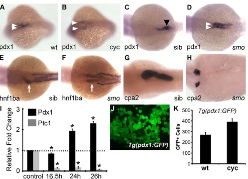
[image:3.612.316.563.59.237.2]effects of Hh signaling on pdx1 expression by qPCR at different time points between 16.5 and 26 hpf in embryos lacking Hh signaling. Since smo1a590mutants are not obviously distinguishable from their siblings prior to 26 hpf, wild-type embryos were treated with the Smo antagonist cyclopamine (Cooper et al., 1998) to analyze earlier time points. Reduction of Hh activity was verified in mutants and cyclopamine-treated embryos by analyzing the expression of the Hh receptor ptc1. At 16.5 hpf, decreased Hh activity resulted in slightly lower pdx1transcript levels, but by 24 hpf and thereafter, expression was ~2-fold higher (Fig. 1I). To verify that these observations were not unique to our mutant, we also examined smohi1640mutants and found increased expression of pdx1 at 26 hpf (data not shown). RNA ISH confirmed these results by revealing a diffuse pdx1 expression pattern and the absence of high-level expressing cells at 16.5 hpf; moreover, the initiation of migration of pdx1-expressing cells was noticeably delayed (Fig. 1A,B, arrowheads). At 26 hpf, the pdx1domains were expanded and remained separated in smo1a590 mutants (Fig. 1C,D, arrowheads), a defect not specific to the pancreas (Fig. 1G,H) as the entire foregut was split as assessed by the expression of the gut marker hnf1ba (Fig. 1E,F, arrows). Consistent with these
Fig. 1. Pdx1 is increased in Hh-deficient embryos by 24 hpf.
(A-D)pdx1 expression at 16.5 hpf (A,B) and 28 hpf (C,D) in wild-type (A,C), cyclopamine-treated (B) and smo1a590mutant (D) zebrafish embryos. At 16.5 hpf, pdx1-expressing cells (white arrowheads) begin converging at the midline (A), and by 28 hpf they have coalesced into a single cluster corresponding to the dorsal pancreatic bud (black arrowhead) (C). In Hh signaling-deficient embryos, the stripes of pdx1 expression are expanded (D) and remain separated (B,D) as pdx1 -expressing cells fail to initiate medial migration. (E,F)hnf1ba expression in the foregut (arrows) at 30 hpf in smo1a590mutants (F) versus wild type (E). (G,H)cpa2 expression in the exocrine pancreas at 3 dpf in smo1a590mutant embryos (H) versus wild type (G). All images in A-H are dorsal views, anterior to the left. (I)qPCR of pdx1and ptc1 expression levels at different developmental stages in embryos lacking Hh signaling (mean ± s.e.m.). Expression of pdx1 was initially reduced at 16.5 hpf but significantly elevated at 24 hpf in cyclopamine-treated embryos as compared with untreated controls and at 26 hpf in smo1a590mutants as compared with wild-type siblings. Asterisks denote a significant change versus control by at least two s.d. (J)A representative image of dissociated cells from a cyclopamine-treated Tg(pdx1:GFP) embryo at 28 hpf. (K)Quantification of GFP+cells in untreated (wt) and
cyclopamine-treated (cyc) Tg(pdx1:GFP) embryos at 28 hpf. The y-axis indicates the number of GFP+cells. All cyclopamine (25M) treatments
were initiated at 2 hpf until harvest.
D
E
V
E
LO
P
M
E
N
observations, we found increased numbers of Tg(pdx1:GFP)-expressing cells at 1 day post-fertilization (dpf) in cyclopamine-treated embryos (393±27; n4) compared with uncyclopamine-treated embryos (272±22; n4) (Fig. 1J,K). As expected, insulin expression was absent or drastically reduced at all stages examined. These results suggest that Hh signaling is important for limiting the size of the pdx1-expressing progenitor cell population.
Decreased Hh signaling during late gastrulation
leads to increased pdx1expression
pdx1 is expressed in the dorsal and ventral pancreatic buds as well as in the intestinal bulb (Field et al., 2003). To address the identity of the ectopic pdx1-expressing cells in Hh-deficient embryos and to define the time window in which Hh negatively regulates pdx1 expression, wild-type embryos were temporarily treated with cyclopamine during specific developmental periods (2-7, 9-12, 14-24 and 14-24-48 hpf) and assayed for pancreatic markers. Cyclopamine treatment 2-7 hpf, the period in which Hh is required for the induction of dorsal pancreatic -cells (diIorio et al., 2002), did not significantly affect pdx1 expression levels at 24 hpf (Fig. 2A,B,Q). However, pdx1-expressing cells failed to migrate. As expected, islet expression of insulin and neurod, a general marker of endocrine precursors, was absent (Fig. 2E,F; data not shown).
Previous studies have reported that Hh is not required for the development of ventral bud-derived endocrine cells (Chung and Stainier, 2008; Zecchin et al., 2004); however, the ventral bud also gives rise to exocrine tissue. Therefore, we examined the expression of the exocrine pancreas specification marker ptf1a (Zecchin et al., 2004) and the acinar differentiation marker cpa2. At 48 hpf, ptf1aexpression was almost absent in the presumptive ventral pancreas area in embryos treated with cyclopamine at 2-7 hpf (Fig. 2I,J). Likewise, cpa2expression at 72 hpf was completely abolished in treated embryos (Fig. 2M,N). Altogether, these results indicate that by the onset of gastrulation, Hh signaling is required for the medial migration and differentiation of pdx1-expressing progenitors into dorsal bud-derived pancreatic endocrine cells as well as exocrine cells.
RA derived from the anterior paraxial mesoderm directly specifies the pancreatic endoderm between 9.5 and 12.5 hpf (Stafford and Prince, 2002). We therefore examined the role of Hh during this stage of RA-mediated pancreas specification. In embryos exposed to cyclopamine at 9-12 hpf, pdx1 expression was increased (Fig. 2Q) and appeared to be expanded in both the rostral and caudal directions (Fig. 2C,D), thereby identifying the timeframe during which Hh signaling negatively regulates pdx1 expression. Interestingly, the bilateral patches of pdx1 were resolved into a single field at the midline, although they failed to condense posteriorly into the proper shape of the endocrine islet. This indicates that the requirement of Hh signaling for the migration of pdx1-expressing progenitors ends by late gastrulation. Most of the embryos that were treated with cyclopamine at 9-12 hpf had a normal-sized islet as well as some ectopic insulin- and neurod-expressing cells anterior to the endogenous islet (Fig. 2G,H; data not shown). Since we did not check intestinal-specific markers, it is possible that anterior intestinal precursors are included in the expanded pdx1-expressing population. Thus, the enlarged pdx1 domain in the embryos treated with cyclopamine at 9-12 hpf is likely to consist of a mixed population of endocrine, exocrine and intestinal precursors. Furthermore, although ptfa1 was clearly expressed in the pancreatic endoderm, in ~50% of the treated embryos it was expressed in bilateral patches (Fig. 2K,L), whereas the rest of the embryos had expression in a single midline
domain (data not shown). By contrast, cpa2 expression was severely reduced (Fig. 2O,P) or absent, indicating that by late gastrulation Hh is no longer required for exocrine fate specification but continues to regulate exocrine pancreas development by promoting the growth and differentiation of exocrine precursors.
[image:4.612.320.557.59.362.2]When Hh signaling was blocked 14-24 hpf, both pdx1 (Fig. 2Q) and insulin (data not shown) expression appeared unaffected, indicating that Hh is not required for the maintenance of pdx1-expressing progenitors. Long after endocrine pancreas development, endodermal shhexpression begins in the anterior
Fig. 2. Effects of transient cyclopamine treatment on exocrine and endocrine pancreas differentiation.(A-P)Dorsal views (anterior to the left) of wild-type zebrafish embryos transiently treated with cyclopamine (25M) either 2-7 hpf (B,F,J,N) or 9-12 hpf (D,H,L,P) versus untreated controls (A,E,I,M and C,G,K,O) and examined at 24 hpf for pdx1 (A-D) and neurod (E-H), at 48 hpf for ptf1a (I-L) and at 3 dpf for cpa2 (M-P) expression. The number of embryos displaying similar expression patterns is indicated in each case. In cyclopamine 2-7h embryos, pdx1-expressing cells fail to migrate towards the midline (A,B) and both islet and exocrine pancreas specification fail as assessed by neurod (E,F, arrows), ptf1a (I,J, arrows) andcpa2 (M,N) expression. In cyclopamine 9-12h embryos, pdx1 expression appeared expanded (C,D) and multiple clusters of neurod-expressing cells were detected at ectopic sites (G,H, arrowheads) anterior to the normal expression domain in the endogenous islet (G,H, arrow) at 24 hpf. At 48 hpf, cyclopamine 9-12h embryos show bilateral expression of ptf1a in exocrine precursors (K,L, arrows), whereas expression of cpa2at 3 dpf in the acinar cells was reduced (O,P). (Q)Relative fold change in pdx1 expression in embryos exposed to cyclopamine transiently during various developmental periods (error bars show s.e.m.). Asterisks indicate a significant change (at least 2 s.d.) compared with control. Embryos treated during 24-48 hpf were harvested at 48 hpf and all others were harvested at 24 hpf.
D
E
V
E
LO
P
M
E
N
foregut at 24 hpf, where it acts to restrict the size of the pancreas by repressing pancreatic gene expression (diIorio et al., 2007). To determine whether elevated pdx1expression in smo1a590mutants is due to a release from anterior inhibition, Hh signaling was inhibited 24-48 hpf. Surprisingly, pdx1expression was slightly reduced in these embryos (Fig. 2Q), although we could not detect any change in insulin expression (data not shown). Altogether, data from these sets of experiments demonstrate that Hh plays temporally distinct roles during both endocrine and exocrine pancreas development.
Increased Hh signaling antagonizes RA-mediated specification of pancreatic endocrine cells
Our loss-of-function experiments so far suggest that, subsequent to its early positive role, Hh plays a separate secondary role during late gastrulation in limiting the number of pdx1-expressing cells induced in the endoderm. As RA specifies the pancreatic endoderm during the same timeframe (Stafford and Prince, 2002), we tested the inhibitory role of Hh during RA-mediated pancreas specification. We used the ability of exogenous RA to ectopically induce pancreatic markers (Stafford and Prince, 2002) as a model system for examining the effects of activating or inhibiting the Hh pathway on pancreas specification. The Smo agonist purmorphamine (Sinha and Chen, 2006) was used to elevate Hh signaling and smo1a590mutants were used to reduce Hh signaling. Transient treatment of wild-type embryos with RA at late gastrulation (9-10 hpf) caused the expansion of pdx1,insulinand neurodinto the anterior endoderm (compare Fig. 3A,D,G with 3B,E,H). Simultaneous treatment with purmorphamine and RA significantly compromised the anterior ectopic induction of all three markers (compare Fig. 3B,E,H with 3C,F,I), indicating that Hh signaling can antagonize RA-mediated specification of endocrine pancreas tissue in the anterior endoderm. As pdx1-expressing cells include the pancreas progenitors as well as anterior intestinal progenitors, this reduction of the pdx1-expressing field in co-treated embryos might also reflect a reduction in intestinal precursors. To confirm that drug-drug interactions had not attenuated the efficacy of exogenous RA, the liver was examined through hhex expression. As with the pancreas, RA treatment 9-10 hpf also induces anterior ectopic liver formation (Stafford and Prince, 2002) (Fig. 3J,K). In embryos treated with RA plus purmorphamine, ectopic hhex expression remained unaffected when compared with embryos treated with RA alone (Fig. 3K,L). Thus, the reduction of endocrine pancreatic tissue caused by purmorphamine was not due to an interference with exogenous RA activity. Consistent with these data, qPCR analysis showed that pdx1 mRNA levels were significantly reduced in RA plus purmorphamine-treated embryos as compared with RA-treated embryos (Fig. 3P). Interestingly, we did not detect a significant change in expression of pdx1 or of endocrine pancreas markers in embryos treated with purmorphamine alone. Furthermore, exocrine specification, as assessed by ptf1a expression, was increased in RA plus purmorphamine-treated embryos (compare Fig. 3N,O with 3M,N), suggesting that at this stage endodermal progenitors remain competent to respond to Hh signals even after Hh is initially required for exocrine specification.
Reciprocal experiments were performed in which smo1a590 mutants and their siblings were treated with RA 9-10 hpf and analyzed for pdx1 expression. RA-treated smo1a590 mutants displayed wider stripes of pdx1 expression than RA-treated wild-type siblings (Fig. 4B,D). It is interesting to note that
pdx1-expressing cells converged at the midline in the posterior foregut of RA-treated siblings, whereas in RA-treated smo1a590embryos pdx1-expressing cells remained bilateral even at 32 hpf (Fig. 4B,D). Collectively, these findings support the model that by late gastrulation, Hh activity switches to a negative role in dorsal endocrine pancreas specification but maintains a positive role in exocrine development.
RA downregulates the expression of ptc1and smo
in endodermal explants
[image:5.612.333.535.57.402.2]Hh proteins act over long ranges to regulate cell specification and proliferation in various developmental processes (Jiang and Hui, 2008). At the end of gastrulation, the endodermal sheet is directly adjacent to the axial mesoderm, which secretes Shh. This raises the question of how Hh activity is excluded from the prepancreatic endoderm. We hypothesized that RA signaling
Fig. 3. Elevated levels of Hh signaling impair RA-mediated induction of ectopic endocrine pancreas in the anterior endoderm.(A-O)Dorsal views (anterior to left) of pdx1 (A-C), insulin (D-F), neurod(G-I) and hhex(J-L) expression at 24 hpf and ptf1a (M-O) expression at 48 hpf, comparing embryos transiently treated from 9-10 hpf with DMSO (A,D,G,J,M), retinoic acid (RA; 1M) (B,E,H,K,N) or RA plus purmorphamine (25M) (C,F,I,L,O). Brackets indicate ectopic expression and arrowheads indicate endogenous expression. The number of embryos displaying similar expression patterns is indicated in each case. (P)qPCR of pdx1 expression at 24 hpf in DMSO-, RA- and RA plus purmorphamine-treated embryos (error bars show s.e.m.). Asterisks indicate a significant change (at least 2 s.d.) compared with control.
D
E
V
E
LO
P
M
E
N
from the paraxial mesoderm may be involved in this process by preventing endodermal cells from either receiving or responding to Hh ligands. To address this possibility, we assessed the effects of RA exclusively on the endoderm using an in vitro explant culture system. To create endoderm explants, we injected sox32 mRNA into Tg(ins:GFP) embryos at the 1-cell stage. sox32 encodes a transcription factor that acts downstream of nodal signaling and is both necessary and sufficient for endoderm formation (Kikuchi et al., 2001; Sakaguchi et al., 2001). Since sox32 overexpression directs non-endodermal cells into the endodermal lineage, embryos do not undergo normal gastrulation and die by 9 hpf (Kinkel and Prince, 2009; Stafford et al., 2006). We therefore explanted endodermal cells from pre-gastrula stage embryos (high or sphere stages). After incubation of explants for 2 hours, they were treated with RA until 24 hpf, followed by a qPCR assay for Hh pathway components. RA treatment induced a reduction in both ptc1 and smo expression levels in endodermal explants (Fig. 5A) and a concomitant increase in the number of Tg(ins:GFP)-expressing cells (121±18; n3) as compared with untreated controls (3±1; n3) (Fig. 5B,C). These observations suggest a possible mechanism for excluding repressive Hh activity from the endoderm during pancreas specification.
Reduced Bmp signaling partially rescues dorsal pancreatic -cells in Hh signaling-deficient embryos
Fate-mapping experiments in the early gastrula embryo have shown that pancreatic progenitors arise from the dorsal Bmp-free zone, suggesting a potential role for Bmps in restricting pancreatic progenitor formation (Warga and Nusslein-Volhard, 1999). To test this idea, Bmp signaling was blocked at successively later time points in wild-type embryos by exposure to dorsomorphin, a compound that inhibits the Bmp type I receptors Alk3, Alk6 and Alk8 (Bmpr1a, Bmpr1b and Acvr1l – Zebrafish Information Network) (Yu et al., 2008), and assessed at 24 hpf by qPCR.
Treatment beginning at early gastrulation (6 hpf) resulted in greater than 2-fold higher expression of both pdx1 and insulin, with decreased effects when the drug was added at later time points (8, 9, 14 hpf) (Fig. 6A).
To determine whether there was a corresponding increase of cells, we performed RNA ISH. Embryos treated with dorsomorphin starting from 6 hpf displayed a strongly enlarged and dysmorphic pdx1 domain (Fig. 6B,C), but only a mildly increased insulin domain (Fig. 6D,E). To confirm this change in islet size, we analyzed -cell number in Tg(ins:GFP)embryos. As shown in Fig. 6F, the number of Tg(ins:GFP)-expressing cells in dorsomorphin-treated embryos (26±1; n11) was modestly increased compared with wild type (21±1; n9), indicating that although early Bmp inhibition robustly induces pdx1-expressing cells, only a small subset of this ectopic population includes dorsal pancreatic -cells. We conclude that early Bmp patterning signals negatively regulate dorsal bud-derived -cell specification.
Dorsomorphin has been observed to have off-target effects against the vascular endothelial growth factor (Vegf) type 2 receptor (Flk1; Kdrl – Zebrafish Information Network) (Hao et al., 2010). To determine whether the inhibition of Vegf signaling contributes to the effects of dorsomorphin on pancreas formation, we used Tg(flk1:GFP)embryos treated with the Flk1 inhibitor SU5416 (Fong et al., 1999). As expected, SU5416 treatment severely disrupted vascular development as previously reported (Cross et al., 2003), but did not significantly affect the expression of pdx1 or insulin at 24 hpf (see Fig. S2A-F in the supplementary material). These observations clearly demonstrate that the pancreatic phenotypes seen in dorsomorphin-treated embryos are not caused by Vegf pathway inhibition and provide further evidence that the vascular endothelium is dispensable for early pancreatic development in zebrafish (Field et al., 2003).
[image:6.612.51.260.58.176.2]We next asked whether the absence of the dorsal pancreas in Hh signaling-deficient embryos might be due to a localized increase of repressive Bmp activity in early endodermal progenitors. To test this, we attempted to rescue dorsal bud-derived -cell formation in
Fig. 4. Decreased Hh signaling leads to increased numbers of RA-induced pdx1-expressing cells.(A-D)Dorsal views (anterior to left) of pdx1 expression at 32 hpf in DMSO-treated wild-type (A), RA-treated wild-type (B), DMSO-treated smo1a590mutant (C) and RA-treated smo1a590mutant (D) zebrafish embryos. RA treatment (1M) from 9-10 hpf induced more pdx1-expressing cells in smo1a590mutants (D) than in wild-type embryos (B). Note the migration defect (arrows) of pdx1 -expressing cells in RA-treated smo1a590mutants (D) as compared with RA-treated wild types (B). The number of embryos displaying similar expression patterns is indicated in each case. Asterisks indicate embryos with mixed genotypes (smo+/+, smo+/–, smo–/–) from smo1a590carrier parents.
Fig. 5. RA downregulates the Hh receptor ptc1in endodermal explants.(A)qPCR of ptc1 and smo expression levels in RA-treated zebrafish endodermal explants (mean ± s.e.m.). Asterisk indicates a significant change (at least 2 s.d.) compared with control. (B,C) Tg(ins:GFP)endodermal explants treated with DMSO (B) or 1M RA (C). To create the endoderm explants, sox32 mRNA was injected into Tg(ins:GFP)embryos at the 1-cell stage (see text).
D
E
V
E
LO
P
M
E
N
[image:6.612.321.546.59.238.2]embryos lacking Hh signaling by reducing Bmp activity. Wild-type embryos were incubated in cyclopamine at 2 hpf, followed by co-incubation with dorsomorphin starting at early (6-7 hpf) or late
gastrulation (10 hpf). Embryos treated starting from 6-7 hpf (Fig. 6D-I) and 10 hpf (data not shown) showed a partial rescue of insulinexpression.
Since it was possible that drug-drug interactions had compromised the inhibitory effects of cyclopamine, we attempted to verify our observations using Tg(hsp70l:dnBmpr-GFP)w30fish, which overexpress dominant-negative bmpr1a upon heat shock treatment, causing inhibition of most, if not all, Bmp signaling (Pyati et al., 2005). Embryos obtained from outcrossing hemizygous Tg(hsp70l:dnBmpr-GFP)w30fish were treated with cyclopamine starting from 2 hpf and heat shocked at 6 hpf, the stage at which dorsomorphin treatment induces the highest levels of insulin and pdx1 expression. Heat shock of untreated Tg(hsp70l:dnBmpr-GFP)w30embryos resulted in increased insulin expression (Fig. 6J,M). Most cyclopamine-treated control embryos showed a complete absence of the islet (n29/44; 66%) (Fig. 6K), although some had a significantly reduced islet consisting of only one or two insulin-expressing cells (n15/44; 34%) (Fig. 6L). After heat shock, insulin expression was detected in 48% of cyclopamine-treated embryos (n94/193) (Fig. 6N-P), which were then scored based on islet size and morphology. The majority of the transgenic embryos showed a small cluster of insulin-expressing cells that was slightly larger than, if not equal to, that of cyclopamine-treated control embryos (n49/94; 52%) (Fig. 6N). Some embryos had an islet that was smaller than normal but significantly larger than that of cyclopamine-treated embryos (n28/94; 30%) (Fig. 6O), whereas others had scattered insulin-expressing cells (n17/94; 18%) (Fig. 6P). None of the heat shocked embryos had islets of normal size, indicating that Bmp inhibition during gastrulation can only partially rescue dorsal bud-derived -cell development in cyclopamine-treated embryos. Altogether, these data suggest that in the early gastrula, Hh signaling has a permissive effect on -cell specification by negatively regulating repressive Bmp activity.
DISCUSSION
Hh signaling and pancreas development
[image:7.612.343.530.57.96.2]Previous studies in the chick and mouse embryo have established that Hh signaling represses Pdx1 induction and pancreatic fate (Apelqvist et al., 1997; Hebrok et al., 1998; Hebrok et al., 2000; Kim and Melton, 1998; Zhang et al., 2001). Using a pharmacological approach, we temporally dissected the functions of Hh signaling during zebrafish pancreas formation (Fig. 7) and
[image:7.612.51.296.65.392.2]Fig. 6. Decrease in Bmp signaling partially rescues dorsal bud-derived -cells in Hh-deficient embryos.(A)qPCR of insulin and pdx1 expression at 24 hpf in wild-type embryos treated with dorsomorphin (15M) or DMSO starting at 6, 8, 9 or 14 hpf until harvest (mean ± s.e.m.). Treatment during gastrulation enhanced both pdx1and insulin transcript levels. Asterisks indicate a significant change (at least 2 s.d.) compared with control. (B,C) Thepdx1 expression domain at 24 hpf is expanded in embryos treated with dorsomorphin (dm) starting at early gastrulation (6 hpf) (C) as compared with untreated embryos (B). White dotted lines outline the dorsal pancreas. (D,E,G-I)insulin expression at 24 hpf in wild-type embryos treated with ethanol plus DMSO (D), dorsomorphin (E), cyclopamine (cyc) (G,H) and cyclopamine plus dorsomorphin (I). Treatment with dorsomorphin began at ~6-7 hpf until harvest. (F)Quantification of Tg(ins:GFP)-expressing cells in 24-hpf embryos treated with dorsomorphin starting at 6 hpf. (J-P)Embryos obtained from outcrosses of hemizygous Tg(hsp70l:dnBmpr-GFP)w30 adult zebrafish were incubated in cyclopamine, heat shocked (HS) at 6 hpf, and examined for insulinexpression at 24 hpf. In cyclopamine-treated control embryos, insulinexpression was either absent (K) or severely reduced (L) as compared with wild type (J). In heat shocked Tg(hsp70l:dnBmpr-GFP)w30embryos, insulin expression and islet size were increased (M). When Bmp activity was reduced by heat shock at 6 hpf in cyclopamine-treated embryos, insulin expression was partially rescued (N-P). Based on islet size and morphology, embryos were scored into three phenotypic classes: small cluster (N), medium cluster (O) or scattered (P). Cyclopamine (25M) treatments started at 2 hpf until harvest. The number of embryos displaying similar expression patterns is indicated in each case. All images are dorsal views, anterior to the left. Scale bars: 100m in B; 50m in D.
Fig. 7. Antagonistic interactions of the Hh, Bmp and RA signaling pathways in dorsal bud-derived endocrine pancreas
development.At the onset of gastrulation, Shh secreted from the dorsal organizer acts as a permissive factor for dorsal bud-derived -cell lineage specification by negatively regulating Bmp activity in endoderm progenitor cells. Low levels of Bmp activity allow intermediate signal(s) (indicated by ?) to activate the differentiation of pdx1-expressing progenitors into pancreatic cells. At the end of gastrulation, Hh switches to a repressive role by antagonizing RA-mediated endocrine pancreas specification; however, Hh still maintains its function as an inducer of exocrine pancreas specification. Inhibitory Hh activity might be excluded from the prepancreatic endoderm by RA itself, by downregulating components of the Hh signal transduction pathway. Black lines, tested interactions; gray lines, possible interactions.
D
E
V
E
LO
P
M
E
N
uncovered a repressive role during late gastrulation. In Hh signaling-deficient embryos, pdx1 expression levels were nearly doubled and there were increased numbers of pdx1-expressing cells, implicating Hh signaling in restricting pancreatic/duodenal tissue formation. We were able to determine the timeframe of this inhibitory Hh signal by transient treatments with cyclopamine during specific developmental periods. The highest level of pdx1 expression was observed when Hh signaling was inhibited from late gastrulation until early somitogenesis, a period when RA signaling specifies the pancreatic endoderm. We investigated whether RA and Hh signaling interact during pancreas specification and found that treatment with purmorphamine at this stage blocks the formation of pancreatic endocrine cells in the anterior endoderm in response to RA signals while enhancing the exocrine cell fate. Reciprocally, we showed that RA treatment induces more pdx1-expressing progenitors in smo mutants than in normal embryos. Together, these findings point to a strong antagonism between the RA and Hh signaling pathways during dorsal bud-derived -cell specification.
deIorio and colleagues found that exposure of zebrafish embryos to cyclopamine from the start of gastrulation eliminates insulin-expressing cells (deIorio et al., 2002). In our transient cyclopamine treatment studies, we show that Hh signaling at early gastrulation is required for the morphogenesis and differentiation of both endocrine and exocrine pancreatic cells at later stages. Following this early positive role, Hh plays a secondary inhibitory role by late gastrulation during RA-mediated specification of the endocrine pancreas; however, Hh continues to positively regulate exocrine differentiation. These findings provide in vivo evidence for multiple sequential roles of Hh signaling in different aspects of pancreas formation.
Exclusion of Hh activity in the pancreatic endoderm may be mediated by RA
Interactions between the RA and Hh signaling pathways have been described within the fin buds, foregut and brain to regulate organ growth and differentiation. For example, in the mouse brain, RA signaling regulates the expression of Hh target genes to optimize cellular responses to Hh (Niederreither and Dolle, 2008), and in zebrafish, administration of RA has been shown to decrease the expression of ptc1 in the developing fins (Laforest et al., 1998). Our in vitro study showed that RA downregulates the expression of Hh pathway components in the prepancreatic endoderm, rendering the endoderm incapable of receiving or responding to repressive Shh ligands. We propose this as a potential molecular mechanism for how inhibitory Hh signals secreted by the adjacent axial mesoderm are blocked from the endoderm during pancreas specification. Our observations are consistent with the report by Chung et al. that endodermal cells do not require functional Smo to differentiate into pancreatic -cells (Chung et al., 2008). Together, these data may explain why purmorphamine treatment did not significantly alter early endogenous endocrine pancreas formation. Future studies would need to address whether RA signaling has similar effects in vivo.
Interaction between Hh and Bmp signaling pathways in -cell development
Studies in various model systems have implicated Bmp signaling in the control of pancreatic organogenesis. Tiso et al. proposed that, in the zebrafish gastrula, graded distribution of Bmp activity along the D/V axis controls A/P patterning of the gut at later stages (Tiso et al., 2002). In their study, bmp2b (swirl) mutants displayed reduced
endocrine progenitors, as indicated by the expression ofneurod and islet1, whereas chordin(chordino) mutants showed slightly expanded islet1expression (Tiso et al., 2002). Furthermore, Song et al. reported that blocking Bmp signal transduction in zebrafish embryos by antisense morpholino knockdown of alk8, a member of the transforming growth factor (Tgf) receptor superfamily, led to a reduction in -cells (Song et al., 2007). By contrast, a recent report in Xenopusidentified a Bmp antagonist that restricts Bmp activity in the dorsal endoderm so as to permit the pancreatic fate (Spagnoli and Brivanlou, 2008). Similar reports of antagonistic Bmp signaling in pancreas development have been reported in mouse (Rossi et al., 2001) and zebrafish (Chung et al., 2008). Chung and colleagues showed that overexpression of bmp2bat early somite stages causes ventral pancreatic and intestinal progenitors to adopt the liver fate (Chung et al., 2008). However, the effect of Bmp signaling at earlier stages of development and its specific role in -cell specification were not examined. To address these issues, we treated embryos with dorsomorphin at various time points and found that suppression of Bmp signaling during gastrulation leads to a significant expansion of the pancreatic/duodenal field, including dorsal bud-derived -cells. This observation is in line with a previous fate-mapping analysis of the early gastrula in which the pancreas was shown to be predominantly derived from endodermal precursors positioned dorsally on the blastoderm margin (Warga and Nusslein-Volhard, 1999), a region where Bmp activity is low due to the expression of the Bmp antagonists chordin, noggin1and follistatin1 (Dal-Pra et al., 2006). It is important to note that although insulin transcript levels were doubled in dorsomorphin-treated embryos, -cell numbers were only mildly increased. Therefore, Bmp inhibition also appears to indirectly stimulate insulin promoter activity in -cells, most likely owing to the increased activity of Pdx1, which directly regulates expression of the insulin gene (Ohlsson et al., 1993). Overall, the opposing effects observed of Bmp signaling in zebrafish -cell development might be due to Bmp playing multiple temporal-specific roles, as genetic mutations and morpholino knockdowns affect Bmp signaling at earlier stages than our transient inhibition studies.
We also showed that decreased Bmp signaling during gastrulation using dorsomorphin or Tg(hsp70l:dnBmpr-GFP)w30 embryos partially restored dorsal bud-derived -cell formation in embryos treated with cyclopamine. This finding suggests that early Hh signaling is required to generate a permissive environment for the specification of dorsal pancreatic -cells by maintaining low Bmp activity in the dorsomarginal domain where pancreas progenitors are derived. Further study is required to address the mechanism of how Hh mediates this function. During the preparation of this manuscript, Chung et al. reported that Bmp signaling negatively regulates the differentiation of endodermal progenitors into dorsal pancreatic -cells (Chung et al., 2009). Altogether, these findings establish that multiple signaling pathways control different aspects of embryonic pancreas development. Understanding their spatially and temporally interactive relationships should help us to design better strategies to generate functional pancreatic -cells in vitro for therapeutic purposes.
Acknowledgements
We thank Nancy Hopkins for smohi1640fish; David Kimelman for
Tg(hsp70l:dnBmpr-GFP)w30fish; Randall T. Peterson for dorsomorphin; Matthew Veldman and Xi Ren for helpful discussions and reading of the manuscript; Anqi Liu for zebrafish maintenance; Gustavo Gomez for assistance with cell counting; and the anonymous reviewers for their helpful suggestions. This work was supported in part by National Institute of Health (NIH-RR13227 and HD061570 to S.L.). Deposited in PMC for release after 12
months.
D
E
V
E
LO
P
M
E
N
Competing interests statement
The authors declare no competing financial interests.
Supplementary material
Supplementary material for this article is available at
http://dev.biologists.org/lookup/suppl/doi:10.1242/dev.050450/-/DC1
References
Apelqvist, A., Ahlgren, U. and Edlund, H.(1997). Sonic hedgehog directs specialised mesoderm differentiation in the intestine and pancreas.Curr. Biol. 7, 801-804.
Biemar, F., Argenton, F., Schmidtke, R., Epperlein, S., Peers, B. and Driever, W.(2001). Pancreas development in zebrafish: early dispersed appearance of endocrine hormone expressing cells and their convergence to form the definitive islet. Dev. Biol. 230, 189-203.
Carpenter, D., Stone, D. M., Brush, J., Ryan, A., Armanini, M., Frantz, G., Rosenthal, A. and de Sauvage, F. J.(1998). Characterization of two patched receptors for the vertebrate hedgehog protein family. Proc. Natl. Acad. Sci. USA
95, 13630-13634.
Chen, W., Burgess, S. and Hopkins, N.(2001). Analysis of the zebrafish smoothened mutant reveals conserved and divergent functions of hedgehog activity. Development128, 2385-2396.
Chen, Y., Pan, F. C., Brandes, N., Afelik, S., Solter, M. and Pieler, T.(2004). Retinoic acid signaling is essential for pancreas development and promotes endocrine at the expense of exocrine cell differentiation in Xenopus. Dev. Biol.
271, 144-160.
Chung, W. S. and Stainier, D. Y.(2008). Intra-endodermal interactions are required for pancreatic beta cell induction. Dev. Cell14, 582-593.
Chung, W. S., Shin, C. H. and Stainier, D. Y.(2008). Bmp2 signaling regulates the hepatic versus pancreatic fate decision. Dev. Cell15, 738-748.
Chung, W. S., Andersson, O., Row, R., Kimelman, D. and Stainier, D. Y. (2009). Suppression of Alk8-mediated Bmp signaling cell-autonomously induces pancreatic {beta}-cells in zebrafish. Proc. Natl. Acad. Sci. USA107, 1142-1147. Cooper, M. K., Porter, J. A., Young, K. E. and Beachy, P. A.(1998).
Teratogen-mediated inhibition of target tissue response to Shh signaling. Science280, 1603-1607.
Cross, L. M., Cook, M. A., Lin, S., Chen, J. N. and Rubinstein, A. L.(2003). Rapid analysis of angiogenesis drugs in a live fluorescent zebrafish assay.
Arterioscler. Thromb. Vasc. Biol. 23, 911-912.
Dal-Pra, S., Furthauer, M., Van-Celst, J., Thisse, B. and Thisse, C.(2006). Noggin1 and Follistatin-like2 function redundantly to Chordin to antagonize BMP activity. Dev. Biol. 298, 514-526.
diIorio, P., Alexa, K., Choe, S. K., Etheridge, L. and Sagerstrom, C. G.(2007). TALE-family homeodomain proteins regulate endodermal sonic hedgehog expression and pattern the anterior endoderm. Dev. Biol. 304, 221-231. diIorio, P. J., Moss, J. B., Sbrogna, J. L., Karlstrom, R. O. and Moss, L. G.
(2002). Sonic hedgehog is required early in pancreatic islet development. Dev. Biol. 244, 75-84.
Duester, G.(2008). Retinoic acid synthesis and signaling during early organogenesis. Cell134, 921-931.
Field, H. A., Dong, P. D., Beis, D. and Stainier, D. Y.(2003). Formation of the digestive system in zebrafish. II. Pancreas morphogenesis. Dev. Biol. 261, 197-208. Fong, T. A., Shawver, L. K., Sun, L., Tang, C., App, H., Powell, T. J., Kim, Y. H.,
Schreck, R., Wang, X., Risau, W. et al.(1999). SU5416 is a potent and selective inhibitor of the vascular endothelial growth factor receptor (Flk-1/KDR) that inhibits tyrosine kinase catalysis, tumor vascularization, and growth of multiple tumor types. Cancer Res.59, 99-106.
Grinblat, Y., Lane, M. E., Sagerstrom, C. and Sive, H.(1999). Analysis of zebrafish development using explant culture assays. Methods Cell Biol. 59, 127-156. Gu, G., Dubauskaite, J. and Melton, D. A.(2002). Direct evidence for the
pancreatic lineage: NGN3+ cells are islet progenitors and are distinct from duct progenitors. Development129, 2447-2457.
Guz, Y., Montminy, M. R., Stein, R., Leonard, J., Gamer, L. W., Wright, C. V. and Teitelman, G.(1995). Expression of murine STF-1, a putative insulin gene transcription factor, in beta cells of pancreas, duodenal epithelium and pancreatic exocrine and endocrine progenitors during ontogeny. Development
121, 11-18.
Hao, J., Ho, J. N., Lewis, J. A., Karim, K. A., Daniels, R. N., Gentry, P. R., Hopkins, C. R., Lindsley, C. W. and Hong, C. C. (2010). In vivo structure-activity relationship study of dorsomorphin analogues identifies selective VEGF and BMP inhibitors. ACS Chem. Biol. 5, 245-253.
Hebrok, M.(2003). Hedgehog signaling in pancreas development. Mech. Dev.
120, 45-57.
Hebrok, M., Kim, S. K. and Melton, D. A.(1998). Notochord repression of endodermal Sonic hedgehog permits pancreas development. Genes Dev.12, 1705-1713.
Hebrok, M., Kim, S. K., St Jacques, B., McMahon, A. P. and Melton, D. A. (2000). Regulation of pancreas development by hedgehog signaling.
Development127, 4905-4913.
Huang, H., Liu, N. and Lin, S.(2001a). Pdx-1 knockdown reduces insulin promoter activity in zebrafish. Genesis30, 134-136.
Huang, H., Vogel, S. S., Liu, N., Melton, D. A. and Lin, S.(2001b). Analysis of pancreatic development in living transgenic zebrafish embryos. Mol. Cell. Endocrinol.177, 117-124.
Jiang, J. and Hui, C. C.(2008). Hedgehog signaling in development and cancer.
Dev. Cell15, 801-812.
Jonsson, J., Carlsson, L., Edlund, T. and Edlund, H.(1994). Insulin-promoter-factor 1 is required for pancreas development in mice. Nature371, 606-609. Kawahira, H., Ma, N. H., Tzanakakis, E. S., McMahon, A. P., Chuang, P. T. and
Hebrok, M.(2003). Combined activities of hedgehog signaling inhibitors regulate pancreas development. Development130, 4871-4879.
Kikuchi, Y., Agathon, A., Alexander, J., Thisse, C., Waldron, S., Yelon, D., Thisse, B. and Stainier, D. Y.(2001). casanova encodes a novel Sox-related protein necessary and sufficient for early endoderm formation in zebrafish.
Genes Dev.15, 1493-1505.
Kim, H. J., Sumanas, S., Palencia-Desai, S., Dong, Y., Chen, J. N. and Lin, S. (2006). Genetic analysis of early endocrine pancreas formation in zebrafish. Mol. Endocrinol.20, 194-203.
Kim, S. K. and Melton, D. A.(1998). Pancreas development is promoted by cyclopamine, a hedgehog signaling inhibitor. Proc. Natl. Acad. Sci. USA95, 13036-13041.
Kim, S. K., Hebrok, M. and Melton, D. A.(1997). Notochord to endoderm signaling is required for pancreas development. Development124, 4243-4252. Kim, S. K., Hebrok, M., Li, E., Oh, S. P., Schrewe, H., Harmon, E. B., Lee, J. S.
and Melton, D. A.(2000). Activin receptor patterning of foregut organogenesis. Genes Dev.14, 1866-1871.
Kinkel, M. D. and Prince, V. E.(2009). On the diabetic menu: zebrafish as a model for pancreas development and function. BioEssays 31, 139-152. Laforest, L., Brown, C. W., Poleo, G., Geraudie, J., Tada, M., Ekker, M. and
Akimenko, M. A.(1998). Involvement of the sonic hedgehog, patched 1 and bmp2 genes in patterning of the zebrafish dermal fin rays. Development125, 4175-4184.
Lawson, Z. and Wheatley, M.(2004). The third extracellular loop of G-protein-coupled receptors: more than just a linker between two important
transmembrane helices. Biochem. Soc. Trans.32, 1048-1050.
Martin, M., Gallego-Llamas, J., Ribes, V., Kedinger, M., Niederreither, K., Chambon, P., Dolle, P. and Gradwohl, G.(2005). Dorsal pancreas agenesis in retinoic acid-deficient Raldh2 mutant mice. Dev. Biol. 284, 399-411. Molotkov, A., Molotkova, N. and Duester, G.(2005). Retinoic acid generated
by Raldh2 in mesoderm is required for mouse dorsal endodermal pancreas development. Dev. Dyn.232, 950-957.
Murtaugh, L. C.(2007). Pancreas and beta-cell development: from the actual to the possible. Development134, 427-438.
Niederreither, K. and Dolle, P.(2008). Retinoic acid in development: towards an integrated view.Nat. Rev. Genet.9, 541-553.
Offield, M. F., Jetton, T. L., Labosky, P. A., Ray, M., Stein, R. W., Magnuson, M. A., Hogan, B. L. and Wright, C. V.(1996). PDX-1 is required for pancreatic outgrowth and differentiation of the rostral duodenum. Development122, 983-995.
Ogden, S. K., Fei, D. L., Schilling, N. S., Ahmed, Y. F., Hwa, J. and Robbins, D. J.(2008). G protein Galphai functions immediately downstream of Smoothened in Hedgehog signalling. Nature456, 967-970.
Ohlsson, H., Karlsson, K. and Edlund, T.(1993). IPF1, a homeodomain-containing transactivator of the insulin gene. EMBO J.12, 4251-4259. Pyati, U. J., Webb, A. E. and Kimelman, D.(2005). Transgenic zebrafish reveal
stage-specific roles for Bmp signaling in ventral and posterior mesoderm development. Development132, 2333-2343.
Ramalho-Santos, M., Melton, D. A. and McMahon, A. P.(2000). Hedgehog signals regulate multiple aspects of gastrointestinal development. Development
127, 2763-2772.
Rohatgi, R. and Scott, M. P.(2007). Patching the gaps in Hedgehog signalling.
Nat. Cell Biol. 9, 1005-1009.
Rossi, J. M., Dunn, N. R., Hogan, B. L. and Zaret, K. S.(2001). Distinct mesodermal signals, including BMPs from the septum transversum mesenchyme, are required in combination for hepatogenesis from the endoderm. Genes Dev.15, 1998-2009.
Roy, S., Qiao, T., Wolff, C. and Ingham, P. W.(2001). Hedgehog signaling pathway is essential for pancreas specification in the zebrafish embryo.Curr. Biol. 11, 1358-1363.
Sakaguchi, T., Kuroiwa, A. and Takeda, H.(2001). A novel sox gene, 226D7, acts downstream of Nodal signaling to specify endoderm precursors in zebrafish.
Mech. Dev.107, 25-38.
Schmittgen, T. D. and Livak, K. J.(2008). Analyzing real-time PCR data by the comparative C(T) method.Nat. Protoc.3, 1101-1108.
Sinha, S. and Chen, J. K.(2006). Purmorphamine activates the Hedgehog pathway by targeting Smoothened.Nat. Chem. Biol. 2, 29-30. Song, J., Kim, H. J., Gong, Z., Liu, N. A. and Lin, S.(2007). Vhnf1 acts
downstream of Bmp, Fgf, and RA signals to regulate endocrine beta cell
development in zebrafish. Dev. Biol. 303, 561-575.
D
Spagnoli, F. M. and Brivanlou, A. H.(2008). The Gata5 target, TGIF2, defines the pancreatic region by modulating BMP signals within the endoderm.
Development135, 451-461.
Stafford, D. and Prince, V. E.(2002). Retinoic acid signaling is required for a critical early step in zebrafish pancreatic development.Curr. Biol. 12, 1215-1220.
Stafford, D., Hornbruch, A., Mueller, P. R. and Prince, V. E.(2004). A conserved role for retinoid signaling in vertebrate pancreas development. Dev. Genes Evol.214, 432-441.
Stafford, D., White, R. J., Kinkel, M. D., Linville, A., Schilling, T. F. and Prince, V. E.(2006). Retinoids signal directly to zebrafish endoderm to specify insulin-expressing beta-cells. Development133, 949-956.
Thisse, C. and Thisse, B.(2008). High-resolution in situ hybridization to whole-mount zebrafish embryos.Nat. Protoc.3, 59-69.
Tiso, N., Filippi, A., Pauls, S., Bortolussi, M. and Argenton, F.(2002). BMP signalling regulates anteroposterior endoderm patterning in zebrafish. Mech. Dev.118, 29-37.
Varga, Z. M., Amores, A., Lewis, K. E., Yan, Y. L., Postlethwait, J. H., Eisen, J. S. and Westerfield, M.(2001). Zebrafish smoothened functions in ventral
neural tube specification and axon tract formation. Development128, 3497-3509.
Warga, R. M. and Nusslein-Volhard, C.(1999). Origin and development of the zebrafish endoderm. Development126, 827-838.
Westerfield, M. (2000). The Zebrafish Book.Eugene, OR: University of Oregon Press.
Yee, N. S., Yusuff, S. and Pack, M.(2001). Zebrafish pdx1 morphant displays defects in pancreas development and digestive organ chirality, and potentially identifies a multipotent pancreas progenitor cell. Genesis30, 137-140. Yu, P. B., Hong, C. C., Sachidanandan, C., Babitt, J. L., Deng, D. Y., Hoyng, S.
A., Lin, H. Y., Bloch, K. D. and Peterson, R. T.(2008). Dorsomorphin inhibits BMP signals required for embryogenesis and iron metabolism.Nat. Chem. Biol.
4, 33-41.
Zecchin, E., Mavropoulos, A., Devos, N., Filippi, A., Tiso, N., Meyer, D., Peers, B., Bortolussi, M. and Argenton, F.(2004). Evolutionary conserved role of ptf1a in the specification of exocrine pancreatic fates. Dev. Biol. 268, 174-184. Zhang, J., Rosenthal, A., de Sauvage, F. J. and Shivdasani, R. A.(2001).
Downregulation of Hedgehog signaling is required for organogenesis of the small intestine in Xenopus. Dev. Biol. 229, 188-202.