TMEM14C is required for erythroid
mitochondrial heme metabolism
Yvette Y. Yien, … , Luanne L. Peters, Barry H. Paw
J Clin Invest. 2014;
124(10)
:4294-4304.
https://doi.org/10.1172/JCI76979
.
The transport and intracellular trafficking of heme biosynthesis intermediates are crucial for
hemoglobin production, which is a critical process in developing red cells. Here, we profiled
gene expression in terminally differentiating murine fetal liver-derived erythroid cells to
identify regulators of heme metabolism. We determined that TMEM14C, an inner
mitochondrial membrane protein that is enriched in vertebrate hematopoietic tissues, is
essential for erythropoiesis and heme synthesis in vivo and in cultured erythroid cells. In
mice, TMEM14C deficiency resulted in porphyrin accumulation in the fetal liver, erythroid
maturation arrest, and embryonic lethality due to profound anemia. Protoporphyrin IX
synthesis in TMEM14C-deficient erythroid cells was blocked, leading to an accumulation of
porphyrin precursors. The heme synthesis defect in TMEM14C-deficient cells was
ameliorated with a protoporphyrin IX analog, indicating that TMEM14C primarily functions in
the terminal steps of the heme synthesis pathway. Together, our data demonstrate that
TMEM14C facilitates the import of protoporphyrinogen IX into the mitochondrial matrix for
heme synthesis and subsequent hemoglobin production. Furthermore, the identification of
TMEM14C as a protoporphyrinogen IX importer provides a genetic tool for further exploring
erythropoiesis and congenital anemias.
Research Article
Hematology
Find the latest version:
Introduction
Heme is a prosthetic group that plays a vital role in redox reac-tions involved in processes such as detoxification, oxygen trans-port, circadian rhythm, microRNA processing, regulation of transcription and translation, and apoptosis (1–4). The majority of heme is synthesized in red blood cells, whose main function is to transport oxygen via the heme-containing oxygen carrier protein, hemoglobin (5).
Despite extensive work on the regulation and mechanisms of heme synthetic enzymes, the mechanisms governing trans-port and intracellular trafficking of heme intermediates, which are crucial for heme synthesis, are poorly understood (6, 7). δ-Aminolevulinate (ALA), the first committed heme synthesis precursor, is synthesized in the mitochondria. ALA is exported
from the mitochondria into the cytosol for subsequent pro-cessing by δ-aminolevulinic acid dehydratase (EC4.2.1.24), porphobilinogen dehydratase (EC2.5.1.61), uroporphyrinogen III (UROgenIII) synthase (EC4.2.1.75), and uroporphyrinogen decarboxylase (EC4.1.1.37) to form UROgenIII and copropor-phyrinogen III (CPgenIII). CPgenIII is then transported back into the mitochondria to synthesize protoporphyrinogen IX (PPgenIX) by coproporphyrinogen oxidase (CPOX; EC1.3.3.3) and then oxidized to form protoporphyrin IX (PPIX) by pro-toporphyrinogen oxidase (PPOX; EC1.3.3.4). PPIX is ultimately metalated with the coordination of Fe(II) by ferrochelatase (FECH; EC4.99.1.1) to form heme. Hence, the transport and traf-ficking of these intermediates represent key regulatory points in the heme synthesis pathway (7–9). Dysregulation of heme intermediate transport can lead to cytotoxic accumulation of tetrapyrrolic synthetic intermediates, which are photoreactive and relatively insoluble when allowed to accumulate, as illus-trated by porphyrias caused by deficiencies in heme synthesis enzymes (10). Anemia may also result from defects in porphyrin trafficking, as heme synthesis is ultimately impaired.
The transport and intracellular trafficking of heme biosynthesis intermediates are crucial for hemoglobin production, which is a critical process in developing red cells. Here, we profiled gene expression in terminally differentiating murine fetal liver-derived erythroid cells to identify regulators of heme metabolism. We determined that TMEM14C, an inner mitochondrial membrane protein that is enriched in vertebrate hematopoietic tissues, is essential for erythropoiesis and heme synthesis in vivo and in cultured erythroid cells. In mice, TMEM14C deficiency resulted in porphyrin accumulation in the fetal liver, erythroid maturation arrest, and embryonic lethality due to profound anemia. Protoporphyrin IX synthesis in TMEM14C-deficient erythroid cells was blocked, leading to an accumulation of porphyrin precursors. The heme synthesis defect in TMEM14C-deficient cells was ameliorated with a protoporphyrin IX analog, indicating that TMEM14C primarily functions in the terminal steps of the heme synthesis pathway. Together, our data demonstrate that TMEM14C facilitates the import of protoporphyrinogen IX into the mitochondrial matrix for heme synthesis and subsequent hemoglobin production. Furthermore, the identification of TMEM14C as a protoporphyrinogen IX importer provides a genetic tool for further exploring erythropoiesis and congenital anemias.
TMEM14C is required for erythroid mitochondrial
heme metabolism
Yvette Y. Yien,1 Raymond F. Robledo,2 Iman J. Schultz,1 Naoko Takahashi-Makise,3 Babette Gwynn,2 Daniel E. Bauer,4,5
Abhishek Dass,4 Gloria Yi,4 Liangtao Li,3 Gordon J. Hildick-Smith,1 Jeffrey D. Cooney,1 Eric L. Pierce,1 Kyla Mohler,6
Tamara A. Dailey,6 Non Miyata,7 Paul D. Kingsley,8 Caterina Garone,9 Shilpa M. Hattangadi,4,5,10 Hui Huang,4 Wen Chen,1
Ellen M. Keenan,1 Dhvanit I. Shah,1 Thorsten M. Schlaeger,4 Salvatore DiMauro,9 Stuart H. Orkin,4,5 Alan B. Cantor,4,5
James Palis,8 Carla M. Koehler,7 Harvey F. Lodish,10 Jerry Kaplan,3 Diane M. Ward,3 Harry A. Dailey,6 John D. Phillips,11
Luanne L. Peters,2 and Barry H. Paw1,4,5
1Division of Hematology, Brigham and Women’s Hospital, Harvard Medical School, Boston, Massachusetts, USA. 2The Jackson Laboratory, Bar Harbor, Maine, USA. 3Department of Pathology,
University of Utah School of Medicine, Salt Lake City, Utah, USA. 4Division of Hematology-Oncology, Boston Children’s Hospital, and 5Department of Pediatric Oncology, Dana-Farber Cancer Institute,
Harvard Medical School, Boston, Massachusetts, USA. 6Biomedical and Health Sciences Institute, Departments of Microbiology, Biochemistry, and Molecular Biology, University of Georgia, Athens,
Georgia, USA. 7Department of Chemistry and Biochemistry, UCLA, Los Angeles, California, USA. 8Department of Pediatrics, Center for Pediatric Biomedical Research, University of Rochester
School of Medicine and Dentistry, Rochester, New York, USA. 9Department of Neurology, Columbia University Medical Center, New York, New York, USA. 10Whitehead Institute and Department of Biology,
Massachusetts Institute of Technology, Cambridge, Massachusetts, USA. 11Division of Hematology, University of Utah School of Medicine, Salt Lake City, Utah, USA.
Authorship note: Yvette Y. Yien, Raymond F. Robledo, and Iman J. Schultz contributed
equally to this work.
Conflict of interest: The authors have declared that no conflict of interest exists. Submitted: May 27, 2014; Accepted: July 17, 2014.
embryoid bodies as well as cultured Friend murine erythroleuke-mia (MEL) cells (22). Our complementary studies, using biochem-ical, cell biology, pharmacologic and genetic methods, consis-tently demonstrate that TMEM14C plays a critical and conserved role in primitive and definitive erythropoiesis and is required for erythroid heme metabolism in vertebrate species. In particular, we show that TMEM14C functions to facilitate the import of PPgenIX into the mitochondria for terminal heme synthesis.
Results
TMEM14C expression is enriched in mammalian erythropoietic tissues.
Maturing erythroid cells synthesize large amounts of heme and acquire exogenous iron to keep pace with the high rate of hemoglo-bin synthesis during erythroid terminal differentiation (23, 24). To identify mitochondrial porphyrin transporters that are coregulated Genes for heme and globin synthesis are coordinately
upreg-ulated during erythroid differentiation (11, 12) by erythroid-spe-cific transcription factors EKLF (also known as KLF1) (13–15) and GATA-1 (16–19). We hypothesized that proteins essential for trans-port of heme synthesis intermediates are also coregulated in dif-ferentiating erythroid cells. In this study, we identified genes that are upregulated in terminally differentiating erythroid cells present in the fetal liver, which synthesize large quantities of heme (20). We discovered that the expression of tmem14c, a gene coding for a transmembrane protein required for zebrafish embryonic erythro-poiesis and heme synthesis (21), was upregulated in terminally dif-ferentiating, definitive primary murine erythroid cells.
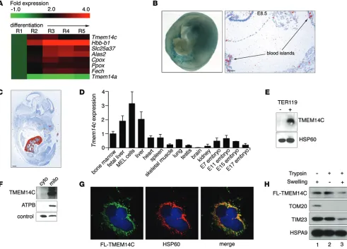
[image:3.585.47.538.57.410.2]To mechanistically dissect the functional role of Tmem14c in erythroid heme synthesis, we performed loss-of-function studies in the mouse, using cultured murine embryonic stem cells and
Figure 1. TMEM14C is enriched in differentiating murine erythroid cells and localizes to the inner mitochondrial membrane. (A) RNAseq analysis of murine
fetal liver cells sorted into 5 progressively differentiated erythroid subpopulations (R1–R5) shows that Tmem14c is upregulated during erythroid differentia-tion. (B) Tmem14c mRNA is expressed in hematopoietic organs, as shown by β-galactosidase staining (blue) of Tmem14cLacZ reporter expression in an E10.5 murine yolk sac (original magnification, ×63) and in situ hybridization of an E8.5 yolk sac (scale bar: 100 μm) and (C) fetal liver at E15.5 (pseudo-red; scale bar:
500 μm). (D) qRT-PCR shows Tmem14c mRNA is highly expressed in erythropoietic tissues and a MEL cell line. Tmem14c expression was normalized to Hprt
levels. (E) Western blot analysis shows specific expression of TMEM14C protein in differentiating TER119+ murine fetal liver erythroid cells. HSP60 serves as
a loading control. (F) Western blot analysis of fractionated Tmem14c-transfected HEK293T cells demonstrates localization of TMEM14C to the mitochondria. The control band indicates a protein that nonspecifically cross-reacts with MFRN1 antibody and migrates at a different molecular weight. (G) Confocal
immu-nofluorescence microscopy (original magnification, ×63) shows that most of the transiently transfected FLAG-TMEM14C (fluorescein) colocalizes (merged, yellow) with HSP60 (rhodamine), a mitochondrial resident protein; nuclei were stained with DAPI (blue). (H) Transiently transfected FLAG-TMEM14C localizes
Supplemental Figure 1A). β-Galactosidase staining of murine embryos carrying a Tmem14c gene trap cassette expressing LacZ under the control of the endogenous Tmem14c promoter con-firmed that Tmem14c was highly expressed in the yolk sac blood cells in the vasculature at E8.5–E10.5 (Figure 1B). Consistent with the RNAseq experiments (Figure 1A), expression of the endogenous TMEM14C protein was specifically enriched in the TER119+ maturing erythroid population of the murine fetal liver (Figure 1E). The enrichment of TMEM14C expression in differ-entiating erythroid cells suggested that it played an important role in terminal erythroid maturation.
TMEM14C is localized to the inner mitochondrial membrane.
To determine the mechanism of TMEM14C function, we charac-terized its subcellular localization. Based on proteomic studies, TMEM14C was predicted to be a mitochondrial protein (21, 26). Western blot analysis of mitochondrial and cytosolic fractions from HEK293T cells that were transiently transfected with TMEM14C confirmed colocalization of TMEM14C with the β-subunit of ATP synthase in the mitochondria (Figure 1F). In addition, con-focal immunofluorescence detection of transiently transfected FLAG-tagged TMEM14C in COS-7 cells showed colocalization of with the heme synthesis machinery during erythroid terminal
dif-ferentiation, we performed RNA sequencing (RNAseq) analysis on murine fetal liver cells that were sorted into fractions corresponding to their differentiation stage (R1–R5) by their surface expression of TER119 and CD71 (20, 25). The expression of Tmem14c, which codes for a predicted mitochondrial transmembrane protein (21, 26), was upregulated in the terminally differentiating (R3–R5) subpopulation in parallel with heme synthetic enzymes relative to progenitor cells (R1–R2) (Figure 1A). The increase in Tmem14c expression during terminal erythroid differentiation was recapitulated in a MEL cell line (Supplemental Figure 1; supplemental material available online with this article; doi:10.1172/JCI76979DS1). In contrast, expression of the related Tmem14a was not induced during erythroid differen-tiation (Figure 1A). The requirement of tmem14c for hemoglobiniza-tion in zebrafish morphants (21) and its coordinated expression with murine heme synthesis enzymes in fetal liver cells suggested that it could play a conserved role in vertebrate erythroid heme synthesis.
[image:4.585.63.503.55.383.2]Analysis of cDNA from murine tissue and in situ hybrid-ization of E8.5–E14.5 mouse embryos revealed that Tmem14c expression is enriched in hematopoietic organs, such as the yolk sac, bone marrow, fetal liver, and spleen (Figure 1, B–D, and
Figure 2. Tmem14c is specifically required for maturation of the primitive erythroid lineage. (A) Design of Tmem14c gene trap construct (E295C12) used to disrupt expression of Tmem14c. LTR, long term repeat; 6x opn, 6x osteopontin enhancer element; SA, splice acceptor sequence; β-Geo*, β-galactosidase gene fused with the neomycin resistance gene; pA, polyadenylation sequence; UTR, untranslated region. (B) Genomic PCR verifies disruption of Tmem14c locus. (C)
Expression of Tmem14c mRNA is abrogated in a Tmem14cgt/gt murine embryonic stem line. (D) The number of hemoglobinized cells is significantly reduced in
In contrast, a large proportion of TMEM14C was degraded when mitochondria were subjected to both hypotonic swelling and trypsin treatment, which exposes inner membrane proteins, such as TIM23, to trypsin degradation, while mitochondrial matrix proteins, such as HSPA9, are resistant. These data demonstrate that the majority of TMEM14C resides in the inner mitochondrial membrane (Figure 1H, lane 3).
TMEM14C is specifically required for terminal erythropoiesis. To
confirm that TMEM14C is required for mammalian erythropoie-sis, we examined hematopoiesis in embryoid bodies derived from a Tmem14c gene trap murine embryonic stem cell line from the German Gene Trap Consortium (Figure 2A). The original embry-onic stem gene trap line (gt/+), E295C12, was grown under high puromycin step-up selection to target the remaining wild-type allele by gene conversion, thus generating a null (gt/gt) clone (refs. 27–29 and Figure 2B). Quantitative RT-PCR (qRT-PCR) analysis (Figure 2C) showed absence of Tmem14c mRNA in the null Tmem14cgt/gt clone compared with that in wild-type and
hete-rozygous control embryonic stem cell clones. TMEM14C with HSP60, a mitochondrial resident protein, with a
Mander’s overlap coefficient greater than 0.7 and a Pearson’s over-lap coefficient of 0.67 (Figure 1G).
[image:5.585.43.548.52.393.2]To assess the submitochondrial localization of TMEM14C, we isolated mitochondria from transiently transfected HeLa cells expressing murine FLAG-TMEM14C and disrupted their outer membrane by hypotonic swelling to create mitoplasts. Hypo-tonic swelling did not disrupt inner mitochondrial and intermi-tochondrial membrane proteins (Figure 1H, lane 1) but liberated proteins in the intermembrane space. Hypotonic swelling also rendered proteins in the intermembrane space and outer mito-chondrial membrane accessible to trypsin digestion (Figure 1H, lane 3). The presence of TMEM14C in the mitochondria after hypotonic swelling showed that it is not an intermembrane space protein (Figure 1H, lane 1). Trypsin treatment of mitochondria degraded outer membrane proteins, such as TOM20, but not inner membrane proteins, like TIM23, and did not affect the pres-ence of TMEM14C in mitochondria, thereby demonstrating that TMEM14C is not an outer membrane protein (Figure 1H, lane 2).
Figure 3. TMEM14C is specifically required for terminal differentiation of primary definitive murine erythroid cells. (A) Primary murine fetal liver cells
developmentally arrested (Figure 3D). Flow cytometry analyses revealed a decrease in the number of terminally differentiat-ing TER119+ erythroid cells (R3–R5) in Tmem14c-deficient fetal livers, whereas the numbers of erythroid progenitors (R1–R2) remained unchanged (Figure 3, E and F). Other hematopoietic lineages were unaffected in the absence of Tmem14c (Supple-mental Figure 2), highlighting its erythroid-restricted role. Ery-throid defects were only present in Tmem14cgt/gt embryos;
het-erozygous mice were viable and fertile and did not exhibit any hematopoietic lineage defects (Figure 3, E and F; Supplemental Table 2; and Supplemental Figure 2).
Since TMEM14C is a mitochondrial protein that is required for hemoglobinization and terminal erythroid maturation, we considered whether it could play a role in heme metabolism. Haploinsufficiency of heme synthesis enzymes, such as FECH (32–34) or UROgenIII synthase (35), results in the accumulation of photoreactive tetrapyrrole biosynthetic intermediates, particu-larly in erythroid and hepatic tissue (36). E12.5 Tmem14cgt/gt fetal
livers autofluoresced under fluorescence illumination, similar to fetal livers from Fech mutant mice (FechTm1Pas FechTm1Pas mice), while
wild-type fetal livers did not, indicating an accumulation of heme intermediates, which autofluoresce due to their highly conjugated tetrapyrrole ring structures (Figure 3G). We therefore concluded that TMEM14C is involved in mitochondrial heme metabolism. In embryoid body cultures, Tmem14c deficiency specifically
resulted in a decrease in the percentage of hemoglobinized cells and erythroid cells (Figure 2, D–F), while myelopoiesis was unaffected (Figure 2G). Erythroid cells derived from the Tmem14cgt/gt embryoid
bodies were developmentally arrested at an early erythroblast stage (Figure 2E). These data show that TMEM14C is specifically required for erythroid terminal maturation or hemoglobin synthesis rather than hematopoietic stem cell biology or lineage determination.
We confirmed that TMEM14C is required for erythropoiesis by silencing Tmem14c in differentiating E14.5 mouse fetal liver cells with shRNA hairpin constructs (30). Fetal liver cells transduced with shRNA to Tmem14c exhibited decreased hemoglobin synthe-sis (Figure 3A), indicating that TMEM14C is continuously required for heme synthesis in the primary definitive erythroid lineage.
We further interrogated the Tmem14c-deficient phenotype in vivo by generating Tmem14cgt/+ mice. A cross between
heterozy-gous animals did not yield any viable homozyheterozy-gous pups, suggest-ing embryonic mortality of Tmem14cgt/gt mice. Tmem14cgt/gt mice
died in utero by E13.5 (Supplemental Table 1), a developmental stage at which definitive fetal liver erythropoiesis becomes the main source of red cells (31). Tmem14cgt/gt embryos had pale
liv-ers compared with livliv-ers of wild-type mouse embryos and were visibly anemic (Figure 3B). Erythroid cells from Tmem14cgt/gt fetal
[image:6.585.38.383.53.433.2]livers, which do not express Tmem14c mRNA (Figure 3C), were
Figure 4. TMEM14C is required for heme synthesis in murine erythroid cells both basally and during terminal differentiation.
(A) qRT-PCR analysis shows a significant
decrease in Tmem14c mRNA in both CRISPR and shRNA-silenced MEL cells compared with their respective controls. (B) Western blot
analysis show deficiency of TMEM14C protein, but not the other heme synthesis enzymes, in CRISPR cells and shRNA-silenced MEL cells. (C) o-dianisidine staining shows a heme syn-thesis defect in CRISPR cells and shRNA-si-lenced cells (original magnification, ×20). (D) 55Fe-Tf metabolic labeling demonstrates
TMEM14C is required for mitochondrial porphyrin metabolism.
To further characterize the role of TMEM14C in erythroid heme and iron metabolism, we depleted Tmem14c in MEL cells using two methods. First, we used CRISPR/Cas-mediated genomic editing to generate stable compound heterozygote knockout (CRISPR) cells (Supplemental Figure 3 and refs. 37, 38). Second, we generated stable Tmem14c knockdown MEL clones using shRNA silencing (shRNA-silenced clones are referred to herein as shRNA). Both cell lines expressed negligible steady-state levels of Tmem14c mRNA and protein (Figure 4, A and B). TMEM14C-deficient and control mitochondria contained similar amounts of PPOX and FECH pro-teins, showing that TMEM14C did not regulate protein levels of the heme synthetic enzymes (Figure 4B). Consistent with observations gathered from tmem14c morphant zebrafish embryos (21), differen-tiating murine embryonic stem–derived erythroid cells (Figure 2), and murine fetal livers (Figure 3), Tmem14c-silenced cells exhibited decreased heme synthesis upon induction of terminal differentia-tion with DMSO in comparison with that of control cells, as shown by o-dianisidine staining for hemoglobinized cells (Figure 4C). Quantitation of heme synthesis rate by 55Fe labeling indicated that TMEM14C was required for heme synthesis both basally and dur-ing terminal erythroid differentiation in MEL cells (Figure 4D). This indicated that the heme synthesis defect in Tmem14c-deficient cells was not secondary to an erythroid differentiation defect but rather that TMEM14C was directly involved in the heme homeostasis.
Because TMEM14C is a mitochondrial protein required for heme synthesis, we assayed for biomarkers reflective of iron homeostasis (39). Tmem14c deficiency did not alter basal cellular
55Fe uptake, although the amount of 55Fe uptake was decreased in differentiating Tmem14c-deficient cells (Figure 5A). The decrease in cellular 55Fe in Tmem14c-deficient MEL cells may reflect the block in erythroid maturation (Figure 4C), with a decreased demand for iron in heme production. Significantly, mitochondrial iron levels, as measured by inductively coupled plasma mass spectrometry and 59Fe labeling, were similar in controls and Tmem14c-deficient cells, both basally and during terminal differentiation (Figure 5B). These data indicated that TMEM14C does not regulate heme synthesis via the control of mitochondrial iron import. In Tmem14c-deficient cells, mitochondrial aconitase (EC4.2.1.3), FECH, and cytosolic xanthine oxidase (EC1.17.3.2) exhibited normal activity, indicating normal [2Fe-2S] cluster assembly in both the mitochondria and cytosol, further excluding a role for TMEM14C in mitochondrial iron metabolism per se (Figure 5C).
[image:7.585.42.330.53.386.2]Flow cytometry analysis of MitoTracker Red–stained cells (see Supplemental Methods) revealed no differences between the CRISPR cells and wild-type cells in the number of MitoTracker–positive cells (Supplemental Figure 4A) or mean cellular MitoTracker fluores-cence (Supplemental Figure 4B), excluding a role for TMEM14C in the regulation of mitochondrial number, apoptosis, or mitochondrial membrane potential. As cellular iron availability is a requirement for normal mitochondrial biogenesis (40), we analyzed HSP60 levels in lysates of uninduced and induced CRISPR and wild-type MEL cells, as a surrogate for mitochondrial protein content. Wild-type and CRISPR cells contained similar levels of HSP60 and control cytoso-lic marker, GAPDH, under both uninduced and induced conditions (Supplemental Figure 4C). These data confirmed that the absence of
Figure 5. TMEM14C is not required for mitochondrial iron homeostasis. (A) Quantitation of cellular iron status by 55Fe
metabolic labeling shows that basal cellular iron import is not decreased in Tmem14c-deficient MEL cells. However, cellular iron content is decreased in Tmem14c-deficient erythroid cells during differentiation. (B) Deficiency in heme synthesis in Tmem14c-silenced cells is not due to a defect in mitochondrial iron content. Inductively coupled plasma analysis of mito-chondrial iron shows that Tmem14c deficiency does not cause a defect in mitochondrial iron content either basally or during erythroid differentiation (left). This was confirmed by 59Fe
metabolic labeling and quantitation of mitochondrial iron from differentiating Tmem14c-silenced cells (right). (C) Normal
TMEM14C did not affect mitochondrial biogenesis or function and excluded the possibility that the heme synthetic defect was second-ary to a general defect in mitochondrial physiology.
We then considered the possibility that TMEM14C might play a regulatory role in mitochondrial heme synthesis. To quantitate the levels of porphyrin intermediates, we performed HPLC analysis on MEL cells differentiated in the presence of DMSO. Whether or not cells were treated with exogenous ALA, an early synthetic precursor in heme synthesis, Tmem14c-silenced cells contained similar levels of intracellular uroporphyrin III as control cells (Figure 6A). ALA-treated CRISPR cells and Tmem14cgt/gt fetal liver cells contained
sig-nificantly more coproporphyrin III than wild-type controls (Figure 6B). In comparison, Tmem14c-deficient cells and Tmem14cgt/gt fetal
liver cells contained significantly less PPIX than wild-type controls (Figure 6C). This contrasted with high levels of PPIX accumulation in the fetal FechTm1Pas FechTm1Pas mouse liver (Figure 6C). Consistent
with decreased PPIX production in Tmem14c-deficient cells and
Tmem14cgt/gt fetal liver, heme levels were decreased (Figure 6D).
The media of ALA-treated CRISPR cells exhibited a trend of ele-vated total porphyrin levels (Figure 6E), indicative of increased
extracellular excretion of cellular porphyrin, consistent with por-phyrin accumulation observed in Tmem14cgt/gt fetal liver tissues
(Figure 3F and Figure 6B). While we were unable to directly assay reduced porphyrinogens in our samples due to their spontaneous oxidation, these data demonstrate that TMEM14C is required for the formation of PPIX from CPgenIII in the mitochondria. In the absence of TMEM14C, this pathway is blocked, causing an accu-mulation of upstream porphyrins.
[image:8.585.39.540.53.382.2]To confirm that TMEM14C plays a role in porphyrin metabolism, we used deuteroporphyrin IX (DP), a synthetic analog of PPIX in which the 2,4 vinyl groups are replaced by hydrogen atoms, to chem-ically complement the heme synthesis defect in shRNA and CRISPR cells. We differentiated control, Tmem14c-deficient, and Snx3-si-lenced cells, which have a primary iron uptake defect (41), and con-currently treated them with either 5 μM Fe-dextran alone or in com-bination with 5 μM DP. We omitted treatment with DP alone, as DP is inherently cytotoxic in the absence of supplemental iron and inhib-ited MEL cell differentiation (data not shown). Cellular heme synthe-sis was assayed by 55Fe-heme incorporation. Consistent with previous data, 55Fe-heme incorporation confirmed that Fe-dextran with DP,
Figure 6. TMEM14C is required for synthesis of PPIX. HPLC analysis of porphyrin intermediates and heme in wild-type, CRISPR, control, and silenced
cell lines and E12.5 fetal liver tissue. Porphyrinogens were oxidized to their corresponding porphyrins during isolation; porphyrin levels are a surrogate for porphyrinogen levels in vivo. (A) Uroporphyrin III (UROIII) levels are normal in differentiating Tmem14c-deficient cells in the absence and presence of exogenous ALA. (B) Coproporphyrin III (CPIII) levels are normal in the absence of exogenous ALA but are mildly elevated in ALA-supplemented
comprised mutations in multiple components of the heme syn-thetic pathway (43, 44). Of note, mutations in Mfrn1 (also known as Slc25a37), required for mitochondrial iron import (28), do not by themselves cause porphyria (27). However, they predispose verte-brates to develop porphyria when cellular ALA levels are increased by gain-of-function mutations in ALAS2 (45) or dietary intake (46).
Our study reveals the potential for defects in porphyrin trans-port to cause or exacerbate porphyria. We identified TMEM14C as a protein that facilitates the transport of terminal heme synthesis intermediates, in particular PPgenIX. TMEM14C is a member of the uncharacterized TMEM14 superfamily of transmembrane pro-teins and has 4 predicted helical domains that are conserved in ver-tebrate species (26), 3 of which span the mitochondrial membrane (47). Development of other organs and nonerythroid hematopoietic lineages is largely normal in tmem14c-deficient zebra fish morphants (21) and mouse embryos (Supplemental Figure 2), indicating that these structurally similar proteins may play analogous, compensa-tory roles to those of TMEM14C in nonerythroid heme synthesis. Consistent with the proposed role of Tmem14c in erythroid heme synthesis, it is not present in Caenorhabditis elegans, a heme auxo-troph devoid of heme synthesis genes (8).
Tmem14c-deficient primary murine fetal liver tissue and
ery-throid cell lines exhibited defective PPIX synthesis and accumu-lated coproporphyrin III (Figure 6, B and C) or excreted excess but not Fe-dextran alone, rescued heme synthesis in
Tmem14c-defi-cient cells to a level similar to that of differentiated control cells. In contrast, DP could not complement the heme synthetic defect in
Snx3- silenced cells, demonstrating the specificity of the heme
syn-thesis rescue by DP (Figure 7A). The increase in heme synsyn-thesis in response to Fe-dextran and DP was not due to an increase in cellular iron content in the Tmem14c-deficient cells (Figure 7B). In combina-tion with the HPLC data, these chemical complementacombina-tion results demonstrate that TMEM14C is specifically required for the terminal steps in the heme synthesis pathway. The presence of TMEM14C in the mitochondrial inner membrane, and the active site orientation of heme synthesis enzymes in its proximity (42), defines a role for TMEM14C in the transport of PPgenIX within the mitochondrial intermembrane space into the mitochondrial matrix for the synthesis of PPIX (Figure 7C). The final step in heme synthesis, the metalation of PPIX by FECH, occurs within the mitochondrial matrix.
Discussion
[image:9.585.78.501.55.344.2]Although porphyrias are currently understood to result from defects in heme synthetic pathway enzymes, the low genetic pen-etrance of mutations in these genes suggests the requirement for additional genetic modifiers that predispose an individual to heme synthesis defects and porphyrin accumulation (43). Thus far, genetic interactions associated with congenital porphyrias have
Figure 7. The heme defect in Tmem14c-deficient MEL cells is complemented by the addition of DP. (A) 55Fe-metabolic labeling of differentiating MEL cells
that were treated with either Fe-dextran (Fe) or Fe-dextran with DP (Fe+DP). Fe-dextran with DP supplementation, but not Fe-dextran alone, significantly increased heme synthesis in Tmem14c-deficient (CRISPR and shRNA) cells to similar quantities as untreated control cells. In contrast, Fe-dextran with DP does not complement the heme synthesis defect in Snx3-deficient cells. *P < 0.05. (B) Quantitation of total cellular iron by 55Fe metabolic labeling.
Fe-dextran and Fe-dextran with DP supplementation did not significantly alter total cellular iron content in control, Tmem14c-deficient, or Snx3-deficient cells. (C) Proposed model for the function of TMEM14C (brown cylinder) as a PPgenIX transporter, enabling access to PPOX and facilitating synthesis of
from CPOX and transports PPgenIX into the mitochondrial matrix (Figure 7C). The proposed role of TMEM14C as PPgenIX importer is consistent with its tightly packed helical structure that is typical of transmembrane transport proteins (47, 56). However, due to spontaneous oxidation of porphyrinogens to porphyrins under our porphyrin isolation conditions, we could not directly measure URO-genIII, CPURO-genIII, or PPgenIX levels to demonstrate this.
In vertebrates, the expression of Tmem14c mRNA is enriched in embryonic (Figure 1B and ref. 21), fetal, and adult hematopoi-etic tissues (Figure 1, C and D). Tmem14c expression is most pro-nounced in terminally differentiating erythroid cells (Figure 1E and Supplemental Figure 1B). Consistent with this observation, the Tmem14c promoter is occupied by the erythroid transcrip-tion factor GATA-1 (19). As many genes regulating heme and iron homeostasis are regulated by iron regulatory proteins and cellular iron status (57, 58), we searched for iron response elements, which are bound by iron regulatory proteins, in the Tmem14c mRNA sequence (http://ccbg.imppc.org/sires/index.html) and found no evidence of iron response elements. We also treated MEL cells with desferrioxamine and Fe-citrate to examine the effects of iron depletion or supplementation on Tmem14c mRNA expression. Neither treatment had an effect on Tmem14c mRNA levels (Sup-plemental Figure 1C), indicating that cellular iron status does not directly regulate steady-state Tmem14c mRNA levels.
Our current data extend the findings that transport proteins play a critical role in heme homeostasis (52, 59–63). Although the National Human Genome Research Institute human genome-wide association studies database (64) and genome-genome-wide asso-ciation studies have yet to link mutations in TMEM14C to hema-tologic disease (65–67), the profound anemic and mild porphyric phenotypes in Tmem14cgt/gt mice indicate that TMEM14C could
function as a genetic modifier for the severity of anemia and porphyria in humans. We predict that further genetic sequenc-ing studies will uncover TMEM14C hypomorphic mutations in individuals suffering from anemias or porphyrias of unknown etiology. Our identification of Tmem14c as an essential regulator of heme synthesis thus provides a novel genetic tool for further studies on normal vertebrate erythropoiesis and pathological states, such as anemia and porphyria.
Methods
Cell lines. DS19 MEL cells were obtained from Arthur Skoultchi (Albert
Einstein College of Medicine, New York, New York, USA). Gene trap mouse embryonic stem cells for TMEM14C (E295C12) were obtained from the German Gene Trap Consortium.
Knockdown of Tmem14c by shRNA hairpins in mouse cells. Tmem14c
(GenBank NM_025387) stable knockdown MEL clones were obtained by stable transfection of an shRNA hairpin (TRCN0000009763, Sigma- Aldrich). Electroporation of the DS19 MEL cells and sta-ble selection of clones was carried out as previously described (37). Knockdown efficiency was assessed by qRT-PCR (Tmem14c probe: Mm00481276_m1, Invitrogen; Hprt probe: Mm01545399_m1, Invit-rogen) and Western blot analyses (Figure 4, A and B).
Fe radiolabeling and radio Fe-heme measurements. 59FeCl
3 (specific
activity: 1 Ci/mmol) and 55FeCl
3 (specific activity: 1.28 Ci/mmol)
(Per-kin Elmer) were loaded onto transferrin as described previously (68). Metabolic labeling was carried out as described previously (28). porphyrin into the cell culture media (Figure 6E). The porphyrin
accumulation in Tmem14cgt/gt fetal liver tissue caused
autofluo-rescence characteristic of murine porphyria models (Figure 3G and refs. 34–36). The apparent absence of autofluorescence in
tmem14c morphant zebrafish embryos (21) could be reconciled by
the inherent chemical instability and photoreactivity of CPgenIII and PPgenIX, excretion of these intermediates into the water and yolk sac, or a combination of these factors. In addition, other structurally similar TMEM14 family proteins like TMEM14A may partially compensate for the loss of TMEM14C function (47), mit-igating porphyrin accumulation in tmem14c morphant embryos.
Due to the localization of TMEM14C in the inner mitochon-drial membrane, we considered and excluded several possibilities for the function of TMEM14C in mitochondrial heme metabolism. We excluded the possibility of a role for TMEM14C in regulating the levels of mitochondrial heme synthesis enzymes (Figure 4B), mitochondrial iron (Figure 5B), and [Fe-S]-cluster assembly (Figure 5C). The observed decrease in cellular iron content in Tmem14c-de-ficient MEL cells was a secondary effect of differentiation defects in these cells, which require less iron for heme production (Figure 5A). Even so, it is likely that normal levels of mitochondrial iron in
Tmem14c-deficient cells are maintained by direct import of iron
from transferrin-containing endosomes, and the imported iron is preferentially used to carry out essential cellular functions (48).
The requirement for TMEM14C in housekeeping erythroid heme synthesis suggests that TMEM14C plays a broader role in ery-throid heme homeostasis, beyond hemoglobin synthesis. We spec-ulate that, in addition to TMEM14C, other TMEM14 family proteins play a major role in maintenance of housekeeping heme synthesis, a process critical for the function of mitochondrial respiratory pro-teins and cell survival (49, 50). The redundant functions of other mitochondrial proteins are underscored by the observation that
Tmem14c-deficient cells have survival rates and mitochondrial
func-tion comparable to that of wild-type cells (Supplemental Figure 4).
Tmem14c-deficient cells synthesized uroporphyrin III and
copro-porphyrin III at levels similar to control cells (Figure 6, A and B). Furthermore, ALA supplementation of cells used in metabolic iron radiolabeling experiments (Figure 4D) and porphyrin HPLC analysis (Figure 6, A–D) did not complement heme synthesis in Tmem14c-de-ficient cells. However, DP, a synthetic protoporphyrin analog, but not iron, dramatically complemented the heme defect in Tmem14c CRISPR cells (Figure 7A). The specificity of our complementation assay is maintained by the marginal rescue of heme in Snx3-deficient cells. These data demonstrate that TMEM14C primarily and directly facilitates the terminal steps of mitochondrial heme synthesis.
1. Martinkova M, Kitanishi K, Shimizu T. Heme-based globin-coupled oxygen sensors: linking oxygen binding to functional regulation of diguanylate cyclase, histidine kinase, and methyl-accepting chemotaxis. J Biol Chem. 2005;288(39):27702–27711.
2. Ajioka RS, Phillips JD, Kushner JP. Biosynthesis of heme in mammals. Biochim Biophys Acta. 2006;1763(7):723–736.
3. Feng D, Lazar MA. Clocks, metabolism, and the epigenome. Mol Cell. 2010;47(2):158–167. 4. Girvan HM, Munro AW. Heme sensor proteins.
J Biol Chem. 2013;288(19):13194–13203.
5. Chen C, Paw BH. Cellular and mitochondrial iron homeostasis in vertebrates. Biochim Biophys Acta. 2012;1823(9):1459–1467.
6. Chung J, Chen C, Paw BH. Heme metabo-lism and erythropoiesis. Curr Opin Hematol. 2012;19(3):156–162.
7. Yien YY, Paw BH. Iron and heme transport and trafficking. In: Culotta VC, Scott RS, eds. Metals
in Cells. Chichester, United Kingdom: John Wiley
and Sons; 2013:113–130.
8. Hamza I, Dailey HA. One ring to rule them all: trafficking of heme and heme synthesis interme-diates in the metazoans. Biochim Biophys Acta. 2012;1823(9):1617–1632.
9. Schultz IJ, Chen C, Paw BH, Hamza I. Iron and porphyrin trafficking in heme biogenesis. J Biol
Chem. 2010;285(35):26753–26759.
10. Sassa S. Modern diagnosis and management of the porphyrias. Br J Haematol. 2006;135(3):281–292. 11. Li L, et al. Ldb1-nucleated transcription complexes
function as primary mediators of global erythroid gene activation. Blood. 2013;121(22):4575–4585. 12. Wontakal SN, et al. A core erythroid
transcrip-tional network is repressed by a master regulator of myelo-lymphoid differentiation. Proc Natl
Acad Sci U S A. 2012;109(10):3822–3837.
13. Yien YY, Bieker JJ. EKLF/KLF1, a tissue-restricted integrator of transcriptional control, chromatin remodeling, and lineage determination. Mol Cell
Biol. 2013;33(1):4–13.
14. Pilon AM, et al. Genome-wide ChIP-Seq
reveals a dramatic shift in the binding of the transcription factor erythroid Krüppel-like fac-tor during erythrocyte differentiation. Blood. 2011;118(17):e139–e148.
15. Tallack MR, et al. A global role for KLF1 in thropoiesis revealed by ChIP-seq in primary ery-throid cells. Genome Res. 2010;20(8):1052–1063. 16. Amigo JD, et al. Identification of distal
cis-reg-ulatory elements at mouse mitoferrin loci using zebrafish transgenesis. Mol Cell Biol. 2011;7(7):1344–1356.
17. Cheng Y, et al. Erythroid GATA1 function revealed by genome-wide analysis of tran-scription factor occupancy, histone modifi-cations, and mRNA expression. Genome Res. 2009;19(12):2172–2184.
18. Fujiwara T, et al. Discovering hematopoietic mechanisms through genome-wide analysis of GATA factor chromatin occupancy. Mol Cell. 2009;36(4):667–681.
19. Yu M, et al. Insights into GATA-1-mediated gene activation versus repression via
genome-R01 DK052830, to J. Kaplan and D.M. Ward; genome-R01 HL088468, to L.L. Peters; R01 DK070838, to B.H. Paw; and P01 HL032262, to A.B. Cantor, H.F. Lodish, S.H. Orkin, and B.H. Paw).
Address correspondence to: Barry H. Paw, Hematology Division, Brigham and Women’s Hospital, 1 Blackfan Circle, Karp 05.211, Boston, Massachusetts 02115, USA. Phone: 617.355.9008; E-mail: bpaw@rics.bwh.harvard.edu. Or to: Luanne L. Peters, The Jack-son Laboratory, 600 Main Street, Bar Harbor, Maine 04609, USA. Phone: 207.288.6391; E-mail: luanne.peters@jax.org.
Iman J. Schultz’s present address is: InteRNA Technologies, Utrecht, The Netherlands.
Gordon J. Hildick-Smith’s present address is: Weill Cornell Medi-cal College, New York, New York, USA.
Jeffrey D. Cooney’s present address is: University of Texas Health Science Center, San Antonio, Texas, USA.
Gloria Yi’s present address is: MGH Institute of Health Profes-sions, Boston, Massachusetts, USA.
Eric L. Pierce’s present address is: Georgia Institute of Technol-ogy, Atlanta, Georgia, USA.
Non Miyata’s present address is: Kyushu University, Fukuoka, Japan.
Wen Chen’s present address is: Texas A&M Health Science Cen-ter, Temple, Texas, USA.
Shilpa M. Hattangadi’s present address is: Yale University Medical School, New Haven, Connecticut, USA.
Hui Huang’s present address is: Columbia University Medical Center, New York, New York, USA.
Chemical complementation assays. MEL cells were differentiated
chemically with 1.5% DMSO for 72 hours. Concurrently, cells were treated with 5 μM Fe-dextran (Sigma-Aldrich) alone or in combination with 5 μM DP (Frontier Scientific). Heme synthesis was assayed by labeling with 55Fe (Supplemental Methods).
Statistics. Statistical analysis was carried out using 2-tailed paired
or unpaired Student’s t test. Significance was set at P < 0.05. All graphs depict the mean ± SEM.
Study approval. The present studies in animals were reviewed and
approved by The Jackson Laboratory Animal Care and Use Committee.
Acknowledgments
wide chromatin occupancy analysis. Mol Cell. 2009;36(4):682–695.
20. Zhang J, Socolovsky M, Gross AW, Lodish HF. Role of Ras signaling in erythroid differentiation of mouse fetal liver cells: functional analysis by a flow cytometry-based novel culture system.
Blood. 2003;102(12):3938–3946.
21. Nilsson R, et al. Discovery of genes essential for heme biosynthesis through large-scale gene expres-sion analysis. Cell Metab. 2009;10(2):119–130. 22. Ney PA, D’Andrea AD. Friend erythroleukemia
revisited. Blood. 2000;96(12):3675–3680. 23. Napier I, Ponka P, Richardson DR. Iron trafficking
in the mitochondrion: novel pathways revealed by disease. Blood. 2005;105(5):1867–1874. 24. Richardson DR, et al. Mitochondrial iron
traf-ficking and the integration of iron metabolism between the mitochondrion and cytosol. Proc
Natl Acad Sci U S A. 2010;107(24):10775–10782.
25. Wong P, Hattangadi SM, Cheng AW, Frampton GM, Young RA, Lodish HF. Gene induction and repression during terminal erythropoiesis are mediated by distinct epigenetic changes. Blood. 2011;118(16):e128–e138.
26. Pagliarini DJ, et al. A mitochondrial protein com-pendium elucidates complex I disease biology.
Cell. 2008;134(1):112–123.
27. Chung J, et al. Iron regulatory protein-1 protects against mitoferrin1-deficient porphyria. J Biol
Chem. 2014;289(11):7835–7843.
28. Shaw GC, et al. Mitoferrin is essential for erythroid iron assimilation. Nature. 2006;440(7080):96–100.
29. Woo AJ, et al. Identification of ZBP-89 as a novel GATA-1-associated transcription factor involved in megakaryocytic and erythroid development.
Mol Cell Biol. 2008;28(8):2675–2689.
30. Hattangadi SM, Burke KA, Lodish HF. Home-odomain-interacting protein kinase 2 plays an important role in normal terminal erythroid dif-ferentiation. Blood. 2010;115(23):4853–4861. 31. Baron MH, Isern J, Fraser ST. The embryonic origins of erythropoiesis in mammals. Blood. 2012;119(21):4828–4837.
32. Childs S, Weinstein BM, Mohideen MA, Donohue S, Bonkovsky H, Fishman MC. Zebrafish dracula encodes ferrochelatase and its mutation provides a model for erythropoietic protoporphyria. Curr
Biol. 2000;10(16):1001–1004.
33. Kiupel M, Montagutelli X, Sundberg JP. The fer-rochelatase deficiency (Fechtm1Pas) mouse mutation:
a mouse model for erythropoietic protoporphyria.
JAX Communication. 1999;1:1–7. http://www.
porfyrier.dk/pdf_filer/communication01.pdf. 34. Tutois S, et al. Erythropoietic protoporphyria in the
house mouse. A recessive inherited ferrochelatase deficiency with anemia, photosensitivity, and liver disease. J Clin Invest. 1991;88(5):1730–1736. 35. Bishop DF, et al. Uroporphyrinogen III synthase
knock-in mice have the human congenital ery-thropoietic porphyria phenotype, including the characteristic light-induced cutaneous lesions.
Am J Hum Genet. 2006;78(4):645–658.
36. Bishop DF, Clavero S, Mohandas N, Desnick RJ. Congenital erythropoietic porphyria: character-ization of murine models of the severe common (C73R/C73R) and later-onset genotypes. Mol
Med. 2011;17(7):748–756.
37. Cong L, et al. Multiplex genome engineer-ing usengineer-ing CRISPR/Cas systems. Science. 2013;339(6121):819–823.
38. Canver MC, et al. Characterization of genomic deletion efficiency mediated by clustered regu-larly interspaced palindromic repeats (CRISPR)/ Cas9 nuclease system in mammalian cells. J Biol
Chem. 2014;289(31):21312–21324.
39. Xu W, Barrientos T, Andrews NC. Iron and copper in mitochondrial diseases. Cell Metab. 2013;17(3):319–328.
40. Rensvold JW, Ong SE, Jeevananthan A, Carr SA, Mootha VK, Pagliarini DJ. Complementary RNA and protein profiling identifies iron as a key regulator of mitochondrial biogenesis. Cell Rep. 2013;3(1):237–245.
41. Chen C, et al. Snx3 regulates recycling of the transferrin receptor and iron assimilation. Cell
Metab. 2013;17(3):343–352.
42. Rhee HW, et al. Proteomic mapping of mitochon-dria in living cells via spatially restricted enzy-matic tagging. Science. 2013;339(6125):1328–1331. 43. Balwani M, Desnick RJ. The porphyrias:
advances in diagnosis and treatment. Blood. 2012;120(23):4496–4504.
44. To-Figueras J, et al. ALAS2 acts as a modifier gene in patients with congenital erythropoietic porphyria. Blood. 2011;118(6):1443–1451. 45. Wang Y, et al. Abnormal mitoferrin-1 expression
in patients with erythropoietic protoporphyria.
Exp Hematol. 2011;39(7):784–794.
46. Troadec MB, et al. Targeted deletion of the mouse Mitoferrin1 gene: from anemia to pro-toporphyria. Blood. 2011;117(20):5494–5502. 47. Klammt C, et al. Facile backbone structure
deter-mination of human membrane proteins by NMR spectroscopy. Nat Methods. 2012;9(8):834–839. 48. Sheftel AD, Zhang AS, Brown C, Shirihai OS,
Ponka P. Direct interorganellar transfer of iron from endosome to mitochondrion. Blood. 2007;110(1):125–132.
49. Kim HJ, Khalimonchuk O, Smith PM, Winge DR. Structure, function and assembly of heme centers in mitochondrial respiratory complexes.
Biochim Biophys Acta. 2012;1823(9):1604–1616.
50. Homedan C, et al. Acute intermittent porphyria causes hepatic mitochondrial energetic fail-ure in a mouse model. Int J Biochem Cell Biol. 2014;51:93–101.
51. Chavan H, Khan MM, Tegos G, Krishnamurthy P. Efficient purification and reconstitution of ATP binding cassette transporter B6 (ABCB6) for functional structural studies. J Biol Chem. 2013;288(31):22658–22669.
52. Krishnamurthy PC, et al. Identification of a mam-malian mitochondrial porphyrin transporter.
Nature. 2006;443(7111):586–589.
53. Krishnamurthy P, Schuetz JD. The role of ABCG2 and ABCB6 in porphyrin metabolism cell sur-vival. Curr Pharm Biotechnol. 2011;12(4):647–655. 54. Ulrich DL, et al. ATP-dependent
mitochon-drial porphyrin importer ABCB6 protects against phenylhydrazine toxicity. J Biol Chem. 2012;287(16):12676–12690.
55. Helias V, et al. ABCB6 is dispensable for erythro-poiesis specifies the new blood group system Langereis. Nat Genet. 2012;44(2):170–173. 56. Maslennikov I, et al. Membrane domain structures
of three classes of histidine kinase receptors by cell-free expression and rapid NMR analysis. Proc
Natl Acad Sci U S A. 2010;107(24):10902–10907.
57. Anderson CP, Shen M, Eisenstein RS, Leibold EA. Mammalian iron metabolism and its control by iron regulatory proteins. Biochim Biophys Acta. 2012;1823(9):1468–1483.
58. Casey JL, et al. Iron-responsive elements: regula-tory RNA sequences that control mRNA levels and translation. Science. 1988;240(4854):924–928. 59. Chiabrando D, et al. The mitochondrial heme
exporter FLVCR1b mediates erythroid differenti-ation. J Clin Invest. 2012;122(12):4569–4579. 60. Desuzinges-Mandon E, Arnaud O, Martinez L, Huche F, Di Pietro A, Falson P. ABCG2 transports transfers heme to albumin through its large extracellular loop. J Biol Chem. 2010;285(43):33123–33133.
61. Koch M, Breithaupt C, Kiefersauer R, Freigang J, Huber R, Messerschmidt A. Crystal structure of protoporphyrinogen IX oxidase: a key enzyme in haem and chlorophyll biosynthesis. EMBO J. 2004;23(8):1720–1728.
62. Ogino T, et al. Serum-dependent export of protoporphyrin IX by ATP-binding cassette transporter G2 in T24 cells. Mol Cell Biochem. 2011;358(1):297–307.
63. Proulx KL, Woodard SI, Dailey HA. In situ conver-sion of coproporphyingen III to heme by murine mitochondria: terminal steps of the heme biosyn-thetic pathway. Protein Sci. 1993;2(7):1092–1098. 64. Hindorff LA, et al. Potential etiologic and
func-tional implications of genome-wide association loci for human diseases and traits. Proc Natl Acad
Sci U S A. 2009;106(23):9362–9367.
65. Ganesh SK, et al. Multiple loci influence erythro-cyte phenotypes in the CHARGE Consortium.
Nat Genet. 2009;41(11):1191–1198.
66. Soranzo N, et al. A genome-wide meta-analysis identifies 22 loci associated with eight hemato-logical parameters in the HaemGen consortium.
Nat Genet. 2009;41(11):1182–1190.
67. van der Harst P, et al. Seventy-five genetic loci influencing the human red blood cell. Nature. 2012;492(7429):369–375.


![Figure 5. TMEM14C is not required for mitochondrial iron homeostasis. ities for [2Fe-2S] cofactor–dependent mitochondrial aconitase, FECH, and cytosolic xanthine oxidase in cells exclude defects in [2Fe-2S] cluster assembly](https://thumb-us.123doks.com/thumbv2/123dok_us/8147126.801330/7.585.42.330.53.386/mitochondrial-homeostasis-cofactor-dependent-mitochondrial-aconitase-cytosolic-xanthine.webp)


