1
CHARACTERISATION OF LIPOPROTEINS IN
STAPHYLOCOCCUS AUREUS
ASHRAF ELGALLALI
Centre for Parasitology and Disease Research,
School of Environment and Life Sciences,
University of Salford,
Salford, UK
I
Table of
Contents
List of tables ... III List of figures ... VI Acknowledgements ... VIII Declaration... IX List of abbreviations ... X Abstract ... XIII
Chapter one ... 1
1. Introduction ... 2
1.1 Staphylococcus aureus ... 2
1.2 Lipoproteins ... 18
1.3 S. aureus genome ... 26
1.5 The Nematode Caenorhabditis elegans ... 37
1.6 Whole transcriptome shotgun sequencing technology (RNA-Seq) ... 47
2.7 Project aims and objectives ... 51
Chapter two ... 52
2. Material and methods ... 53
2.1 Bacterial strains and growth conditions... 53
2.2 Oxacillin/cefoxitin disc diffusion test... 53
2.3 Proteomic analysis of S. aureus lipoproteins ... 54
2.4 In-gel Trypsin digestion ... 56
2.5 Protein identification by LC/MS/MS ... 57
2.6 Bioinformatics of S. aureus genomic DNA... 60
2.7 Isolation of RNA and assessment quality/concentration ... 69
2.8 Caenorhabditis elegans as a model of bacterial infection ... 74
II
Chapter three... 82
3. Genetic analysis of Staphylococcus aureus lipoproteins ... 83
3.1 Introduction ... 83
3.2 Results ... 85
3.3 Discussion ... 116
Chapter four ... 120
4. Proteomics identification of S. aureus lipoproteins under different growth conditions ... 121
4.1 Introduction ... 121
4.2 Results ... 124
4.3 Discussion ... 133
Chapter five ... 136
5. Transcription of S. aureus lipoprotein genes studied using quantitative RT-PCR, RNA sequencing and during pathogenesis in C. elegans ... 137
5.1 Introduction ... 137
5.2 Results ... 139
5.3 Discussion ... 206
Chapter six ... 215
6. General summary and discussion ... 216
7. References ... 221
Appendices ... 254
Appendix 1 ... 255
III
List of tables
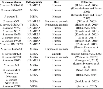
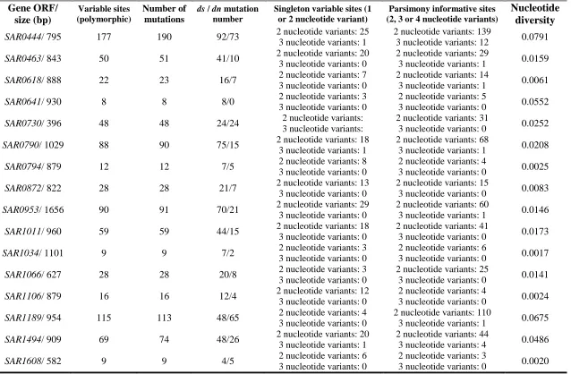
Table 1. SDS-PAGE loading gel concentrations ... 55 Table 2. Mixture of 20 μM stock solutions made up of 4 peptides of known sizes used as internal calibration ... 58 Table 3. S. aureus strains used in BLAST comparative analysis ... 62 Table 4. Oligonucleotide primers, base sequences, genes locations, and predicted sizes of PCR products for S. aureus lipoproteins ... 63 Table 5. Primers/probes, genes base sequences and predicted sizes of qPCR products for S. aureus lipoprotein genes ... 73 Table 6. Details of RNA samples from three independent experiment of treated and control cells ... 77 Table 7. List of 50 lipoprotein genes tested by PCR in three S. aureus strains (MRSA252, RN4282 and T1) ... 90 Table 8. Phylogenetic analysis of 44 lipoprotein genes for 20 S. aureus strains, calculated by DnaSP program ... 91 Table 9. Comparison of lipoprotein function in S. aureus MRSA252 and other S. aureus strains according to the UniProt database... 94 Table 10. An overview of genes encoding S. aureus lipoprotein and their regulating
operons description, the promoter sequences and 4 binding sites were predicted using the BPROM web-based software (Solovyev and Salamov, 2011). Operons were predicted using Genome 2D (genome2D.molgenrig.nl) and Artemis
(http://www.sanger.ac.uk/science/tools/artemis) (Rutherford et al., 2000). ... 99 Table 11. Promoter sequences of S. aureus MRSA252 lipoproteins ... 102 Table 12. List of S. aureus MRSA252 lipoproteins including details of their genes and lipoprotein signal peptides, the carboxy-terminal region (C-region or lipobox), the
hydrophobic (H-) region and amino-terminal (N-) region. ... 107 Table 13. Zone diameter and MIC interpretive for S. aureus tested with 30 μg of cefoxitin ... 115 Table 14. List of identified lipoprotein of S. aureus MRSA252 in 1D SDS-PAGE 1D ... 126 Table 15. List of identified lipoprotein of S. aureus MRSA252 in 2-DE ... 126 Table 16. List of S. aureus MRSA252 lipoproteins identified in gel-free in-solution
IV
Table 18. Expression fold level of 5 lipoprotein genes in 3 S. aureus strains examined by quantitative real-time PCR ... 140 Table 19. Transcriptome comparison of lipoprotein genes in S. aureus 8325-4 at different phases of growth ... 145 Table 20. Comparison of S. aureus MRSA252 lipoprotein genes transcriptome in the C. elegans infection model at 16 and 40 h ... 154 Table 21. Major groups of up-regulated genes in S. aureus at 16 h of C. elegans infection model and their predicted function ... 160 Table 22. Top 100 up-regulated genes in S. aureus infection of C. elegans at 16 h
comparing to 16 h control sample ... 160 Table 23. Major groups of down-regulated genes in S. aureus at 16 h of C. elegans
infection compared to non-infected control sample and their predicted function ... 163 Table 24. Top 100 down-regulated genes in S. aureus infection of C. elegans at 16 h comparing to to non-infected control sample ... 163 Table 25. Major groups of up-regulated genes of S. aureus at 40 h of C. elegans infection comparing to non-infected control sample and their predicted function ... 166 Table 26. List of the most up-regulated genes of S. aureus at 40 h after infection of C. elegans comparing to 16 h control sample ... 166 Table 27. Major groups of down-regulated genes in S. aureus at 40 h of C. elegans
infection comparing to 16 h control sample model and their predicted function ... 169 Table 28. The most down-regulated genes in S. aureus at 40h of C. elegans infection comparing to 16 h control sample ... 169 Table 29. Major groups of up-regulated genes in S. aureus at 40 h of C. elegans infection compared to 16 h of infection sample and their predicted function ... 172 Table 30. Top 100 up-regulated genes in S. aureus infection of C. elegans at 40 h
comparing to 16 h of infection sample ... 172 Table 31. Major groups of down-regulated genes in S. aureus at 40 h of C. elegans
V
VI
List of figures
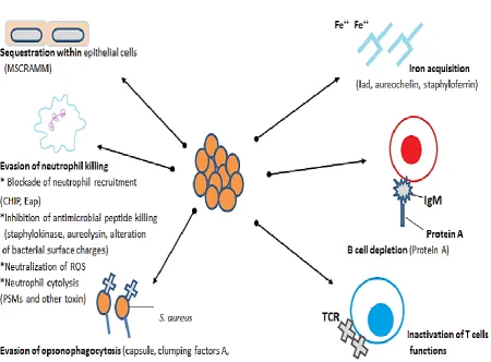
Figure 1. S. aureus survival strategies during infection stages ... 11
Figure 2. (A) S. aureus cell surface and secreted proteins in a different bacterial growth phases. (B) Shows a cross section of the bacterial cell envelope ... 14
Figure 3. The structure of the lipid modification in lipoproteins ... 18
Figure 4. Tripartite structure of lipoprotein signal sequence... 21
Figure 5. Biosynthesis pathway of bacterial lipoprotein in Gram-positive (two-step) and (A to D) biosynthesis pathway of Gram-negative bacteria (three-step) ... 24
Figure 6. Life cycle of C. elegans at 22°C ... 39
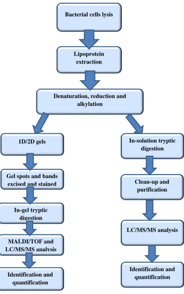
Figure 7. Workflows of in-gel and in-solution protein digestion procedures ... 59
Figure 8. RT-PCR assay amplification efficiency of gene SAR0216 ... 72
Figure 9. Schematic diagram illustrating an overview of C. elegans killing assay ... 79
Figure 10. A 1.5% agarose gel image showing PCR products of 6 lipoprotein genes fragments ... 87
Figure 11. A 1.5% agarose gel image showing PCR products of 4 lipoprotein genes fragments ... 87
Figure 12. The distribution and location of lipoprotein genes mapped on the reference circular genome of S. aureus MRSA252 and labeled according to their position of the strands. ... 98
Figure 13. Amino acid numbers and position in lipobox of lipoproteins in 20 S. aureus strains ... 106
Figure 14. Phylogenetic relationships of lipoprotein among 20 S. aureus strain ... 114
Figure 15. The growth curves of S. aureus MRSA252 at 37°C in Tryptone soya broth .. 130
Figure 16. Comparisons of differential expression fold patterns of lipoprotein genes as determined by real-time quantitative PCR assay ... 142
Figure 17. Comparisons of differential expression patterns of lipoprotein genes as determined by real-time quantitative PCR assay ... 143
Figure 18. C. elegans survival in TS agar at 20°C with S. aureus MRSA252 ... 147
Figure 19. N2 Bristol wild-type (WT) C. elegans survival in NGM agar at 20°C with E. coli OP50 ... 148
Figure 20. Images of C. elegans fed on S. aureus ... 149
Figure 21. Images of healthy C. elegans fed on E. coli OP50 ... 150
VII
VIII
Acknowledgements
I am extremely indebted to Allah for the good health and wellbeing that were necessary to complete this thesis.
I would sincerely like to express my special appreciation and thanks to my supervisor, Prof. Howard Foster for his incredible endless support and his continuous willingness and assistance to finish my PhD, for his patience, motivation and enormous knowledge, without his guidance and encouragement this thesis would not be completed.
Very special thanks to my family: my parents, brother and sisters for supporting me, without their motivation and encouragement I would not complete this thesis.
I would especially like to thank my friends, I am extremely grateful and indebted to them for sharing expertise, and valuable guidance and encouragement extended to me.
IX
Declaration
X
List of abbreviations
aa ……….…. amino acid
Agr ………. Accessory gene regulator
CA-MRSA……….…….… Community-acquired methicillin-resistant S. aureus CHIP……….…. Chemotaxis inhibitory protein
CPM ………... Counts Per million Mapped reads DE ……….. Differentially Expressed
DOLOP ……….. Database of Bacterial Lipoproteins DNase ………. Deoxyribonuclease
DTT ……… Dithiotreitol
EAp……….… Extracellular Adherence protein E-value ………Expect value
FC ………Fold Change
FDR ……… False Discovery Rate
FPKM ………. Fragments per Kilobase Transcript per Million mapped reads Fur ………. Ferric uptake regulator
GFP ……….... Green fluorescent protein
HA-MRSA……….…. Hospital-acquired methicillin-resistant Staphylococcus aureus IM lipoproteins ……..……. Inner-Membrane lipoproteins
IAA ………. Iodoacetamide ICU ….……… Intensive Care Units ISA ………..………Iso-Sensitest agar
Isd………....…… Iron-regulated surface determinant pro-IL-1β ……….pro-Interleukin-1β
XI LPXTG motif ………….. (Leu-Pro-any-Thr-Gly) Lsp ………... Lipoprotein signal peptidase
MALP-2 ………... Macrophage activating lipopeptide-2 MRSA ………...…... Methicillin-resistant Staphylococcus aureus
MSCRAMM……... Microbial surface components recognizing adhesive matrix molecules
NJ ………...…….. Neighbor joining tree
NOD………..…… Nucleotide-binding oligomerization domain OM lipoproteins ……... Outer Membrane lipoproteins
ORFs ………..…..… Open reading frames OTU …………..………... Operational taxonomic unit
PAMP…………..……….. Pathogen-associated molecular pattern PBP……… Penicillin-binding protein
PMNs cells….………..….. Poly morphonuclear leukocyte PRR……… Pattern recognition receptor PSM………..…. Phenol Soluble Modulin PV ………..…... P-value
PVL ………..….…Panton-Valentin leucocidin RNase ……….…...ribonuclease
SAB ………...… Staphylococcus aureus bacteraemia SBPs ………. Substrate Binding Proteins
SCCmec ……….... Staphylococcal chromosomal cassette mec SDS ….……….……...….. Sodium Dodecyl Sulfate
SAR ………...…Staphylococcal Accessory Regulator SNPs ………... Single-nucleotide polymorphism SP………...Signal peptide
XII srtA ………... surface protein sorting A
Tat ………..….. Twin arginine translocation pathway TCR……….…..….T cell receptor
TMED …..…….…….….. Tetramethylethylenediamine TLR….…..……….…Toll-like receptor
TSB….………...…….….. Tryptic Soy Broth TST ………... Toxic Shock Toxin-1
VRSA ...…... vancomycin resistant Staphylococcus aureus VISA ……….…… vancomycin intermediate Staphylococcus aureus WT……….… Wild type
XIII
Abstract
The Gram-positive bacterium Staphylococcus aureus is an extremely successful opportunistic bacterium capable of causing a wide range of hospital-acquired and community-acquired infections, and is becoming increasingly virulent and resistant to antibiotics. In order to investigate this pathogen, various methods have been used to analyse the pathogenic behaviour including genomics, transcriptomics and proteomics. S. aureus expresses approximately 55-70 lipoproteins with only about half with known functions. Little is known about the biochemical functions of many individual lipoproteins and their proteomics has not been investigated in detail. Lipoproteins have a broad ranging functionality and perform various roles in bacterial activity and attract a particular interest to investigate their virulence and survival influences in the course of host infection. The initial part of this study was to find out whether the lipoproteins of S. aureus have similar genetic characteristics among all strains. PCR and Quantitative Real-Time PCR
experiments were performed to analyse the genetic and the expression levels for some lipoprotein genes. The majority of PCR results showed high similarity in lipoprotein genetic structure among the examined strains. Phylogenetic trees from concatenated lipoprotein genes alignment were generated to represent the lipoprotein genes distribution of S. aureus strains. To identify and characterise proteomic of S. aureus lipoproteins a comprehensive quantitative proteome profiling of S. aureus lipoproteins using gel-free /in-solution trypsin digestion system followed by LC-MS/MS quantification identified 38 lipoproteins that represent two-thirds of the S. aureus MRSA252 lipoprotein. In addition, S. aureus-mediated infections with live C. elegans were performed on solid assays to investigate the host-pathogen relationships. S. aureus MRSA252 exhibited a high level of nematocidal activity with average time for half of the worms to die of ~ 2 d and infected C. elegans showed visible signs of illness. To evaluate lipoprotein transcripts expression level and microbe/host-specific pathogenic factors RNA of both S. aureus and C. elegans were characterised after isolation from the infected C. elegans and subjected to RNA
Sequencing, the large-scale data has provided useful information on pathogen and host activities during infection. RNA sequencing analysis showed different types of regulations and interactions of lipoprotein transcripts during host exposures to indicate 3 transcripts significantly were up-regulated and 11 down-regulated. RNA sequencing analysis showed that 62 lipoprotein transcripts were expressed during C. elegans infection model.
XIV
lipoproteins that were expressed in the non-infection condition representing approx. two-thirds of the S.aureus MRSA252 lipoproteins. The results suggest that some lipoproteins were involved in pathogenesis of C. elegans but their function were not clear. More
1
Chapter one
2
1. Introduction
1.1 Staphylococcus aureus
1.1.1 General introduction
Staphylococcus aureus has been recognized as one of the most common pathogenic Gram-positive bacteria around the world with a serious prevalence of hospital- and community-associated (CA) infections. It causes a range of infectious diseases such as sepsis,
endocarditis and pneumonia. In addition, it can cause uncomplicated skin infections impetigo, also soft tissue infections, with or without abscess formation (Noguchi et al., 2006). Furthermore, S.aureus can cause toxin-mediated diseases such as toxic shock syndrome, scalded skin syndrome and food poisoning (Dinges et al., 2000). S. aureus are pathogenic bacteria that have become a great public health concern in recent years due to their ability to produce an array of virulence factors and drug-resistant variants. Before the availability of antibiotics, S. aureus infections were most often non-fatal but after the discovery and use of penicillin the prognosis of staphylococcal infections significantly improved. The first reported isolation of methicillin-resistant staphylococcus aureus in 1961 in the United Kingdom was shortly after methicillin introduced into clinical use (Barber, 1961). Since that time infections caused by methicillin resistant S. aureus
3
introduction of legislation and introduction of healthcare-associated improvement programmes in 2006 was significantly associated with the reduction of MRSA BSIs (Duerden et al., 2015). The MRSA BSI rates per 100, 000 population have slightly decreased from 1.8 to 1.7 to 1.4 and to 1.5 in 2012, 2013, 2014 and 2015, respectively (Health Protection Agency, 2015). S. aureus related infections include skin and soft tissue infections, pneumonia, infective endocarditis and bloodstream infections in United States are over half a million cases and more than 10,000 deaths per year (Klevens et al., 2007, Otto, 2010). Methicillin resistance in S. aureus is due to the acquisition of a mobile genetic element, their size ranging from 21/67 kb in size, termed the staphylococcal cassette chromosome mec (SCCmec) (Katayama et al., 2000).
Glycopeptide antibiotics including vancomycin prevent maturation of bacterial cell wall by binding to the terminal D-alanyl-D-alanine residues of peptidoglycan precursors,
consequently blocking the bacterial enzymes involved in the late steps of peptidoglycan synthesis (Anderson et al., 1965). Infections with MRSA strains are usually treated with vancomycin, but the first vancomycin intermediate-level resistant isolates (VISA) were discovered in 1997 (Hiramatsu et al., 1997). There are early reports of high-level
vancomycin resistant isolates (VRSA) that have acquired the vanA resistance gene from vancomycin resistant enterococci (Control and Prevention, 1997, Hierholzer et al., 1995). Reports revealed that 40%-60% of all hospital S. aureus infections in Europe, United States and Japan were resistant to methicillin (Lindsay and Holden, 2006). Resistant MRSA strains express a special enzyme known as penicillin-binding protein 2a (PBP2a) which is not inactivated by β-lactams and keeps generating the cross-linked peptidoglycan in their presence (Ubukata et al., 1985). Platensimycin antibiotic has been shown to be effective against some strains of MRSA and VRSA (Wang et al., 2007). There are some limited effective antibiotics that cure MRSA infections and some are to be developed in the near future (Dryden, 2014).
1.1.2 S. aureus microbiology and taxonomy
4
positive results of coagulase (Willis et al., 1966), mannitol-fermentation and deoxyribonuclease tests (Raymond and Traub, 1970). It is catalase positive (unlike
streptococci), a facultative anaerobic organism. S. aureus is extremely heat sensitive and is inactivated at a temperature > 46ºC while their enterotoxins are heat-stable proteins which survive heat treatment and low pH conditions (Evenson et al., 1988). Both MRSA and methicillin-susceptible S. aureus have been found to survive for many weeks in a completely dry environment (Beard-Pegler et al., 1988).
The S. aureus cell wall is a strong protective coat and relatively amorphous form, about 20-50nm in thickness (Shockman and Barren, 1983), composed from repeating
disaccharide N-acetylmuramic acid-(β1–4)-N-acetylglucosamine, cross-linking is via a pentglycine interpeptide bridge (Ghuysen and Strominger, 1963). The staphylococcal cell wall also contains secondary polymers (proteins, carbohydrates and teichoic acids) that are immobilised in the peptidoglycan scaffold (Navarre and Schneewind, 1999).
Peptidoglycan is the major component of the cell wall and comprises up to 50% of the cell wall mass (Waldvogel, 1995). However, there is another essential component group of phosphate containing polymers known as teichoic acids. S. aureus has two types of
teichoic acids, cell wall teichoic acid and cell membrane associated lipoteichoic acid, both of them make up about 40% of cell wall mass (Knox and Wicken, 1973), which give a negative charge to the bacterial cell surface and take part in the acquisition and localisation of metal ions especially in divalent cations and the activities of autolytic enzymes
(Wilkinson, 1997). Over 90% of S. aureus clinical isolated strains have been shown to possess capsular polysaccharides; production of this capsule is reported to decrease phagocytosis in vitro and to increase S. aureus virulence in a mouse bacteraemia model (Thakker et al., 1998). S. aureus is typically an extracellular pathogen able to survival and persist in different host tissues (Brouillette et al., 2003), it is also capable of internalising within non-professional phagocytic cells such as epithelial and endothelial cells for a short time, this strategy helps the pathogen to evade host defence mechanisms and the action of antimicrobial agents that mainly act in the extracellular space (Sinha et al., 1999).
1.1.3 Transmission of S. aureus
5
2014), with asymptomatic colonisation being more frequent than infection. Throat colonization was more common than nasal colonization (Marshall and Spelman, 2007). Between 25% -50% of healthy persons may be persistently or transiently colonised, however, colonisation rate is higher amongst individuals who are immuno-compromised, for example diabetic patients, HIV-infected patients, patients who require haemodialysis and people with skin diseases (Sydnor and Perl, 2011). S. aureus also colonises and infects many mammals including domestic animals such as dogs and cats, some birds e.g.
chickens and turkeys, and farm animals such as cows, pigs and goats (Baptiste et al., 2005). Infection with S. aureus is a common cause of dairy cow mastitis with significant economic impact; moreover these animals may act as good reservoirs for human
colonisation (McCarthy and Lindsay, 2010).
The majority of S. aureus patients with symptomatic infections are invaded with their own colonising strains (Kluytmans et al., 1997). Infection might be also acquired from another person or from environmental exposure. S. aureus transmission in the community sector is associated with skin-to-skin contact and contaminated household environment (Desai et al., 2011), bacteria can survive for long periods on household fomites for up to 2 months (Baggett et al., 2004). Reports on MRSA environmental contamination have shown that 5%-8% among households without MRSA disease however in households with a CA-MRSA-infected person or a healthcare labourer were 26%-32% (Scott et al., 2008,
Uhlemann et al., 2011). Airborne spread and transmission through contacts with contaminated materials may also be involved (Cooper et al., 2004).
Hospital transmission (patient to patient) is the most likely outcome from transient colonisation of the hands of hospital workers, who then transfer strains between patients. Spread of bacteria in aerosols from the respiratory or nasal discharges from heavily
6
Hygiene and medical interventions in United Kingdom have led to a significant reduction in incidence of health care-associated methicillin resistant S. aureus MRSA related
infections (Mandatory Surveillance MRSA). S. aureus MRSA shows attributes that are not always found in most clinically isolated bacteria, it has the ability to express a range of virulence factors and therefore is always considered medically relevant when encountered in clinical specimens. S. aureus is able to develop and expand resistance to a wide range of antimicrobial treatments and it is a major pathogen in both hospital and community
infections (Styers et al., 2006).
1.1.4 Epidemiology of S. aureus infections
Development of new molecular typing approaches has allowed the study of population structure and epidemiology of bacterial pathogens, but these approaches are variable in practical for large population samples. The evolution of methicillin-resistant
Staphylococcus aureus (MRSA) has changed the clinical and molecular epidemiology of S. aureus infections in the past three decades, these observations have showed the
importance of epidemiological variations of S.aureus infections and the molecular characterisation of pathogenic resistant strains. Bacterial adaptability and the significant speed of bacterial evolution together with environmental challenges give this bacterium an ability to create genetic variation for their survival. Little information is available about the epidemiology of S. aureus in the non-western parts of the world; due to the increasing number of people traveling worldwide this epidemiology has changed.
In general, numbers of both community-acquired and hospital-acquired staphylococcal infection has increased with increasing levels of antibiotic resistance and the emergence of epidemic strains (Chatterjee and Otto, 2013). S. aureus pathogenicity indicates that
virulence is a multi-factorial process as single gene inactivation experiments could not prevent S. aureus pathogenic ability (Fedtke et al., 2004). The national data on
7
between 2005 and 2008 confirmed a 34 % decreased of MRSA-related bloodstream infections incidence (Kallen et al., 2010). S. aureus bacteraemia (SAB) infection overall rates may have stabilized over the past two decades, with incidence of SAB vary from 10 to 30 cases per 100,000 person per a year in industrialized community (Laupland et al., 2013). A little is known about the incidence of SAB in the nonindustrialized, in contrast, incidence of community-acquired SAB between 2004 and 2010 in northeast Thailand was approx. 3 per 100,000 people-years (Kanoksil et al., 2013). The incidence of MRSA cases among 3,662 S. aureus isolated in four hospitals in Siberian Russia between 2007 and 2011, prevalence of HA-MRSA was 22% while the CA-MRSA was 2.9% (Khokhlova et al., 2015).
From a clinical point of view, new approaches to reduce the S. aureus infections include minimizing the duration of hospital stay, improved surveillance systems, use of antibiotic to eliminate nasal carriage before elective surgery and more strict hand hygiene regulations (Skov et al., 2012). English National Point Prevalence Survey on Healthcare-associated Infections and Antimicrobial Use indicated a sharp decrease of healthcare-associated MRSA bacteraemia from 1.8% of cases with MRSA bacteraemia in 2006 to 0.1% in 2011 (Mandatory Surveillance MRSA). Over time, CA-MRSA epidemic became more
complicated in various geographic locations and urgently need to improve the understanding of S. aureus epidemiology. Schaumburg et al.investigated the
8
Since S. aureus toxic shock syndrome (TSS) was first described by Todd et al. in 1978, TSS was linked with superabsorbent tampons in menstruating women (Herzer, 2001), with highest annual infection rate of 13.7 per 100,000 menstruating women (Osterholm and Forfang, 1982). Introduction of more hygienic advice to all susceptible women to use suitable tampons reduced the annual incidences of S. aureus TSS to 1 per 100,000
menstruating women and 0.3 per nonmenstruating women (Hajjeh et al., 1999), after that, the incidence of S. aureus TSS has stayed stable with the annual incidences around 0.69 per 100,000 menstruatingwomen and 0.32 per 100,000 of total populations (DeVries et al., 2011).
Vaccine candidates for S. aureus have been tested in clinical trials using both active and passive immunization modalities, but in attempts to develop a vaccine in future there are several issues should be considered, firstly, antigens used should be expressed by a majority of S. aureus strains, second, these antigens have been proved to stimulate the immune response of preclinical animal models of infection, finally, in passive
immunization animal experiments the produced antibodies against these antigens must be protective (Kuklin et al., 2006). Four cell wall-anchored surface proteins of S. aureus as antigens in a murine model were assembled into a combined vaccine has given high levels of protection against invasive infections (Stranger-Jones et al., 2006). The majority of antibacterial subunit-based vaccines components have two categories, either a secreted toxins or abundantly expressed surface exposed molecules (Grandi, 2010). IsdA and IsdH S. aureus surface proteins that are expressed during infection were used in cotton rat model of nasal colonisation, rats vaccinated with IsdA or IsdH developed protection against nasal carriage (Clarke et al., 2006).
A number of excellent strategies to developing an effective S. aureus vaccine, including (1) a sensible fund needed to be invested in clinical trials. (2) A multiple mixed bacterial antigens will be preferable to become the main scientific approach for new vaccines. (3) Cell-mediated immune response biological function during infection should be evaluated in an appropriate patient population (Patti, 2011). An effective combined vaccine provided stable protection against S. aureus contains five conserved antigens known to have
9 1.1.5 Pathogenesis of S. aureus
Staphylococcus aureus is an adaptable microbe that can express a range of virulence factors, including adhesins, enzymes, toxins and capsular polysaccharides. These virulence factors are controlled by a set of staphylococcal regulatory networks, including eight main accessory gene regulators system (Bien et al., 2011).
S. aureus is one of the pyogenic pathogenic bacteria with an ability to induce abscess in both local and metastatic infections. There are five stages in the pathogenesis of S. aureus infections; (1) colonisation, (2) local infection, (3) systemic dissemination and/or sepsis, (4) metastatic infection, and (5) toxinosis. Once the organism breach skin or mucous membranes and reach underlying host tissues, they can cause an illness in any part of the infected tissue, causing a range of diseases varying from minor skin infections to life- threatening systemic infections, such as endocarditis and haemolytic pneumonia.
Adherence is the first step for bacterial colonisation of a new host, this process mediated by several adhesins. By expressing a range of surface bound proteins known as microbial surface components recognising adhesive matrix molecules (MSCRAMMs), these
molecules recognize the most prominent components of the extracellular matrix and blood plasma including, collagen-binding protein, fibrinogen, fibronectin-binding proteins A and B, elastin binding protein, prothrombin binding protein and von Willebrand factor binding protein have been well characterised (Clarke and Foster, 2006, Foster and Höök, 1998). S. aureus has characteristic survival strategies during infection as shown in figure 1.
S. aureus produces various toxins that are classified on the basis of their mechanisms of action; it has three well known types of toxin: (i) cytotoxins, with main functions to lyse host cells to provide nutrients required for bacterial growth, (ii) leukotoxins (Luk), (iii) pyrogenic-toxin superantigens and (v) exfoliative toxins (Fueyo et al., 2005). The
pyrogenic-toxin superantigens are small sized proteins related and sharing some degrees of amino acid sequence homology, binding to major histocompatibility complex (MHC) class II proteins and produce an extensive T-cell proliferation and cytokine release (Marrack and Kappler, 1990). Different forms of enterotoxin molecule are responsible for the illness caused by these proteins such as toxic shock syndrome and food poisoning.
10
range of exoproteins such as exotoxins and enzymes, the main function of these
exoproteins may be to convert host tissue cells into nutrients required for bacterial growth (Dinges et al., 2000). These cytolytic proteins form β-barrel pores in host cell plasma membrane and lyse the target cells cause leakage of cellular content (Foster, 2005). One of these important controversial virulence factor is PVL a cytotoxin bicomponent β-pore-forming toxin which produced by 2–3% of clinical S. aureus isolate (Kuehnert et al., 2006), this factor was universally found in all CA-MRSA clones (David and Daum, 2010), clinical data linked the bicomponent cytolysin PVL with necrotising skin infections and pneumonia (Gillet et al., 2002). Pathogenic strains differ in post-invasion strategies to give different disease signs, for example, the extremely pathogenic S. aureus strains 6850 and ST239 invade host cells and release different toxins and virulence factors regulated by agr global regulator system, while, other strains persist within intact cells without causing inflammatory affects and failed to express agr system virulence factors (Grundmeier et al., 2010). Additional sets of exotoxins produced S. aureus which include the pyrogenic toxin superantigens (PTSAgs) toxic shock syndrome toxin-1 (TSST-1) and staphylococcal enterotoxins are able to stimulate proliferation of T-lymphocytes (Holtfreter and Broker, 2005). In toxin-mediated staphylococcal disease, since bacterial toxin (in this case one of several enterotoxins) has been elaborated, food poisoning can occur even in the absence of viable bacteria, however, in staphylococcal toxic shock syndrome (TSS) if toxin is
elaborated at colonised sites, for example, in the presence of a superabsorbent tampons, this is sufficient to produce this syndrome (in this case menstrual TSS). Staphylococcal Scalded Skin Toxin SSS causes a skin disease mainly affecting children and
immunocompromised patients. SSS toxin consists of two serotypes, exfoliatin A and B, which have superantigenic activity and therefore induce selective polyclonal expansion of T-cells restricted to certain Vβs T-cell receptors (Ladhani, 2003).
Infections with S. aureus occur often as a consequence of inoculation into an uncovered wound; however, in the upper respiratory tracts, viral infection damages mucosal layer and predisposes host to S. aureus pneumonia (McCullers, 2006). Early stages of S. aureus contact with host tissues without the mucosal layer or damaged skin triggers up-regulation of virulence genes (Novick, 2003). S. aureus main component peptidoglycan and
11
trigger the pro-inflammatory signalling leading to different immune cell activation (Pisetsky, 2007).
[image:26.595.87.538.290.622.2]A recent murine infection model study to determine the surface proteome of USA300 MRSA S.aureus found that the majority of in vivo expressed surface associated proteins were lipoproteins, which were part of ABC-type transport systems involved in nutrient acquisition especially metal ion uptake proteins (Diep et al., 2014a).
12
1.1.6 Detection of methicillin/oxacillin/cefoxitin resistance in S. aureus by using cefoxitin as test agent
Molecular and non-molecular methods are currently available to detect the presence of MRSA in clinical samples, correct detection of MRSA strains is very importance to ensure effective treatmentand to prevent further transmission,methicillin/oxacillin-resistant staphylococci are heterogeneous in their showing a resistance to β-lactam antibiotics, while experimental conditions play a major role as well on the expression of these genes and altering the detection of resistance.
S. aureus susceptible and resistant strains produce four major penicillin-binding proteins (PBP 1, 2, 3 and 4) (Georgopapadakou and Liu, 1980), the fundamental cause of most methicillin resistance is production of an extra penicillin-binding protein PBP2a (PBP2') which determines the methicillin resistance strains (Hartman and Tomasz, 1984, Reynolds and Brown, 1985). PBP2a is a high molecular weight class B PBP (Ghuysen, 1994), PBP2a is located in the bacterial cell wall and has a low binding affinity for most of the semi-synthetic penicillins such as methicillin and oxacillin (Appelbaum, 2007), but the structural basis for this low affinity is not fully understood. PBPs proteins are mediated by the mecA gene, however, mecA homologous genes with 80% nucleotide similarity were found in Staphylococcus sciuri (Hanssen and Ericson Sollid, 2006), also homologous genes with 91% nucleotide similarity were detected in Staphylococcus vitulinus (Schnellmann et al., 2006). There are some other genes that may affect expression of methicillin resistance in S. aureus but these genes were detected in both susceptible and resistant strains (Labischinskia et al., 1998). Also some strains appear to have a low level of resistance with variations to existing of PBPs (Bignardi et al., 1996). Strains that produce extra penicillinase have shown low level of resistance in some conditions (Mcdougal and Thornsberry, 1986). Cross resistance between methicillin and other β-lactam antibiotics was identified in MRSA strains (Chambers, 1997).
13
recommended resistance and susceptibility breakpoints for the 30 μg cefoxitin disk test to detect mecA-mediated resistance in S. aureus from ≤19 mm and ≥20 mm to ≤ 21 mm and ≥ 22 mm, respectively (Broekema et al., 2009). The international guidelines for using
oxacillin to define methicillin resistance strains as resistance ≥4 mg/L and susceptible ≤ 2 mg/L (Howe and Andrews, 2012).
1.1.7 Cell envelope proteins of staphylococcal and their functions
Staphylococci have a number of unique apparatuses to immobilise proteins on their surface, either via covalently linked by their C-terminal to cell wall peptidoglycan or with non-covalent binding of proteins to either the peptidoglycan or secondary wall polymers e.g. teichoic acids (Navarre and Schneewind, 1999). These proteins play a role in bacterial pathogenicity by establishing successful colonisation, invasion of host tissue and surviving in the host environment including; adhesion, antiphagocytic influence, destruction of host cell surface components and hydrolysis of molecules for nutrient utilisation (Lee and Fischetti, 2006). Some of these proteins are shown in figure 2. Staphylococci have no pili or fimbrial structures and employ surface protein-mediated adhesion to host cells as a mechanism to escape from immune defenses also to survive within the infected host (Telford et al., 2006). Many cell wall proteins are also responsible to organise synthesis and maturation of bacterial peptidoglycan at particular sites during cell growth and division (Höltje, 1998).
Surface proteins of staphylococci that are anchored in the cell wall surface consist of at least two topogenic sequences, i.e., an N-terminal signal peptide and C-terminal cell wall sorting signal (Abrahmsen et al., 1985). Sortases enzymes promote the covalent anchoring of surface proteins to the cell wall envelope, this enzyme catalyse transpeptidation reaction within the first cleaving surface protein substrate at the cell wall sorting signal, the
14
of their extracellular localization another class of in-vivo expressed surface-associated proteins of S. aureus are lipoproteins (Diep et al., 2014a, Sutcliffe and Harrington, 2002). A new bioinformatics experimental tool can be used to predict bacterial proteins exposed on the surface of the organism that commonly involved in host-pathogen interaction to identify antibacterial targets for both therapy and vaccination (Giombini et al., 2010).
Figure 2. (A) S. aureus cell surface and secreted proteins in a different bacterial growth phases. (B) Shows a cross section of the bacterial cell envelope
[image:29.595.89.532.243.528.2]15 1.1.8 LPXTG motif (Leu-Pro-any-Thr-Gly)
16 1.1.9 Host responses to S. aureus infection
Different bacterial subcellular components have been reported to activate the host cells response, such as bacterial envelope elements e.g. lipoproteins, peptidoglycan, teichoic acids or secreted compounds such as enterotoxins or toxic shock syndrome toxin (Stoll et al., 2005). Invading bacteria replicate in infected tissues and induce proinflammatory responses with the release of cytokines and chemokines which are necessary for recruiting immune cells to the site of infection. Invasions of host immune cells occurs together with partial liquefaction and necrosis of tissue and production of peripheral fibrin walls to avoid microbial spread and preparation to eliminate the necrotic tissue (Jonsson et al., 1985). Detection of the invading pathogen is the first step of host immune system, the innate immune response is capable of recognising pathogens and provides a relative first line of defence. This recognition requires pattern recognition receptors (PRRs), including Toll-like receptors (TLRs) and intracellular nucleotide-binding oligomerization domain receptors (NLRs) (Akira et al., 2006). S. aureus virulence is related to different bacterial surface components (e.g., polysaccharide capsule and protein A), as well as surface bound proteins for example, clumping factor and fibronectin binding proteins and also
extracellular proteins (e.g. coagulase, hemolysins, enterotoxins, TSST-1, exfoliatins and Panton-Valentine leukocidin (PVL) (Archer, 1998).
The ability of bacteria to cause disease is due to evasion of host defence; this includes resistance to antimicrobial peptides and killing by phagocytic cells (Levy, 1996). In the lungs, defences include mucociliary clearance of the respiratory system epithelium and increasing the production of antimicrobial peptides, surfactant proteins, chemokines and cytokines mediating immune cells which help to prevent colonisation by pathogens (Bals and Hiemstra, 2004). Host epithelial cells forms the initial line of defence against
17
control adaptive immunity (Iwasaki and Medzhitov, 2004). Eleven human TLRs and 13 mouse TLRs have been identified so far, each TLR is able to identify pathogen associated molecular patterns derived from different microbes (Akira et al., 2006). Bacterial
lipoprotein act as trigger molecules to activate the host innate immune responses via TLR2 and subsequently TLR signals participate in direct regulation of adaptive immunity
(Iwasaki and Medzhitov, 2010). Synthetic lipoprotein analogs such as Pam3Cys lipopeptides, Pam3CSK4 from Escherichia coli and dipalmitoyl MALP-2 from Mycoplasma fermentans have shown similar proinflammatory properties of bacterial lipoproteins (Takeuchi et al., 2000). Further experiments led to clear evidence in which triacylated lipopeptides recognise through TLR2/TLR1, while diacylated lipopeptides recognise through TLR2/TLR6 (Akira, 2003).
Lipoproteins are the dominant immunobiologically active compound in S. aureus by their influence to induce cytokine release (Hashimoto et al., 2006b). An evidence shows that released lipoprotein were important for induction of pro-inflammatory cytokine
interleukin-1β (IL-1β) and the activation of Nlrp3 inflammasome the member of
18 1.2 Lipoproteins
1.2.1 Lipoprotein biosynthesis and localisation
In general, cellular proteins can exist in different forms, either in soluble form in the cellular spaces (cytoplasm in both monoderm and diderm bacteria or periplasm in diderms only), or anchored to cell membranes (cytoplasm membrane in monoderms, inner- or outer membrane in diderms), some are anchored to cell wall (in monoderms), whereas, other proteins can be translocated into host cells or released into the extracellular spaces (Desvaux et al., 2009). The attachment of lipids to cellular proteins is an important post- translational modification occurring in both prokaryotes and eukaryotes. In 1969, some modifications were discovered in the major outer membrane protein of E. coli within the lipid N-acyl-S-diacylglyceryl cysteine at the N-terminal (Braun and Rehn, 1969).
Afterwards, many related and unrelated bacterial proteins with the same lipid modification were discovered and are generally known as lipoproteins. The attached lipid to the outer membrane protein was later known as a diacylglyceryl group, this moiety attached by thioether linkage to the sulfhydryl group of N-terminal cysteine and the α-amino group of diacylglyceryl modified cysteine is fatty acylated as in figure 3 (Hantke and Braun, 1973). Another study in the Gram-positive bacterium Acholeplasma laidlawii have established a major class of membrane lipoproteins which share type II signal peptide sequences with a conserved lipid-modified cysteine residue at the N-terminus to enable this protein to anchor onto the periplasmic leaflet of the plasma membrane or outer membrane in Gram-negative bacteria (Dahl et al., 1985). Bioinformatic studies on the available bacterial genomes sequences indicated that lipoprotein genes constitute approximately 1–3% of their total genes (Babu et al., 2006).
Figure 3. The structure of the lipid modification in lipoproteins
19
Bacterial cellular activities required a diverse class of membrane proteins that can work well in aqueous medium, whilst anchored to the hydrophobic membrane of the cell envelope. Bacteria have evolved several strategies for their various membrane proteins: e.g. (1) proteins with a hydrophobic surface, which along with other noncovalent and even ionic interactions, associate with the membrane; (2) transmembrane proteins carrying peptide segments in their helical or beta sheeted structure cross the membrane to provide anchorage and help the parts of the transmembrane segments perform the relevant roles; (3) some proteins have lipid modification with exo or endo fatty acids, as well as other lipid moieties, that provide a hydrophobic anchor either at one end or on the surface of such proteins (Babu et al., 2006). Bacterial lipoproteins have been classified according to their functional nature as antigens, adhesins, binding proteins, enzymes, transporters, toxin, structural proteins and hypothetical lipoproteins (Babu and Sankaran, 2002). Cell envelope lipoproteins of Gram-positive bacteria are anchored into the outer leaflet of the plasma membranes, a lipid modification takes place by covalent addition of a
diacylglyceride to an indispensable cysteine residue located in the C-terminal region of a signal peptide as described for the prototypical Braun’s lipoprotein of E. coli (Braun and Wu, 1994). lipoprotein are effective within a subcellular part located between the inner aspect of plasma membrane and outer aspect of the peptidoglycan and other layers of the cell wall (Hutchings et al., 2009). In the absence of an outer membrane in Gram-positive bacteria, proteins must be attached to the plasma membrane to be retained within the cell envelope. For this reason, several lipoprotein have functions similar to the periplasmic or surface proteins of Gram-negative bacteria (Rahman et al., 2008).
20
functions of individual lipoprotein but their proteomics has not been investigated as much in Gram-positive bacteria as they have in Gram-negative bacteria. Determination of the accurate lipidated structures of lipoprotein is crucial for elucidating the molecular basis of interactions between the host and microorganism.
1.2.2 Lipobox of S. aureus lipoproteins and their modification
Most bacterial proteins that are synthesised within the cell and transferred to extra cytoplasmic space or growth medium have an N-terminal signal peptide or signal sequence. Analysis of the signal sequences of several lipoproteins revealed common structural features that are recognised prior to lipid modification. The signal sequence is divided into three regions: secretory signal peptides structures contain a short positively charged N-region, a hydrophobic H-region that spans the membrane and a C-region has small and uncharged residues around the cleavage site which is recognised by the peptidase to cleave the peptide and produce a mature protein (Heijne, 1983). Signal peptides are mainly divided into secretory signal peptides that are cleaved by Signal Peptidase I and others cleaved by Signal Peptidase II, which is characteristic of the membrane-bound lipoproteins (Sankaran and Wu, 1995). Lipoproteins are initially
translated as preprolipoproteins in both Gram-negative and Gram-positive bacteria, which have an N-terminal signal peptide, containing ~ 20 amino acids with distinctive
characteristic elements of the signal peptides of secreted proteins (Inouye et al., 1977). The C-region of lipoprotein signal peptides contain a four-amino-acid motif called the lipobox (Sankaran and Wu, 1993). Proteins intended to be lipidated carry a conserved sequence at the C-region end of signal peptides with Cysteine (+1 position) to which the
21
Figure 4. Tripartite structure of lipoprotein signal sequence Modified from Babu et al. (2006).
The sequence of the C-region of signal peptides (lipobox) with sequences [LVI]
[ASTVI][GAS]C, is modified through the covalent attachment of a diacylglycerol moiety to the thiol group on the side chain of the essential cysteine residue, including a regular four amino-acid sequence at the C-terminal end of signal peptide sequence –Leu_3– Ser/Ala_2–Ala/Gly_1–Cys_1 ending with the modifiable cysteine (Babu and Sankaran, 2002). A statistical survey on amino acids of lipobox predicted lipoproteins from 234 completely sequence of different bacterial genomes preceding the lipid-modifiable Cys (+1 position) shows that these are well conserved, about 70% of the models showed that the -3 position was Leu (71%), Val (9%) and Ile (6%). Also, occasionally Ala, Phe, Gly, Cys, or Met residues were found in the -3 position but with low frequencies (<5%). The -2
position was more variable and could contain uncharged polar and nonpolar residues Ala (30%), Ser (28%), Thr (12%), Val (10%) and Ile (8%). Gly, Leu and Met were observed with low frequencies in this position, usually, the -1 position was occupied equally by Gly (45%) or Ala (39%); but Ser was found in 16% of all results (Babu et al., 2006).
22
1.2.3 Post-translational lipid modification of lipoproteins
Sec-associated YidC appears to assist preprolipoproteins pass the cytoplasmic membrane as unfolded proteins via the general secretory (Sec) pathway (Fröderberg et al., 2004), or with the help of a SecA1 and SecA2 (Feltcher et al., 2013), however, other secretion pathways are used to transport lipoprotein across the membrane in some high GC Gram-positive bacteria via the twin-arginine translocation pathway (Sheldon and Heinrichs, 2012). The biosynthetic pathway of bacterial lipoprotein involves three sequentially acting enzymes: preprolipoprotein diacylglyceryl transferase (Lgt), prolipoprotein signal
peptidase (Lsp) and apolipoprotein N-acyltransferase (Lnt). The first step of biosynthetic pathway involves the lipoprotein diacylglyceryl transferase (Lgt), which attaches the diacylglyceryl group from phosphatidylglycerol to the thiol of Cys, (first amino acid after the signal peptide) via a thioether linkage resulting in a prolipoprotein. The second enzyme Lsp (lipoprotein signal peptidase), recognises the diacylglyceryl modification and cleaves between the amino acid at position -1 and the lipid-modified cysteine residue, and leaves cysteine of the lipobox shown as new amino-terminal residue (Tokunaga et al., 1982). Lipoproteins from Gram-negative bacteria are further modified by lipoprotein N acyltransferase (Lnt) which adds an N-acyl group to the diacylglyceryl cysteine and
produces a mature triacylated lipoprotein triacylated lipoprotein (Sankaran and Wu, 1994). These enzymes are important in lipoprotein biosynthesis in Gram-negative bacteria, but, in Gram-positive bacteria Lgt and Lsp have been found to be essential in some of the high GC-content species tested, but not in low GC-content species such as S. aureus (Nakayama et al., 2012). Bacterial lipoproteins are structurally split into two types; diacylated
Gram-23
positive bacteria (actinomycetes) contain homologues of Lnt the final enzyme involved in the maturation of a lipoprotein (Vidal-Ingigliardi et al., 2007). Triacylated lipoproteins were detected in low-GC Gram-positive S. aureus (Asanuma et al., 2011, Kurokawa et al., 2009). Also, putative Lnt genes were identified in Gram-positive mycobacteria (Tschumi et al., 2009). Recent biochemical evidence that N-acylation-free diacyl lipoprotein accumulated when bacteria were grown in acidic or high salt concentration media
(Kurokawa. et al., 2012), all these results suggest that bacterial lipoprotein biosynthesis is changeable in response to growth conditions and variable between bacterial strains. Both Lgt and Lsp enzymes are extensively conserved in eubacteria, but Lnt has not been confirmed in all low G-C content Gram-positive bacteria (Asanuma et al., 2011, Stoll et al., 2005). Consequently, whether lipoproteins of S. aureus are di- or triacylated is still not confirmed. Lipoprotein in low GC-content bacteria such as Firmicutes and Tenericutes had been considered as diacylated with the absence of E. coli Lnt gene in their genome
(Kovacs-Simon et al., 2011, Tschumi et al., 2009, Zückert, 2014), and lipoprotein in Tenericutes was shown to have diacyl form (Shibata et al., 2000), also contained the N-acylated triacyl form (Serebryakova et al., 2011). MS/MS analysis on lipoprotein structures of two related low G-C Gram-positive bacteria M. genitalium and M.
24
Figure 5. Biosynthesis pathway of bacterial lipoprotein in Gram-positive (two-step) and (A to D) biosynthesis pathway of Gram-negative bacteria (three-step)
(A) The precursor of lipoprotein is preprolipoprotein translocated by the Sec or Tat machinery into the outer leaflet of the plasma membrane, (B) thiol group of invariant cysteine in lipobox is modified by a diacylglyceryl moiety by Lpp Lgt transfers a diacylglyceryl, generating a thioether linkage. (C) Lsp cleaves the signal peptide at N-terminus leaving the cysteine as new amino-terminal residue forming the mature
25
Preprolipoproteins are transported across the cytoplasmic membrane by one of two
different pathways. Firstly; the general secretory pathway (Sec), which is the predominant route of protein transport for those proteins carrying a secretory signal peptide by the action of Sec translocase which recognises proteins bearing N-terminal signal peptides and transfers them across the membrane in an unfolded conformation (Driessen and Nouwen, 2008). The Sec pathway exports unfolded proteins using the energy generated by ATP hydrolysis (Natale et al., 2008). The majority of preprolipoproteins in Gram-negative Escherichia coli are exported by Sec machinery (Sugai and Wu, 1992), Sec was shown to influence the Lsp mediated mechanism of lipid modified prolipoprotein that are formed by Lgt (Kosic et al., 1993).
Secondly; via the twin arginine protein translocase pathway (Tat), this recognises signal peptides that carry a distinctive form of two consecutive Arginines (R-R) in the N-region (Lee et al., 2006). Tat translocase exports folded proteins possessing a twin-arginine motif and is reliant on the proton motive force (Natale et al., 2008). Translocation of lipoproteins by the (Tat) system in the high-GC Gram-positive bacteria Actinomycetes has been
demonstrated (Kovacs-Simon et al., 2011). Also further modification has been reported on lipoprotein for attachment to the cell wall or anchoring in the cytoplasmic membrane (Schmaler et al., 2010). Bioinformatic analysis study in high-GC-content Gram-positive Streptomyces species has indicated that a significant fraction of preprolipoproteins were exported via Tat with up to 20% of putative lipoprotein (Zückert, 2014). Meanwhile no information are available on Tat dependent lipoproteins in the low-GC-content Gram positive bacteria, even some Firmicutes genomes shown lack of Tat pathway (Dilks et al., 2003), it has not been proved that lipoproteins can be transported via the Tat pathway, therefore mature lipoproteins in the membrane appear to be exported via the Sec pathway. 1.2.4 Roles of lipoproteins in bacterial pathogenesis
The significance of lipoproteins from the point of their roles in bacterial pathogenesis is of interest, as these lipid-modified proteins play a variety of roles in host-pathogen
interactions. S. aureusLgt mutant strains were measured for production of
26
inhibitor to SPase II; bacteria were treated with globomycin and SPase II deficient mutant strains showed accumulation of lipid-modified prolipoproteins (Hayashi and Wu, 1990). Also, lipoproteins released from gram-negative Enterobacteriaceae played a role in activating the inflammatory response and avoiding the host defence by inducing cytokine production in the macrophage (Zhang et al., 1998). Moreover, a 19 kDa lipoprotein of Mycobacteria that elicits antibody and T cell responses in humans and mice, induced innate immune response in dendritic cells and neutrophils (Neufert et al., 2001). Modified antigens of lipoproteins are good candidates in vaccine development, for instance,
lipoprotein 20 is an outer membrane lipoprotein that is an excellent vaccine candidate antigen against Helicobacter pylori (Keenan et al., 2000), while the vaccine of Lyme disease was generated from OspA and DbpA lipoproteins of the spirochete Borrelia burgdorferi, which was found to be effective in several animal models (Chang et al., 1995, Hanson et al., 1998). Expression of PPIase PrsA lipoprotein in S. aureus affects both glycopeptide and oxacillin resistance in MSSA and MRSA strains (Jousselin et al., 2012). Five lipoproteins were involved in nutrient acquisition in murine identified model (Diep et al., 2014a). Lipoprotein FhuD2 immunisations has shown protective immunity against S. aureus in murine infection model and proved as an effective vaccine candidate (Mishra et al., 2012). Mariotti, et al. confirmed the efficiency of apo-FhuD2 as a protective antigen, vaccination with FhuD2 or FhuD2 formulated with hydroxamate siderophores were protective in a murine S. aureus infection model (Mariotti et al., 2013).
1.3 S. aureus genome
27
proteins and antimicrobial resistance that may increase its virulence and give resistance to all antibiotics families; expression of all these genes is regulated by specific and very sensitive mechanisms (Fitzgerald et al., 2001). Different methods such as: (i) multilocus enzyme electrophoresis (MLEE); (ii) random amplified polymorphic DNA polymerase chain reaction (RAPD-PCR); (iii) restriction fragment length polymorphism (RFLP); (iv) pulsed-field gel electrophoresis (PFGE); (v) staphylococcal protein A (spa) typing ;and (vi) multi-locus sequence typing (MLST) have been used to investigate the molecular epidemiology of staphylococci (Ben Nejma et al., 2014, Qu et al., 2014).
The completed genome sequences of numerous strains of S. aureus suggested that there was: (i) a high degree of nucleotide sequence similarity among the different strains; (ii) acquisition of genetic information by horizontal transfer between bacterial species had occurred and (iii) there was a unique number of pathogenicity or genomic islands and mobile genetic elements that contain clusters of enterotoxin and exotoxin genes or
antimicrobial resistance determinants (Baba et al., 2008). The first microarray comparative genomics studies to characterise S. aureus genomic diversity and virulence gene
distribution among 36 strains covered 92% of genes in S. aureus COL genome, identified that 22% of S. aureus compared genomes was variable (Fitzgerald et al., 2001).
Whole genome comparative analytical study of 12 S. aureus strains revealed unique features, among all of the sequenced strains 32.8 to 33% G-C contents did not vary significantly, with chromosome lengths range from 2.74 to 2.91 Mbp, genomic islands found included prophages (range of 1 to 4) and pathogenicity islands (PI) were found in different numbers (Baba et al., 2008). Genome comparative studies on S. aureus revealed that the genome was composed of a complex combination of genes, many of them have been acquired by lateral genetic transfer (LGT) determine virulence important properties and antimicrobial resistance, but insufficient information is known about transfer
mechanisms (Hiramatsu et al., 2004). The common and unique genomic island νSaα in S. aureus encode tandem paralog referred to as lipoproteins cluster (lipoprotein-like [lpl]) comprises 10 lipoprotein genes with a lipo-box containing signal sequence (Babu et al., 2006). The νSaα mobile genetic elements suggested to be acquired independently through intra-species genetic transfer between S. aureus strains (Baba et al., 2008).
Lindsay et al.(2006) used multi-strains whole genome microarrays to compare 61
28
individuals, 10 human lineages were found to dominate and some minor lineages, each lineage possessed a unique combination of surface proteins and regulators called core-variable (CV) genes, which match to the clonal complexes (CCs) specified by MLST typing, this lineage has not evolved independently, but as a consequence of multiple recombinations of CV genes between them, also, all strains hold a range of mobile genetic elements (MGE) and make up approximately 15% to 20% of the genome and include bacteriophages, integrated plasmids, pathogenicity islands, staphylococcal cassette
chromosomes (SCC), genomic islands and transposons (Lindsay et al., 2006). All of these emerge to play a role as putative virulence genes and resistance, but variations within lineages indicate regular horizontal transfer (Witney et al., 2005). A few extant clones are linked with the majority of infections and some of lineages are non-randomly related with distinct human and animal infections, whether a limited number of lineages are responsible for a large part of S. aureus infections implies that interclone variance in relative virulence is large and raises the possibility that difference in genome content can lead to significant roles in pathogenicity (Fitzgerald et al., 2001).
A total of 56 UK isolates of animal associated S. aureus and 161 human S. aureus isolates from healthy individuals and community acquired infections in UK, determine that animal-associated S. aureus group was related to ten lineages, with approximately 60% of them assigned to only four lineages, most mastitis cases were caused by bovine strains, however a few human lineages caused mastitis, nearly 54% of horse-associated S. aureus isolates belonged to human associated lineages clusters (Sung et al., 2008).
S. aureus has many well characterised global regulators of virulence determinant production; such as accessory gene regulator agr (Peng et al., 1988), staphylococcal accessory regulator sarA (Cheung and Projan, 1994), sae (Giraudo et al., 1994), sigB (Bischoff et al., 2001), arl (Fournier and Hooper, 2000), and some Sar proteins repress one or more of sarA homologues genes (Arvidson and Tegmark, 2001), each of these
29
sarS expression level (Cheung et al., 2001). This indication suggests that agr down-regulates spa expression when down-regulating expression of sarS activator (Tegmark et al., 2000). Virulence gene regulators can affect the expression of target genes directly or indirectly.
Between 45 and 66 genes encoding S. aureus lipoproteins were predicted out of almost 2,500 open reading frames of S. aureus genome (Babu et al., 2006, Sibbald et al., 2006), but most of the lipoprotein remained as predicted lipoprotein or with unknown functions (Schmaler et al., 2010). One of the problems in determining gene functions in
pathogenesis is that inactivation of limited genes does not completely remove the S. aureus pathogenicity, suggesting that virulence is a multi-factorial process (Said-Salim et al., 2003).
1.3.1 Staphylococcus aureus global regulation genes
Virulence genes expression in general tends to be influenced by a different factors including, the concentration of autoinducing peptides and by bacterial density, pH and CO2, and each of these signals controls different regulatory systems. The ability of S.
aureus to produce a number of virulence factors including those related to life threatening infections, antibiotic resistance and survival in distinct adverse environments and antibiotic resistance genes is controlled either by two component systems (e.g., agr, saeRS, srrAB, arlRS, lytSR) and/or by transcriptional regulators (e.g., sarA, sigB, sar family genes, tcaRA) (Ballal et al., 2009, Gordon et al., 2013).
1.3.2 Agr (accessory gene regulator)
30
~120 genes, to show 76 genes up-regulated and 44 were down-regulated (Dunman et al., 2001). The effector of agr at the transcriptional and translational levels is a RNA molecule, RNAIII-agr intergenic region, which modulates virulence factor expression (Novick et al., 1993).
The agr locus consists of 5 genes, agrA, agrC, agrD, agrB and hld, however its consist of two divergent transcripts RNAII and RNAIII with sizes of 3 kb and 0.8 kb, respectively, which are under the control of two main promoters P2 and P3 (Peng et al., 1988). RNAII transcript driven by P2 promoter and encodes four gene operons, agrBDCA, RNAIII effective molecule is responsible for up-regulation of extracellular protein synthesis and down-regulation of cell wall associated protein production during post exponential phase (Chien et al., 1999).
The consistent regulation of extracellular and cell wall virulence factors during growth has hinted at the contribution of global regulatory system in S. aureus, e.g. in the exponential phase, cell-wall adhesive functions proteins are actively synthesized to correspond with the tissue binding and colonization stage of infection, these proteins known as MSCRAMMs (microbial surface components recognizing adhesive matrix molecules), while post-exponential phase, the expression of cell wall proteins is decreased and the synthesis of extracellular toxins and enzymes is increased (Cheung et al., 2004). Both agr and sarA global regulators have been shown to co-ordinately regulate the transition from
exponential to post exponential bacterial protein expression (Novick, 2003). Some virulence gene can be under the influence of several regulators or regulatory systems, which influence each other to ensure expression of the target gene within appropriate conditions. Expression of these factors is controlled by multiple regulatory systems such as the accessory gene regulator (agr) and staphylococcal accessory regulator (sarA) loci, S. aureus accessory regulatory protein (SarA) influencing both exoprotein and cell surface protein expression (Cheung et al., 1992).
1.3.3 Sar protein family structure
Sar locus encoded within a 1.2-kb DNA fragment, encompasses three overlapping
31
has been shown to promote production of many extracellular and cell wall-associated proteins, while inhibiting the transcription of protein A and protease genes, also it is required for full agr expression in S. aureus (Cheung et al., 1997). SarA in S. aureus is constitutively produced during cell growth phase, meanwhile expression of each Sar transcript occurs in a growth phase dependent manner; the highest expression level of sarA and sarB are mainly transcribed during the log phase, in contrast, sarC is predominantly expressed during stationary phase of growth (Bayer et al., 1996). SarA protein regulate some target genes by directly binding to gene promoters or indirectly by downstream effects on regulons e.g. binding to agr promoter or by stabilising mRNA during
exponential phase (Roberts et al., 2006) . SarA is the major regulatory molecule of this locus and mediates its elect both directly by binding to target gene promoters (e.g. agr, hla and spa) and indirectly via the downstream elect on other regulons that sarA locus controls the expression of over 100 target genes (Dunman et al., 2001).
Locus sarA encodes a 372 bp open reading frame with three upstream promoters (P2, P3, and P1) driving three overlapping transcripts, each of them coding for the 14.5 kDa SarA protein (Cheung. et al., 1997). The 372 bp ORF sarA together with extensive 800 bp upstream sequence is present within the main transcript sarB (Chien and Cheung, 1998). The crystal structure of a SarA DNA complex has been explained and indicates that SarA mediates DNA supercoiling form (Roberts et al., 2006). SarA similar to SarR structures, it is diametric winged helix character including each monomer consisting of 5 α-helices, 3 β-strands and several loops (α1α2-β1α3α4-β2β3-α5), SarA dimer possesses a central helical core and two winged helix motifs, each winged helix motif has a helix-turn-helix motif (α3α4) and a β-hairpin turn wing (β2β3), both of them employed as putative DNA binding domains (Schumacher et al., 2001).
1.3.4 Staphylococcal mechanisms and genes encoding lipoproteins for iron and manganese uptake
Uptake of nutrients from the environment by bacteria is an essential process that supplies bacteria with different elements and allows them to recognise the environmental