GENE EXPRESSION OF DNA REPAIR PROTEINS IN
COLORECTAL CANCER AND MEDULLOBLASTOMA
PhD Thesis
2019
KUBURAT TEMITOPE ODUFUWA
School of Environment and Life Sciences University of Salford
Submitted in Partial Fulfilment of the Requirements of the Degree of Doctor of Philosophy
DECLARATION
i CONTENTS
CONTENTS ... i
LIST OF FIGURES ... v
LIST OF TABLES ... viii
ABSTRACT ... ix
ABBREVIATIONS ... x
ACKNOWLEDGEMENTS ... xiii
... 1
1.1. General introduction... 1
1.2. DNA Damage ... 3
1.3. DNA Repair Mechanisms ... 5
1.3.1. Mismatch Repair ... 7
1.3.2. Base Excision Repair ... 8
1.3.3. Nucleotide Excision Repair. ... 11
1.4. Colorectal Cancer ... 15
1.4.1. Genetic Predisposition to Colorectal Cancer ... 16
1.4.2. Colorectal Cancer Treatment and Oxaliplatin ... 18
1.4.3. Colorectal Cancer Treatment and Development of Targeted Therapy ... 19
1.5. Medulloblastoma ... 22
1.5.1. Histology and Pathobiology of Medulloblastoma ... 26
1.5.2. Molecular Subgroups of Medulloblastoma ... 27
1.5.3. Risk-Based Classification of Medulloblastoma Patients ... 35
1.5.4. Current Medulloblastoma Therapies ... 37
1.5.5. Biologically-Informed Medulloblastoma Treatment Strategies: Targeted Therapies ... 38
ii
1.6. Mesothelioma ... 42
1.6.1. Histology and Prognostic Features of Mesothelioma ... 42
1.6.2. Molecular Pathogenesis of Malignant Mesothelioma ... 44
1.6.3. Asbestos-Induced Molecular and Genetic Damage ... 45
1.6.4. Activation of Oncogenic Cascades ... 49
1.6.5. Inactivation of Tumour Suppressor Genes ... 50
1.6.6. Chemotherapeutic Treatment of Mesothelioma ... 52
1.6.7. New Approaches to Mesothelioma Treatment: Targeted Therapy ... 54
1.7. Stem Cells ... 59
1.7.1. Molecular Signatures and Genomic Regulations in Embryonic Stem Cells... 61
1.7.2. Core Transcriptional Regulators in Embryonic Stem Cells ... 63
1.7.3. Regulation of Somatic Cell Reprogramming... 66
1.7.4. Embryonic Stem Cell Signatures in Cancer ... 68
1.7.5. DNA Repair in Embryonic Stem Cells ... 68
1.8. Research Aims ... 71
1.9. Research Objectives ... 72
... 73
2.0. MATERIALS AND METHODS ... 73
2.1. Materials ... 73
2.1.1. Tissue Samples Collection and Designation ... 75
2.2. Methods ... 75
2.2.1. Tissue Preparation and RNA Extraction... 75
2.2.2. RNA Extraction from Tissue Using Trizol Reagent ... 76
2.2.3. Determination of the Concentration of RNA ... 77
2.2.4. Agarose Gel Electrophoresis of the RNA Product ... 77
2.2.5. Complementary DNA (cDNA) Synthesis by Reverse Transcription ... 77
iii
2.2.7. Amplification of Genes of Interest in Colon Cancer and Normal Colon Tissues
by Reverse Transcription PCR (RT-PCR). ... 79
2.2.8. Amplification of Genes of Interest in the Biobank Tumour Samples by RT-PCR ... 80
2.2.9. Quantitative PCR (qPCR) ... 81
2.3. Cell Culture ... 84
2.3.1. Cells ... 84
2.3.2. Cell Culture Methods ... 84
2.3.3. Cell Seeding Conditions ... 85
2.3.4. Cell Viability Assays (MTT Assay) ... 85
2.3.5. Gene Expression Analysis in Cell Lines ... 86
2.3.6. Preparation of cancer stem cells. ... 87
2.3.7. Flow Cytometry Analysis of Cancer Stem Cells Derived from Mesothelioma Cell Line Mero-25. ... 87
2.4. Protein Analysis ... 88
2.4.1. Protein extraction and Separation by SDS-PAGE ... 88
2.4.2. Western blotting ... 89
... 90
RESULTS ... 90
3.1. Preliminary experiments ... 90
3.1.1. Colon tissue samples ... 93
3.2. Reverse Transcripion-PCR ... 97
3.2.1. Primer Specificity ... 97
3.2.2. Quantitative PCR ... 104
3.2.3. Analysis of the Expression Patterns of DNA Repair Genes in Different Colon Tumour Samples Relative to Gapdh. ... 114
3.3. Medulloblastoma: DAOY Normal and Cisplatin-Resistant Cell Lines. ... 122
iv
3.4.1. Assessment of Yield and Integrity of RNA Extracted from Medulloblastoma
DAOY normal and cisplatin - resistant cell lines. ... 124
3.4.2. Confirmation of Target Product and Primer Specificity. ... 125
3.4.3. Expression pattern of selected DNA repair genes in medulloblastoma DAOY cells. ... 127
3.4.4. Protein Analyses ... 128
3.5. Mesothelioma ... 130
3.5.1. Assessment of Integrity of RNA Extracted from Mesothelioma Cells. ... 130
3.5.2. RT-PCR from Mero25 – derived RNA. ... 130
3.6. Human Embryonic Stem Cells ... 133
3.6.1. Assessment of RNA Integrity ... 133
3.6.2. Reverse-transcription PCR. ... 133
... 135
4.1. DISCUSSION ... 135
4.2. CONCLUSIONS ... 140
REFERENCES ... 141
v LIST OF FIGURES
Figure 1.1. A current model for BER in mammalian cells.. ... 9 Figure 1.2. A representation of nucleotide excision repair... 13 Figure 1.3. Molecular structures of (A) cisplatin and (B) oxaliplatin. ... 18 Figure 1.4. A schematic representation of the molecular mechanisms underlying the process of tumour development due to asbestos fibres. High-mobility group box 1 (HMGB1) protein; reactive oxygen species (ROS); transforming growth factor-β (TGF-β), and vascular endothelial growth factor (VEGF). ... 48 Figure 1.5. Schematic representation of the key molecular and genetic alterations involved in the development of malignant mesothelioma and possible strategies for therapeutic intervention. ... 57 Figure 1.6. A comparative representation of published regulatory networks for embryonic stem cells, showing pluripotency network reconstructions with respect to published reports (A & B) and a combination of both networks to reveal areas of negative and positive interactions (C). ... 64 Figure 1.7. A representation of the organization of pluripotency gene regulatory networks in hierarchical fashion. showing signalling pathways (green coloured), the mediators of signalling pathway (yellow colouration), constituents of the transcriptional pluripotency networks (in red shades), and lowest levels of the pluripotency network (shown in violet shades). ... 65 Figure 3.1 Agarose gel electrophoresis of RNA extracted from HCT116 and Mero25 ... 90 Figure 3.2 Agarose gel electrophoresis of RT-PCR products of Gapdh, Ercc1, Ogg1, Nthl1
and Mlh1 genes for HCT116 and Mero-25 cell lines. ... 91 Figure 3.3 Agarose gel electrophoresis of RT-PCR products of Gapdh, Neil1, Neil2 and
Neil3 for HCT116 and Mero-25 cells. ... 92
Figure 3.4 Agarose gel electrophoresis of RNA extracted from different colon tumour tissue samples and matched normal colon tissue. ... 95 Figure 3.5. Agarose gel electrophoresis of RNA extracted from the 16 colon tissue samples obtained from the biobank (see Table 3.2 for full details). ... 96 Figure 3.6. Agarose gel electrophoresis of RT-PCR products corresponding to Gapdh and
Neil3 from tissue samples 10N and 10T. ... 97
vi
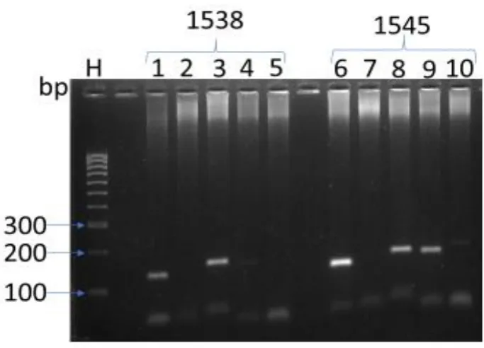
Figure 3.8. Agarose gel electrophoresis of RT-PCR products of tissue samples 1538 & 1545
for Gapdh, Neil1, Neil3, Ercc1 and Nthl1. ... 99
Figure 3.9. Agarose gel electrophoresis of RT-PCR products of tissue samples 1604T & 1610 for Gapdh, Neil1, Neil3, Ercc1 and Nthl1. ... 100
Figure 3.10. Agarose gel electrophoresis of RT-PCR products of tissue samples 1573 & 1573T for Gapdh,Neil1, Neil3,Ercc1 and Nthl1. ... 101
Figure 3.11. Agarose gel electrophoresis of RT-PCR products of tissue samples 1581 and 1620 for Gapdh, Neil1, Neil3, Ercc1 and Nthl1. ... 102
Figure 3.12. Agarose gel electrophoresis of RT-PCR products of tissue samples 1580, 1581T & 1597 for Gapdh, Neil1, Neil3, Ercc1 and Nthl1. ... 103
Figure 3.13. Agarose gel electrophoresis of RT-PCR products of tissue samples 1626, 1627 & 1630 for Gapdh, Neil1, Neil3, Ercc1 and Nthl1. ... 104
Figure 3.14. Analysis of Gapdh expression in tissue sample 10T. ... 105
Figure 3.15. Analysis of Neil3 expression in tissue sample 10T. ... 106
Figure 3.16. Analysis of Ercc1 expression in tissue sample 13N. ... 107
Figure 3.17. Analysis of Mlh1 expression in tissue sample 34T. ... 108
Figure 3.18 Gene expression levels of six DNA repair genes in colon tumour tissue compared to matched normal tissue ... 110
Figure 3.19 Gene expression levels of three DNA repair genes in colon tumour tissue compared to matched normal tissue ... 112
Figure 3.20. Gene expression levels of four DNA repair genes in biobank colon tumour tissue compared to normal colon tissue samples ... 114
Figure 3.21. Gene expression levels of four DNA repair genes in biobank normal colon tissue samples. ... 115
Figure 3.22. Gene expression levels of four DNA repair genes in biobank colon tumour tissue samples. ... 116
Figure 3.23. Gene expression levels of Neil1 in sixteen colon tissue samples... 117
Figure 3.24. Gene expression levels of Neil3 in sixteen colon tissue samples... 118
Figure 3.25. Gene expression levels of Ercc1 in sixteen colon tissue samples. ... 119
Figure 3.26. Gene expression levels of Nthl1 in sixteen colon tissue samples. ... 120
Figure 3.27. Growth response of DAOY normal and cisplatin - resistant cell lines to cisplatin. ... 123
vii
Figure 3.29. Growth response of DAOY normal and cisplatin - resistant cell lines to tert -butyl hydroperoxide. ... 124 Figure 3.30. Agarose gel electrophoresis of RNA extracted from DAOY medulloblastoma cells (Normal & cisplatin - resistant). ... 125 Figure 3.31. Agarose gel electrophoresis of RT-PCR products for Gapdh, Neil3 and Ercc1
from DAOY normal and cisplatin-resistant cells. ... 126 Figure 3.32. Increased gene expression levels of Neil1, Neil3 and Ercc1 in cisplatin resistant DAOY cell lines. (A) and (B) are representations of the same data, with outlyers removed in (B). ... 127 Figure 3.33. Western blot analysis of NEIL3 and β-actin in DAOY normal (DAOY N) and cisplatin - resistant (DAOY R) cell lines. ... 129 Figure 3.34. Western blot analysis of ERCC1 in DAOY normal and cisplatin - resistant (DAOY R) cell lines. ... 129 Figure 3.35. Agarose gel electrophoresis of RNA extracted from Mero25 cells. ... 130 Figure 3.36. RT-PCR of Gapdh, Neil3, and Ercc1 from Mero25 mesothelioma cells. .... 131 Figure 3.37. RT-PCR of putative cancer stem cells from Mero25 mesothelioma cells. ... 131 Figure 3.38. Flow cytometry analysis of (A) Mero25 and (B) Mero25 - derived cancer stem cells. ... 132 Figure 3.39. Agarose gel electrophoresis of RNA extracted from human ES cells. ... 133 Figure 3.40. Agarose gel electrophoresis of RT-PCR products of Gapdh, Neil3 and Ercc1
viii LIST OF TABLES
Table 1.1. Molecular Subgroups of Medolloblastoma. ... 28
Table 1.2. A synopsis of the key genomic and clinical features of the medulloblastoma molecular subgroups. ... 34
Table 1.3. Medulloblastoma patient risk stratification using molecular and survival outcome as criteria... 36
Table 1.4. Presentation of the genomic studies of self-renewal and pluripotency characteristics of embryonic stem cells. ... 61
Table 2.1. Agarose Gel Electrophoresis Buffers ... 73
Table 2.2. Reagents used in Protein Analysis. ... 74
Table 2.3. qPCR Gene Specific Primers... 78
Table 2.4. Composition of the PCR reaction mixture. ... 80
Table 2.5. PCR Reaction Conditions. ... 80
Table 2.6. One Taq PCR reaction mixture. ... 81
Table 2.7. PCR Reaction conditions for OneTaq Hot Start Reactions. ... 81
Table 2.8. Loading of the 10-fold serially diluted cDNA in triplicate in a 96-well plate. .. 82
Table 2.9. Sample Loading Pattern for qPCR in a 96-well plate. ... 83
Table 2.10. qPCR conditions ... 83
Table 2.11. Composition of SDS-polyacrylamide gels. ... 88
Table 3.1 Nomenclature of matched tumour and normal colon samples ... 93
Table 3.2 Description of the sixteen biobank colon tissue samples ... 94
Table 3.3. Summary of the expression patterns of selected DNA repair genes in colon tumours versus matched normal colon tissue. ... 113
Table 3.4. Summary of the expression patterns of four selected DNA repair genes in colon tumours versus matched normal colon tissue (Biobank samples). ... 114
Table 3.5 Summary of the expression patterns of DNA repair genes for the sixteen colon tissue samples obtained from the biobank. Figures represent fold difference to the expression of Gapdh. ... 121
ix ABSTRACT
DNA repair plays a critical role in maintaining the integrity of the genome and the dysregulation of key DNA repair genes has been implicated in the development, progression and chemotherapeutic resistance of different cancer types. Consequently, many studies have made attempts to identify and quantify the expression of various DNA repair genes and their products in different cancers. It is on a similar note that this research was conceived, to evaluate the expression patterns of the base excision repair genes Neil1, Neil2, Neil3, Ogg1,
and Nthl1, the nucleotide excision repair gene Ercc1 and the mismatch repair gene Mlh1 in
colorectal cancer (CRC) tumours and matched normal colon tissue. The project was then extended to analyse a further sixteen colon samples that focused on Neil3, Nthl1 and Ercc1, that encode DNA repair proteins that have been implicated in chemotherapy resitance mechanisms. To learn more about mechanisms of genotoxic agent resistance, Neil3 and
Ercc1 were analysed at the transcriptome and proteome level in DAOY medulloblastoma
cells and cisplatin – resistant DAOY cells. Additionally, attempts were made to generate mesothelioma-derived cancer stem cells and preliminary gene expression analyses were undertaken on human embryonic stem cells. Thus, RNA was extracted, complementary DNA synthesized, and RT-PCR performed. Gene expression levels were determined by quantitative PCR using the Sybr green method and analysed using the comparative Ct method and glyceraldehyde 3-phosphate dehydrogenase (Gapdh) as the standard. Of the matched samples investigated, 75% showed increased expression of one or more of the DNA repair genes analysed, however, there was no clear pattern of expression and a wide range of expression levels observed for individual genes in both normal and tumour tissues. For example, the gene encoding the DNA glycosylase Nthl1 was the most frequently highly expressed in both normal and tumour samples with about 75% showing high expression of
the Nthl1 gene with expression levels ranging from 3.3 to over 1400-fold higher than Gapdh.
DAOY cells were grown in cisplatin and gene expression of Neil3 and Ercc1 analysed in the resulting cisplatin - resistant cells. Results indicated that the expression of both these genes may be increased in the resistant cell line and that NEIL3 protein was also increased. Cancer stem cells were derived from parental mesothelioma cells but were still too small a fraction of the cell population to be analysed by these methods. The expression of Gapdh,
Neil3 and Ercc1 was determined in a series of preliminary experiments on human embryonic
x ABBREVIATIONS
8-oxoG: 8-oxoguanine
AP: apurinic/apyrimidinic APC: adenomatous polyposis coli APE1: AP-endonuclease-1
BER: base excision repair
BLAST: basic local alignment search tool bp: base pair
CAK: CDK-activating kinase cDNA: complementary DNA CDK: cyclin dependent kinase CETN2: centrin 2
CIMP: CpG island methylator phenotype CRC: colorectal cancer
CSA & B: Cockayne syndrome complementation group A and B CSC: Cancer stem cells
Ct: threshold cycle
DDR: DNA damage response DNA: deoxyribonucleic acid DSB: double-strand break
EDTA: ethylenediaminetetraacetic acid EGFR: epidermal growth factor receptor EPCAM: epithelial cell adhesion molecule
ERCC1: excision repair cross-complementation group 1
ES: embryonic stem FBS: foetal bovine serum
GAPDH: glyceraldehyde-3-phosphate dehydrogenase GG-NER: global genome nucleotide excision repair hMLH1: human MutL homolog
hNEIL3: human NEIL3
HNPCC: hereditary nonpolyposis colorectal cancer ICL: interstrand crosslink
xi MLH1: MutL homolog 1
MMR: mismatch repair
MSI: microsatellite instability
MSI-H: high-level microsatellite instability
MTT: 3-(4,5-dimethylthiazol-2-yl)-2,5-diphenyltetrazolium bromide MUTYH: human mutY DNA glycosylase
NCBI: National Center for Biotechnology Information, Nei: endonuclease VIII
NEIL1/2/3: endonuclease VIII-like 1/2/3 NER: nucleotide excision repair NK: natural killer
NTHL1: endonuclease III homolog NUDT1: nudix hydroxylase 1
OGG1: 8-oxoguanine DNA glycosylase PARP-1: poly (ADP-ribose)polymerase 1 PCR: polymerase chain reaction PD-1: programmed cell death 1 PD-L1: programmed cell death ligand 1 PMS2: postmeiotic segregation increase 2 PNK: polynucleotide kinase
POL: polymerase
Polβ: DNA polymerase β Polδ: DNA polymerase δ Polε: DNA polymerase ε
PUA: polyunsaturated aldehyde, qPCR: quantitative PCR
RNA: ribonucleic acid RNAPII: RNA polymerase II ROS: reactive oxygen species RPA: replication protein A RT-PCR: reverse-transcription PCR siRNA: small interfering RNA SSB: single-strand break
xii
TC-NER: transcription-coupled nucleotide excision repair VEGF: vascular endothelial growth factor
UV: ultraviolet
USP7: ubiquitin specific-processing protease 7 UVSSA: UV-stimulated scaffold protein A, XP: xeroderma pigmentosum
XRCC1: X-ray repair cross complementing protein 1
xiii ACKNOWLEDGEMENTS
To God be the glory for the successful completion of the PhD research. He, being my Alpha and Omega, provided everything I required to embark on the programme, uphold me till the end and granted me special favour to successfully complete it.
I would like to express my deep and sincere gratitude to my supervisor, Dr Rhoderick Elder, for his continuous and unwavering guidance, encouragement and support during the project. He believed in my abilities and encouraged me to use all the available time, facilities and energy at my disposal to conduct the research successful. Other members of the Cockroft Building, School of Environment & Life Sciences were of tremendous assistance to me in one way or another.
My colleagues at the University of Salford stood by me and did not allow me to be discouraged at any stage of the programme. Pastor Toyin Oludipe and his amiable wife (Dr. (Mrs) Dorcas Oludipe) were pillars of support for me throughout the programme. My Manchester family members provided the enabling friendly environment that I much needed during the programme.
The following family friends and brethren played important roles while I was looking for opportunities to travel abroad for the programme and stood by me and my family throughout my four year sojourn in Manchester; Dr. and Mrs J.S. Ashidi, Dr. and Mrs J.A. Fayemi, Dr. and Mrs Niyi Adebanjo, Prof. (Mrs) H.T. Benedict, Prof. Joke Jibowo, Engineer Wale Akindehinde.
My academic mentors laid the solid foundation which God enabled me to build upon with the completion of this PhD research. Amongst these wonderful mentors are Prof. O.A Daini, Prof. G. A. Adenuga. Prof. O.O. Adebawo, Prof. Olowookere, Prof. O.O. Osilesi, Prof. (Mrs) Magbageola (UNILAG) and Dr. O.O. Osuntoki (UNILAG)
xiv
Without the strong support and encouragement of my family, I would not have embarked on this research at all. My siblings and cousins: Mrs Modupe Abdul- Lawal and Children, Barrister and Mrs Fatai Olusegun Lawal, Mr. Sola Oyenuga, Mr. and Mrs Wale Adeniyi, Mr. and Mrs Seun Adeniyi and Mrs Kehinde Sofoyeke were very supportive in different ways throughout the programmes. Only God can reward your kindness.
1 1.1. General introduction
In a multicellular organism, the constituent cells undergo cell division as a reproductive mechanism, resulting in the formation of well organised and collaborative entities known as tissues. These cells are subject to a strictly regulated form of collaboration, characterized by selflessness and apparently devoid of competition. Hence, at any point in time, each cell is either, resting, growing, differentiating, dividing, or dying, as needed; and for the benefit of the other cells that made up the organism. Thus, a form of social control network oversees cellular behaviours, including cell-to-cell communication, such as relay, receipt and interpretation of sets of intracellular, as well as extracellular signals in an elaborate manner (Tlsty and Coussens, 2006). Disregard to the sense of community or any attempt to evade the cell cycle system that controls the timing of cell division by a cell, or group of cells, could pose serious problems for the organism. Additionally, molecular disturbances such as genetic mutations, may selectively confer undue advantage on a given cell or group of cells that may result in the cells growing more rapidly, differentiating and dividing more rapidly and evading death signals (Hanahan & Weinberg, 2000). Consequently, the selectively advantaged cells may become the progenitor of a vigorously growing mutant clone, initiating selfishness and encouraging competition. Through repeated mutations, the mutant clones could engage in serious competition, and natural selection could usher in dangerous cell population, and subsequent tumour development. This is known as the mutator phenotype and is probably the main theory of cancer development (Loeb, 2001).
2
specialized competitive advantage, resulting in the successive and progressive change of normal human cells into cancer cells (Loeb, 2001). The heterogeneous nature of cancer accounts for the prevalence of many different types, each displaying different combinations of characteristic cellular and genetic changes. Even within a single type of cancer, heterogeneity and tumour subsets that are uniquely defined can be identified (De Sousa et al., 2013).
The increasing incidence of cancers is alarming, and cancer has become one of the major factors responsible for disease-related deaths globally (Kanavos, 2006). This high death rate could be attributed to the inherent abilities of cancer cells to resist chemotherapy, metastatic ability and high degree of immortality with concomitant recurrence capacity. Current treatments include conventional therapies such as surgery, radiotherapy and chemotherapy (Banerjee et al., 2017; Huang et al., 2017). In the past decade, immuno-gene therapy has been introduced as the fourth treatment modality, due to its robustness and promises (Parney & Chang, 2003). Unfortunately, irrespective of the availability of these variety of options for the treatment of cancer, it is still daunting to define an effective treatment regimen to cure patients especially those whose cancers have metastized to distant organs. This can be linked to resistance to therapy and relapse associated with most cancers (Zahreddine & Borden, 2013).
3
that render cells hyperresponsive to growth factor ligands and structurally alter receptor molecules that facilitate ligand-independent cell division (Cheng et al., 2008).
In the last decades, evaluation of several human tumours has implicated somatic and germline mutations as additional downstream pathways by which cancer cells sustain immortality (Stratton et al., 2009; Davies and Samuels, 2010). To contribute to the global quest to conquer cancer, this PhD project sought to investigate the expression patterns of selected DNA repair genes in different cancer types, including colorectal cancer (CRC) and medulloblastoma. However, the thesis begins with a brief review of DNA damage, some of which is pre-mutagenic and therefore thought to be a prerequisite for carcinogenesis. This is followed by a review of DNA repair mechanisms that are found in mammalian cells. Many of the proteins involved in DNA repair are tumour suppressor genes and individuals lacking particular DNA repair functions are often more cancer prone. Next, a synopsis of each of the cancer types employed in this research is presented, followed by an introduction to stem cells. While preliminary experiments presented here were carried out on human embryonic stem (ES) cells, the concept of cancer stem cells is the focus of much research to find more effective treatments (Dawood et al., 2014).
1.2. DNA Damage
4
While the inherent lability of the DNA molecule can be beneficial, such as in the process of natural selection in evolution, it can also be deleterious to the cell and organism, resulting in altered pathology. Thus, deleterious DNA changes can cause genomic instability and challenge the stability and integrity of the organism (Pierce, 2017).
Several endogenous and exogenous agents responsible for insults on DNA have been described. The most common exogenous or environmental DNA-damaging agents are ultraviolet (UV) radiation, chemical agents and chemotherapeutic drugs. Some of these will result in disease conditions such as cancer. For instance, it is well known that skin cancers are the result of exposure to UV light (Seebode et al., 2016) and lung cancer is largely a consequence of cigarette smoke inhalation (Doll and Bradford-Hill, 1950). Related to this, it has also been reported that colon epithelial cells are also prone to exogenous mutagens, many resulting from normal metabolism of ingested material (Greenman et al., 2007). Apart from the afore-mentioned exogenous sources of DNA damage, cellular DNA is also under constant attack from endogenous agents such as reactive oxygen species (ROS) produced as a consequence of normal aerobic metabolism or resulting from inflammatory cytokines that leads to a state of oxidative stress (Federico et al., 2007). However, another source of DNA damage comes from the replicative DNA polymerases. During DNA replication, the enzyme DNA polymerase adds a nucleotide to the strand of the DNA (Pierce, 2017). However, the DNA polymerase can add incorrect nucleotides during DNA replication and although the main replicative DNA polymerases have a 3ʹ - 5ʹ "proofreading" exonunclease activity and can recognize and correct many of these errors, inefficient corrections could lead to mutations that can in turn result in disease conditions such as cancer (Pierce, 2017).
5
including, pancreatic (Vaquero et al., 2004) prostate (Kumar et al., 2008) breast (Hecht et al., 2016) and colon (Acharya et al., 2010) show an increased level of ROS. Besides its role in cancer, ROS has been implicated in several human diseases including the ageing process, cardiovascular disease, diabetes, sterility, autoimmune diseases and neurological diseases (Sedelnikova et al., 2010). The mechanism of action of ROS is by induction of several covalent modifications to DNA including in single base lesions, DNA strand breaks, intra- and inter-strand cross-links.
An example of oxidative DNA damage caused by ROS is 8-oxo-7,8-dihydroguanine (8-oxoG), which although not toxic, is highly mutagenic (Suzuki & Kamiya, 2016). 8-oxoG is known to mismatch with adenine and by so doing results in ‘GC’ to ‘TA’ transversions. Several independent reports have confirmed the involvement of this mis-pairing in somatic mutations in lung, CRC, breast, gastric, and ovarian cancer (Fortini et al., 2003). Besides causing mutation, ROS is involved in activating transcription factors such as NF-kB, activator protein-1 (AP-1) and hypoxia inducible factor-1 (HIF-1α), whose role in cancer cell growth and survival, angiogenesis, invasion, and metastasis have been well established (Gupta et al., 2012).
1.3. DNA Repair Mechanisms
6
base excision repair (BER), (iii) nucleotide excision repair (NER), (iv) Non-homologous end-joining (NHEJ) and, (v) homologous recombination repair (HRR). Recently, Abbotts et al., (2014) reported that loss or truncation of the efficiency of one or more of the DNA repair pathways can accelerate the accumulation of additional mutations by up to 1000-fold (Abbotts et al., 2014). Thus, unrepaired DNA damage is the major source of potentially mutagenic lesions that drive carcinogenesis (Friedberg et al., 2005).
Loss of any one of these pathways can result in serious consequences for the organism. For example, individuals lacking NER are hypersensitive to sunlight and exhibit either accelerated ageing or cancer predisposition (Friedberg et al., 2005). Similarly, several inherited colorectal cancer (CRC) syndromes are associated with genetic defects in both MMR and BER (Weren et al., 2015). On the other hand, high expression of DNA repair proteins in cancer cells can confer resistance to certain chemotherapeutic agents used in cancer treatment, such as the platinum-based compounds and alkylating agents.Thus, it has been reported that the excision repair cross-complementation group 1 (Ercc1) gene is upregulated in colon cancer cells treated with oxaliplatin, and that the small interfering RNA (siRNA) knockdown of Ercc1 in these cells sensitises them to the chemotherapeutic effects of oxaliplatin (Seethram et al., 2010). The BER DNA glycosylase NEIL3 is also highly expressed in most cancer cell lines and metastatic melanoma compared to normal tissue cells, where its expression is generally restricted to rapidly dividing cells in the thymus and testes (Section 1.3.2; Kauffmann et al., 2008; Hildrestrand et al., 2009). As more biochemical information becomes available on the activity of this enzyme (Martin et al.,
2017; Albelazi et al., 2019), it is of increasing interest to determine the levels of expression
of Neil3 in tumour samples and the likely effect this may have on conferring resistance to
genotoxic chemotherapeutic agents.
7 1.3.1.Mismatch Repair
DNA mismatch repair proteins function by recognizing base mismatches and other lesions
(e.g. nucleotide insertion or deletion) that result from DNA polymerase slippage during
DNA replication (Lodish, 2004). As a result, it is sometimes called, post-replication repair and if it fails, the errors induced during DNA replication are fixed in the genome (mutation). As mentioned previously, during DNA replication the enzyme DNA polymerase adds a nucleotide to the growing strand of DNA at the replication fork. MMR is involved in the post-replicative repair of the errors made by DNA polymerases that have escaped proofreading (Umar & Kunkel, 1996). The MMR system will recognise base-base mismatches, insertions or deletions occurring in the double-stranded DNA (Harfe & Jinks-Robertson, 2000). Following this recognition, MMR proteins then initiate the process of resection and specifically degrade the affected region of the newly synthesised strand. A DNA polymerase will then correctly resynthesise the daughter strand of the DNA in a template dependent manner (Lodish, 2004).
Several genes are important in the normal function of MMR including, in human cells: MutS homolog 2 and 6 (Msh2 and Msh6), MutL homolog 1 and 3 (Mlh1 and Mlh3) and post-meiotic segregation increased 1 and 2 (Pms1 and Pms2: Harfe & Jinks-Robertson, 2000). A mutation in any of these genes can result in microsatellite instability (MSI) and predisposes the individual to certain cancers, including colorectal and ovarian cancers (Abbotts et al., 2014).
Thus, germline mutations in Mlh1, Msh2 or Pms2, or even deletions in the epithelial cell adhesion molecule (Epcam) gene that cause allele-specific Msh2 inactivation has been linked to hereditary nonpolyposis colorectal cancers (HNPCC; Lynch syndrome) that results in early onset CRC. It has been reported that inactivating mutations in any of the MMR genes can be found in up to 70% of HNPCC and that, of these, over 90% occur in the hMsh2
or hMlh1 genes and display a high level microsatellite instability (MSI-H) phenotype
(Wheeler et al., 2000).
hyper-8
methylation of the promoter region are at a higher risk (up to 60%) of developing CRC. In order to reverse Mlh1-methylation in colon cancer, the group of Fujita and colleagues (2007) demonstrated that the de-methylating agent 5-aza-2ʹ-deoxycytidine (5-aza-dC) can induce
Mlh1 expression and sensitise cancer cells to 5-fluorouracil (Fujita et al., 2007). Consequently, the expression profile of Mlh1 will be analysed in the course of this project, particularly in solid colon cancer tumour tissues, with the view to ascertaining the expression pattern at the transcriptional level.
1.3.2.Base Excision Repair
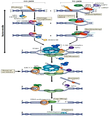
Amongst the DNA repair mechanisms, BER is the most versatile repair mechanism and is involved in repairing the majority of DNA damage arising from both endogenous and exogenous sources (Svilar et al., 2011). These include single-strand breaks (SSB), depurination and deamination, alkylation and oxidation derived base damage. DNA base damage resulting from exposure to environmental factors, ROS as well as alkylation-induced damage especially those from alkylating agents including chemotherapy and radiotherapy are also repaired by BER (Maynard et al., 2009; Whitaker et al., 2017). The broad functionality of BER results mainly from a group of enzymes called DNA glycosylases that recognise and excise a plethora of chemically modified bases from DNA (Mullins et al., 2019). Thus, DNA glycosylases recognize and initiate BER by removing an overlapping subset of damaged bases, leaving an abasic site that is further processed by short-patch BER or long-patch BER that uses different proteins to complete the repair process (Whitaker et al., 2017). Additionally, poly (ADP-ribose) polymerase 1 (PARP-1) and PARP-2 are known to facilitate BER by binding to DNA ends at SSBs and synthesizing poly (ADP-ribose) polymers on acceptor proteins and the DNA itself (Dantzer et al., 1999; Talhaoui et al., 2016). The poly(ADP-ribose) destabilizes the nucleosome structure allowing BER proteins access to the damage site. The role of BER in protecting the colorectal tissue against oxidative DNA damage, caused by high levels of oxygen radicals either generated by bacteria or dietary carcinogens cannot be overemphasized. Steps involved in BER are described in Figure 1.1.
9
[image:25.595.121.461.375.637.2]distincy superfamilies, (i) the uracil DNA glycosylases (UDG), (ii) the helix-hairpin-helix DNA glycosylases (HhH), (iii) the alkylpurine DNA glycosylases (APNG or MPG) and, (iv) the endonuclease VIII – like DNA glycosylases (NEIL). These enzymes can be further divided into two classes, depending on whether they are monofunctional (UDG and APNG/MPG), or bi-functional (HhH and NEIL) (Jacobs & Schär, 2012). The monofunctional DNA glycosylases (UDG and APNG/MPG) have only DNA glycosylase activity, catalysing the breakage of the glycosylic bond between the deoxyribose sugar and the damaged base, while bifunctional DNA glycosylases also have an associated apurinic/apyrimidinic (AP) lyase activity (Jacobs & Schär, 2012). The five bifunctional DNA glycosylases in mammalian cells, 8-oxoguanine DNA glycosylase (Ogg1), endonuclease III homolog (Nthl1), and the three endonuclease VIII paralogs (NEIL1, NEIL2 and NEIL3) all recognise and excise oxidised bases from either double-stranded or single-stranded DNA (Jacobs & Schär, 2012).
Figure 1.1. A current model for BER in mammalian cells. (Elder, unpublished).
The DNA glycosylases NEIL1, NEIL2 and NEIL3 are mammalian homologs of the
Escherichia coli Nei protein and have been shown to excise oxidised purine and pyrimidine
10
Amongst these enzymes, NEIL3 is the largest, uniquely containing a long C-terminal region ending with tandem GRF-zinc finger domains (Albelazi et al., 2019). Both NEIL1 and NEIL3 are reported to be cell cycle regulated, with expression peaking in S-phase and S/G2-phase respectively, while NEIL2 is constitutively expressed throughout the different stages of cell cycle (Neurauter et al., 2012; Hazra & Mitra, 2006; Chakraborty et al., 2015). Thus, while evidence from biochemical studies and knockout mice support the idea of an involvement of NEIL2 in transcription-coupled BER (Chakraborty et al., 2015), recent biochemical evidence reinforces previous reports supporting the involvement of Neil1 and NEIL3 at the replication fork (Albelazi et al., 2019; Neurauter et al., 2012). Furthermore, NEIL3 also shows a unique restricted expression pattern in normal cells, being expressed only in highly dividing cells such as those in the developing brain, the thymus and testes (Hildrestrand et al., 2009; Morland et al., 2002). However, high levels of NEIL3 have been observed in metastatic melanoma (Kauffmann et al., 2008) and in cancer cells generally (Hildrestrand et al., 2009; Duweb, 2015). Recently, it was demonstrated that abnormal expression of the Neil DNA glycosylase genes is associated with somatic mutation in several human cancers (Shinmura et al., 2016).
An ongoing study in our laboratory reported that Neil3 is highly expressed in the CRC cell line HCT116. However, following siRNA treatment, this expression can be substantially reduced. Further knockdown of Neil3 can sensitize tumour cells to oxaliplatin treatment (Taylor et al., 2015). These results reveal a potentially novel activity for Neil3 and indicate that it could be a major resistance mechanism to certain chemotherapeutic DNA damaging agents in solid tumours. Consequently, it is one of the objectives of this thesis, to investigate the expression profiles of Neil3 gene in solid tumours derived from different colorectal cancer patients.
11
sites, again reinforcing the importance of unrepaired oxidative base damage in carcinogenesis and of the DNA glycosylases and BER in maintaining genetic integrity.
1.3.3.Nucleotide Excision Repair.
Mammalian nucleotide excision repair (NER) is a constitutive DNA repair mechanism and its impairment can result in several disease conditions including cancer and premature ageing (Marteijn et al., 2014). It is usually involved in the repair of DNA lesions usually bulky adducts that destroy the normal double-helical conformation of duplex DNA, irrespective of whether the insult is induced by endogenous or exogenous agents (Friedberg
et al., 2005). Bulky adducts are products of different DNA damaging agents including UV
12
13
Figure 1.2. A representation of nucleotide excision repair (de Laat et al., 1999)
14
in the displacement of the stalled RNA polymerase via a transient interaction (Kamiuchi et al., 2002). Upon stalling at a lesion, it has been documented that the affinity of CSB for RNA polymerase II increases, leading to the formation of CSA-CSB complex. The formed complex is then involved in the reversal of the translocation (backtracking) of RNA polymerase II, thus rendering the DNA lesion accessible for repair. After this step, the GG-NER and the TC-GG-NER converge to continue the repair process of the damaged DNA (Figure 1.2; de Laat et al., 1999).
The dissociation of RAD23B in the GG-NER mechanism and the backtracking step in the TC-NER mechanism will trigger the deployment of the transcription factor II H (TFIIH) complex, that is known to have helicase activities. The presence of two TFIIH basal helicase subunits including XPB and XPD results in the opening of the DNA duplex site where damage has occurred. However, if XPD fails to detect any damage, the DNA repair process can be aborted. This is because the major role of XPD is for damage verification. If there is successful verification of damage by XPD, the helicase activities will proceed successfully. This process will result in a bubble formation, allowing the engagement of XPA and RPA (replication protein A), as well as the assembly of the complexes necessary to initiate incision (Compe & Egly, 2012).
The XPF-ERCC1 complex is a structure activated endonuclease that incises the DNA strand on the 5ʹ side of the helix distorting lesion. Similarly, XPG performs a similar function 3ʹ to the lesion (Figure 1.2). This step leaves a single strand gap of 22-30 nucleotides and it has been suggested that this step is necessary in triggering, a DNA damage signalling reaction (Marteijn et al., 2014).
In cancer, several studies have shown that the ERCC1-XPF complex is responsible for conferring resistance to platinum based drugs (Seetharam et al., 2010; Baba et al., 2012) and in particular it has been demonstrated that the over expression of the Ercc1 gene is associated with oxaliplatin resistance in metastatic colon cancer (Choueiri et al., 2015). Thus, it has been shown that its knock down by siRNA-mediated gene silencing can sensitize the CRC cell lines to oxaliplatin, thereby implicating the role of Ercc1 in conferring resistance to this crosslinking agent (Seetharam et al., 2010).
It is based on the above premise that this PhD thesis seeks to profile the expression of Ercc1
15 1.4. Colorectal Cancer
Colorectal cancer (CRC) or colorectal adenocarcinoma is one of the most predominant malignant neoplasms and it contributes significantly to cancer-related deaths worldwide (Ferlay et al., 2015). In 2014, the number of newly diagnosed patients in the United States alone reached nearly 140,000, ranking this disease in second place as a cause of death due to cancer in adults (Siegel et al., 2014). Worldwide, it occupies third place and second place respectively, as the leading cause of deaths relating to cancer in men and women (Ferlay et al., 2015). Although 55% of the cases are found across the industrialized world, Australia and New Zealand record the highest rates of CRC with Africa showing the lowest rates (Ries
et al., 2017). The discrepancy in the documented incidences and death rate can be attributed
to poor diagnosis or improper data registry (Ferlay et al., 2015). Diagnosis of CRC at the stage when it has not metastized to distant organ usually signals a good prognosis and about 50% of patients have a 5-year survival. However, patients at the metastatic stage have only 12% survival rate at 5 years (Ferlay et al., 2015). CRC may be asymptomatic for several years and the American Cancer Society has recently recommended screening from 45 years of age (Mannucci et al., 2019). Detecting blood in the stool and unexplained weight loss have previously been reported to be the only symptoms warranting further exploration for polyps and CRC (Adelstein et al., 2011).
While there is no single, distinct cause of CRC, several risk factors leading to its development have been described (Kupfer & Ellis, 2017). This neoplasm is sporadic with the majority caused by diet, lifestyle, age and only about 15% to 35% linked to hereditary factors (Burt, 2007; Mishra & Hall, 2012). Evidence shows that patients with history of genetic instability have a greater chance of getting the disease and such patients are especially likely to show germline mutations relative to the patients that have spontaneous CRC (Gallagher et al., 2010).
Based on their origin, CRC has been traditionally categorized into two biological subgroups, namely a minority (15%) that show microsatellite instability (MSI), which is primarily predominant at the right colon and known to be frequently linked to the CpG island methylator phenotype (CIMP; Popat et al., 2005). Additionally, predictive and prognostic information indicate that they exhibit hyper-mutation including mutation at both the KRAS
16
(Marisa et al., 2013; Roepman et al., 2014). It is worthwhile to mention that the above classifications are largely based on gene expression profiling; thus, might solely focus on single mutations or epigenetic alterations. Therefore, with the advances in genomic technology, scientists are now focussing on whole exome and genome sequencing to cover a wide range of genome analysis. This technique can sequence the entire human coding DNA looking at both the coding and non-coding regions. This will help to provide additional information on all the alterations that might have occurred at a single nucleotide including copy number and structural variants. Recently, screening with high-throughput gene expression profiling including next generation sequencing or expression arrays (microarray) has demonstrated that some CRC types overlap with the above-mentioned groups and cannot be established only by single mutations or epigenetic profiling (Sinicrope et al., 2016). The development of CRC is seen to be a multistep process that involves the development of benign polyps that have the capability to evolve into carcinoma in situ by the accumulation of somatic mutations (Shussman and Wexner, 2014). Factors such as age, diet, lifestyle, and family history are associated with the development of polyps and CRC (Rasool et al., 2013). Even though there is a good correlation between polyps and CRC development, three different subtypes of polyps have been described, distinguished on the basis of histology, such as tubular/villous adenoma, hyperplastic polyps and sessile/traditional serrated adenomas (Kalimuthu et al., 2016). Similarly, there is a suggested correlation between the risk of cancer development with the number and size of previously developed polyps (Shussman & Wexner, 2014). This means that multiple colonic polyp development with malignant potential will amount to an increased lifetime risk of developing CRC.
1.4.1.Genetic Predisposition to Colorectal Cancer
17
including hyper-mutated, non-mutated and CIMP subtypes (Rodriguez-Salas et al., 2017). The hyper-mutated tumours have been reported to account for up to 16% of all CRC (Dienstmann et al., 2017). While only one-quarter display somatic mismatch repair (MMR) gene and DNA polymerase alterations, three-quarters of them show high-frequency MSI (MSI-H) (Cancer Genome Atlas Network., 2012). Mutations in the germline account for 2 - 5% of CRC and may be as a result of autosomal dominant syndrome (Gatalica et al., 2017). The most prevalent and most studied is termed hereditary non-polyposis colorectal cancer (HNPCC) or Lynch syndrome (Lynch et al., 2015). When compared to the age of the patients, evidence shows that most patients with sporadic CRC are older patients, while patients with Lynch syndrome and other genetic predispositions are usually younger (Mauri
et al., 2019). This can be due to loss of Mlh1 expression, the increase of which is directly
proportional with age (Kakar et al., 2003). Moreover, sporadic colorectal tumours are also characterized by high BRAF (V600E) mutation with loss of MLH1 and PMS2 proteins (Kakar et al., 2003).
Furthermore, microsatellite instability (MSI) positive CRC can be found at the proximal bowel exhibiting poor differentiation. This can be due to the presence of dense lymphocytic infiltration, suggesting strong anti-tumoural immune responses. Moreover, this is an indicative of positive prognosis (Nosho et al., 2010). On the other hand, the non-mutated subtype accounts for 84% of CRC; with the majority characterised by many somatic copy number changes and aneuploidy; exhibiting genetic alterations at the KRAS and PIK3CA
genes. This subtype is also known to possess loss of heterozygosity of several tumour-suppressor genes including APC and TP53 (Cancer Genome Atlas Network, 2012).
18
Additionally, two recessive cancer - predisposing genes MUTYH and Nthl1 that both code for proteins that act in base excision repair (BER) have been confirmed to be associated with increased polyposis and adenomatous polyposis with a high risk of CRC respectively (Weren et al., 2018). Indeed, carriers of biallelic mutations in Nthl1 have been shown to have an increaseed risk of other cancer types, including breast cancer (Kuiper & Hoogerbrugge, 2015).
1.4.2.Colorectal Cancer Treatment and Oxaliplatin
Systemic chemotherapy has been used to control cancer and alleviate its related symptoms at the metastatic stage. For metastatic CRC, it has been reported that a combination of the antimetabolite fluoropyrimidines (intravenous 5-fluorouracil and oral capecitabine), the DNA topoisomerase I inhibitor, irinotecan and the genotoxic platinum – based agent oxaliplatin (FOLFOXIRI) showed an improved survival of these patients (Leal, 2017). However, achieving complete remission at this stage is still daunting, as resistance accounting for nearly 30-50% is still a major obstacle (O’Connell et al., 2008).
Oxaliplatin (Figure 1.3B) is a third-generation platinum – based alkylating agent forming primarily N-alkylation products at the N7 of guanine. Similar to cisplatin (cis -diamminedichloroplatinum (II); Figure 1.3A), this leads to both intra- and inter-strand cross links (ICLs) in the DNA molecule, effectively disrupting DNA replication and transcription and leading to cell death. For cisplatin, the high extracellular chloride ion concentration maintains the molecule in an inactive state and only when it is transported inside the cell, where the chloride ion concentration is 5 to 30 times lower, are the chloride groups displaced by water molecules to create an effective alkylating agent. The most prevalent products are 1,2-d(GpG) intrastrand crosslinks that make up 90% of the DNA adducts, 1,2-d(ApG) intrastrand crosslinks and ICLs. In the cell, the activated cisplatin has a half-life of around two hours, while the protecting chelating ligands of oxaliplatin give this agent a much longer
A B
19
half-life, with solutions of oxaliplatin and the related carboplatin being stable in water for a period of weeks to months (Johnstone et al., 2016).
The repair of cisplatin – induced intrastrand crosslinks is thought to be completed by NER and the resistance to DNA repair, and therefore the cytotoxic effectiveness of this agent, is due to the bending of the DNA at the adduct and the resulting binding of the DNA by high-mobility group box proteins that have a great affinity for cisplatin modified DNA and thus shield the lesion from the NER proteins (Awuah et al., 2017). In CRC, increased expression
of Ercc1, which encodes one half of the ERCC1/XPF lesion specific endonuclease, has been
correlated with oxaliplatin resistance (Galluzzi et al., 2012). However, mouse cells lacking the BER DNA glycosylase NEIL3 also showed resistance to cisplatin (Rolseth et al., 2013). Further evidence of a role for DNA glycosylases in ICL repair came from biochemical studies by Couvé and colleagues (2009), which indicated that NEIL1 could excise psoralen – induced ICLs from DNA and more recent work has shown that both NEIL1 and NEIL3 can resolve psoralen - induced ICLs in three- and four-stranded DNA structures (Martin et al., 2017) and also that NEIL3 can release ICLs at DNA replication forks (Semlow et al., 2016). Therefore, as NEIL3 has been reported to be highly expressed in cancer cells and metastatic tumours (Kauffmann et al., 2008; Hildrestrand et al., 2009) and recent work from my laboratory at the University of Salford had also indicated a role of NEIL3 in the resistance to oxaliplatin (Taylor et al., 2015), it was important to determine the levels of NEIL3 in the CRC tissues and cell lines analysed in this work.
1.4.3.Colorectal Cancer Treatment and Development of Targeted Therapy
20
Due to the shortcoming of conventional therapies, scientists are focusing on targeting metastatic CRC at the molecular level. Cancers including CRC can be characterized based on the biomarkers they present. This means that profiling tumours can be a watershed event in optimizing therapy to suit an individual patient’s need. The key is in identifying reliable biomarkers by performing baseline assessment of tumour gene expression and/or immune profile for the best chance of therapeutic success. With the recent innovations in molecular testing techniques that allow for high throughput genomic analysis, patients can be selected for targeted therapy based on their tumour biology and dispositions (Ohhara et al., 2016). So far, techniques involving next generation sequencing and even a much newer technology for detecting a mutation in circulating tumour DNA have been described (Perakis et al., 2017). Since these technologies can detect somatic modifications and mutations such as insertions/deletions, copy number variation and rearrangement and base substitutions, molecular intervention strategies can be tailored to target the key molecules involved in CRC proliferation, envasion and metastasis (Diaz & Bardelli, 2014; Sinicrope et al., 2016). By implication, targeted therapy directed against the wrong mutation or given to a patient with tumour of unrelated characteristics will not benefit the patient.
Several biomarkers for CRC including KRAS, NRAS, BRAF mutation, DNA mismatch repair (MMR), MSI, and CpG island hypermethylation have been evaluated (Sinicrope et al., 2016). Consequent upon reports emanating from such findings, treatment modalities such as the use of small molecule inhibitors, antibodies (Baudino, 2015), immunotherapy (Lynch & Murphy, 2016) and RNA-based technologies such as siRNA or small hairpin RNA (Seetharam et al., 2010) have been evaluated. Although some of these techniques are still at the preclinical stage, the majority have made their way to the clinic, recording some promising outcomes.
Recent reports show that better patient survival can be achieved by recombinant humanized monoclonal IgG antibody targeting either the EGFR or VEGF pathway. The result of the clinical trial using the above antibodies as summarized by Ohhara et al., (2016) indicated that one anti VEGF antibody; bevacizumab, and two EGFR targeting antibodies; cetuximab and panitumumab resulted in significant anti-CRC metastatic control in combination with cytotoxic therapy (Ohhara et al., 2016). The clinical trial report showed that they can be used in first line, second line or even in salvage settings to enhance overall patient survival beyond 40 months from the period of initial diagnosis (Van Cutsem et al., 2011; Heinemann
21
state can benefit from it due to inherent toxicity associated with chemotherapy. Additionally, patients that have KRAS/NRAS mutations are not subject to this therapy as this mutation lies downstream of EGFR. In such instance, mutation at the KRAS/NRAS triggers the transcription of the ligand for EGFR; transforming growth factor-alpha (TGF-α). This will in turn create an autocrine signaling loop that contributes to tumoural resistance to anti-EGFR monoclonal antibodies (mAbs) including cetuximab and panitumuab (Lièvre et al., 2006).
Immunotherapy for the treatment of cancer has come a long way and has been accepted as the fourth treatment modality besides surgery, chemotherapy and radiotherapy. This treatment modality is currently approved for many solid tumours due to its efficacy in controlling cancer with minimal overall toxicity. The functions of immune cells in the development and progression of tumour has been well documented (Hanahan & Weinberg, 2011) indicating that several cancer types show phenotypic immune cell characteristics. It has been demonstrated that CRC with MSI can be characterized by the presence of a particular immunogenic phenotype. It was further established that this subtype has increasing lymphocytic infiltration which can possibly be due to the creation of tumour-specific neo-antigens during accumulation of mutations (Schwitalle et al., 2008). When primary tumour tissues from patients were further characterized an increasing presence of Th1 transcription factors was recorded. This is translated to the presence of activated cytotoxic CD8T cells, Th1 cells producing high levels of IFN-y as well as T-BET expressing T cells (Llosa et al., 2015). The presence of immune cell infiltration in the CRC is indicative of a positive prognosis. However, the immune microenvironment of CRC is composed of immune checkpoints that are cytotoxic to activated T cells. In a similar vein, the presence of apoptotic cell death ligands, such as programmed cell death ligand 1 (PD-L1), programmed cell death 1 (PD-1), T lymphocyte associated antigen 4 (CTLA4), lymphocyte activation gene 3 (LAG3) and indoleamine 2, 3-dioxygenses (IDO) have been shown to be the hallmark characteristic of several cancers including CRC (Llosa et al., 2015).
22
mechanism of tumour regression using this strategy lies in activation of tumour infiltration lymphocyte around the tumour border. Recently, Brahmer and colleagues (2009) showed in their phase II clinical trial that, blocking PD1/PD-L1 interaction in CRC patients with MDX-1106 resulted in complete response for a period more than 21 months (Sui et al., 2015). Looking at the individual treatment modalities, one can see their strengths and weaknesses. Since long-term clinical benefit to more patients is the ultimate goal, future cancer therapy is likely to focus on combinatorial approaches involving targeted inhibitors, immunotherapy, chemotherapy, surgery, radiation as well as novel therapies to achieve success.
1.5. Medulloblastoma
Medulloblastoma is a malignant, embryonal, heterogeneous, and highly aggressive tumour of the central nervous system with a preferential manifestation in children and a marked metastatic tendency via the cerebrospinal fluid (CSF) (Louis et al., 2007; Gibson et al., 2010; Robinson et al., 2012; Gajjar and Robinson, 2014). The development of medulloblastoma is mostly sporadic, originating from the interior fossa because of aberrant cerebellar development (Marino, 2005). In very rare cases, medulloblastoma has been reported to be associated with heritable disorders like LiFraumeni, Turcot or Gorlin syndrome (Parsons et al., 2011; Johansson et al., 2016). More than 70% of reported cases of medulloblastoma occur in patients under the age of 15 years, with the incidence peak being 3 to 6 years (Peris-Bonet et al., 2006). However, medulloblastoma is much less frequent in adults, accounting for less than 3% of primary tumours of the central nervous system (Smoll and Drummond, 2012).
23
higher in children that are not more than 9 years of age, indicating a frequency of less than 8 in every million. On the other hand, 6 persons per million was reported in infants, an indication of a reduction in the rate of incidence, whereas, in children whose age range is between 10 to 14 years, the incidence rate was reportedly lowest, represented by 4 persons in every million (Peris-Bonet et al., 2006). In the case of individuals that were above the age of 14 years but not more than 19 years of age, higher incidence rate was reported at an annual frequency of 2.33 persons per million; with incidence rate declining beyond 19 years of age up to the age of 40 years, depicting an alignment with the embryonal origin of medulloblastoma (Giordana et al., 1999). Between 1978 and 1997, the incidence of medulloblastoma was on the rise, with a record 1.3% increase during this period (Peris-Bonet et al., 2006). In parts of North-America, the occurrence of medulloblastoma was reported to be 5.07 per million children aged 0 to 19 years (Kohler et al., 2011). Comparatively, from 1114 diagnosis of brain tumours at the Egyptian Children’s Cancer Hospital from 2007 to 2013, medulloblastoma represented 23.2% of the overall number of diagnosed cases; this shows an agreement with the reported cases of medulloblastoma in North America and Europe (Ezzat et al., 2016).
Children diagnosed with medulloblastoma in Europe, between the years 2000 – 2007, showed 81% survival for 1 year, 63% for 3 years and 56% survival was reported for 5 years. Essentially, worst prognosis was found among infants, where 5-year survival was reported to be 33%, but for children aged 1 to 4 years, a relatively improved survival of 47%; whereas, marked prognosis was reported for children aged 5 to 14 years of age, at survival rate of 67% (Kohler et al., 2011). From year 1999 and 2007, survival of patients with medulloblastoma remained stable (Gatta et al., 2009), while in the nineties, survival significantly improved and the possibility of the patients dying dropped by 30% (Gatta et al., 2014).
24
survival rate at 64% was reported, while it was relatively lowest in countries within Eastern Europe at 53% (Gatta et al., 2014).
25
during pregnancy (Pogoda et al., 2009). The researchers observed that cured meats, eggs/dairy, and oil products were the main foods regularly linked with increased risk; whereas, foods that are mostly associated with lowered risk were fresh fish, yellow-orange vegetables, and grains (Pogoda et al., 2009).
26
1.5.1.Histology and Pathobiology of Medulloblastoma
To date, the actual cellular origin of medulloblastoma remains subject to debate (Gibson et al., 2010). Several reports have shown that the origin of medulloblastoma could be from two different embryonal cell groups: cells from the ventricular zone, which differentiate into various cells of the cerebellum; and cells from the external germinal layer (EGL), which differentiate into cerebella granule cells (Kuzan-Fischer et al., 2018). These cell groups are related to different molecular subtypes of medulloblastoma and it has been established that ventricular zone cells give rise to the wingless (WNT) subtype, whereas sonic hedgehog (SHH) medulloblastoma is produced from the EGL cells (Fan and Eberhart, 2008; Rusert et al., 2014; Kuzan-Fischer et al., 2018). In the subsequent sections, these molecular subtypes will be reviewed in a more elaborate manner.
The 2007 WHO classification of tumours of the central nervous system recognises five major variants of medulloblastoma, including the classic medulloblastoma, desmoplastic/nodular, medulloblastoma with extensive nodularity (MBEN), anaplastic and large cell (Giangaspero et al., 2007; Massimino et al., 2016; Kuzan-Fischer et al., 2018). From these five forms, large-cell medulloblastoma as well as anaplastic variant of the disease have significant overlapping characteristics; consequently, several studies have attempted to group them into large cell/anaplastic (LC/A) medulloblastoma (Gilbertson and Ellison, 2008). The incidence rate of the combined LC/A type of medulloblastoma has been reported to oscillate between 10% and 22%; whereas, anaplastic medulloblastoma is established only in event of severe and diffuse anaplasia, comprising up to 50% of reported cases (Giangaspero et al., 2007). Nodular/desmoplastic medulloblastoma constitute about 7%, while MBEN comprises up to 3% of the entire reported cases of the disease, while classic subtype of medulloblastoma make up the remaining (Gilbertson and Ellison, 2008; Massimino et al., 2016).
27
the production of a dense intercellular reticulin-positive network of fibres (McManamy et al., 2007).
Medulloblastoma with extensive nodularity (MBEN) is known to be predominant in infants and it has characteristic better prognosis (Eberhart et al., 2002a); and it is distinct from the closely-related nodular/desmoplastic subtype by possessing an expansive lobular conformation because of the unusual elongation of the reticulin-free zones and enriched with neuropil-like tissue (McManamy et al., 2003). Such zones are filled with small cells that have characteristic spherical nuclei that bear close semblance to the cells of a central neurocytoma and exhibit a streaming pattern; coupled with the marked reduction of the internodular component in some areas (McManamy et al., 2007). Structurally, the large cell medulloblastoma is made up of monomorphic cells with large, round, vesicular nuclei, prominent nucleoli and variably abundant eosinophilic cytoplasm. Groups of these large cells tend to combine with morphologically different cells with characteristic nuclear polymorphism and nuclear conformation; this morphological variant has been termed anaplastic (Massimino et al., 2016). Large cell and anaplastic histological forms of medulloblastoma have been reported to show considerable cytological overlap and many studies have attempted to describe the histological alternation between non-anaplastic to anaplastic subtypes over time. However, some studies have reported alternated transition intra-tumour, as deduced from the presence of varying degrees of cytological atypia or anaplasia in any given tumour (Eberhart et al., 2002a; Massimino et al., 2016).
Clinically, different reports have significantly shown good prognosis for the nodular/desmoplastic medulloblastoma at least in certain age groups as well as risk groups, particularly in children at younger age (Rutkowski et al., 2005; McManamy et al., 2007). Additionally, classic form of medulloblastoma has been reported to show significantly better prognostic outcome relative to the LC/A histological variant (Massimino et al., 2013).
1.5.2.Molecular Subgroups of Medulloblastoma
28
(sonic hedgehog), Group 3 medulloblastoma, and Group 4 medulloblastoma (Taylor et al., 2012), as represented in Table 1.1. These four molecular subgroups of medulloblastoma were isolated using series of genomics and molecular studies They are known and identified by properly defined genetic, molecular, clinical, histopathological, and prognostic features (Northcott et al., 2012; Ramaswamy et al., 2014; Schneider et al., 2105; Ramaswamy and Taylor, 2017; Kuzan-Fischer et al., 2018). From recent research findings derived from genetic, transcriptional and epigenetic data, it has been suggested the need to further categorise medulloblastoma into subtypes based on molecularly characteristics and such sub-classification is most likely to have positive impact on patient stratification in future clinical trials (Northcott et al., 2017; Schwalbe et al., 2017; Cavalli et al., 2017).
Table 1.1. Molecular Subgroups of Medolloblastoma.
WNT SHH GROUP 3 GROUP 4
Age Group Children & Adults
Infants, Children & Adults
Infants & Children
Infants, Children & Adults Metastasis Rarely
M+
Uncommonly M+ Very frequently M+
Frequently M+
Prognosis Very good
Infants good, others intermediate
Poor Intermediate
Genetics CTNNB 1 mutation
PTCH1/EMO/SUFU mutation / GL12 amplification /
29 1.5.2.1.WNT (Wingless) Medulloblastoma
This is the least prevalent medulloblastoma molecular subgroup, constituting approximately 11% of the total reported incidences of medulloblastoma (Kool et al., 2012). Though, wingless form of medulloblastoma has been reported to occur at all ages, children are predominantly affected, with the highest frequency of occurrence recorded in children of 10 to 12 years (Taylor et al., 2012). Unlike other molecular subgroups of medulloblastoma, WNT forms have been reported to have a female preponderance, based on gender ratio. Typically, WNT tumours are known to occur in the mid region of the brain, affecting the IV ventricle and extending to the brain stem (Gajjar and Robinson, 2014).


![Fındık Kurdu [Balaninus(= Curculio) Nucum L Colertera: Curculionidae)] ’na Karşı Organik Kökenli Preparatlarla
Mücadele İmkanlarının Araştırılması](data:image/gif;base64,R0lGODlhAQABAIAAAP///wAAACH5BAEAAAAALAAAAAABAAEAAAICRAEAOw==)